Abstract
There are two major serotypes of vesicular stomatitis virus (VSV), Indiana (VSIV) and New Jersey (VSNJV). We recovered recombinant VSIVs from engineered cDNAs that contained either (i) one copy of the VSIV G gene (VSIV-GI); (ii) two copies of the G gene, one from each serotype (VSIV-GNJGI); or (iii) a single copy of the GNJ gene instead of the GI gene (VSIV-GNJ). The recombinant viruses expressed the appropriate glycoproteins, incorporated them into virions, and were neutralized by antibodies specific for VSIV (VSIV-GI), VSNJV (VSIV-GNJ), or both (VSIV-GNJGI), according to the glycoprotein(s) they expressed. All recombinant viruses grew to similar titers in cell culture. In mice, VSIV-GNJ and VSIV-GNJGI were attenuated. However, in swine, a natural host for VSV, the GNJ glycoprotein-containing viruses caused more severe lesions and replicated to higher titers than the parental virus, VSIV-GI. These observations implicate the glycoprotein as a determinant of VSV virulence in a natural host and emphasize the differences in VSV pathogenesis between mice and swine.
Vesicular stomatitis viruses (VSV) are members of the genus Vesiculovirus of the family Rhabdoviridae. They belong to the order Mononegavirales, which includes all the nonsegmented negative-strand RNA viruses. The 11-kb VSV genomic RNA is transcribed into five capped and polyadenylated mRNAs by the viral RNA-dependent RNA polymerase. The mRNAs encode five structural proteins: the nucleocapsid protein, N; the phosphoprotein, P, which is a cofactor of the RNA-dependent RNA polymerase, L; the matrix protein, M; and the attachment glycoprotein, G. The genes are arranged in the order 3′-N-P-M-G-L-5′, and gene expression is obligatorily sequential from a single 3′ promoter (1, 2). Due to transcriptional attenuation at each gene junction, the amount of mRNA transcribed is a function of gene position relative to the promoter. Promoter-proximal genes are transcribed at higher levels than more distal genes (23, 54).
VSV produces an acute disease in cattle, horses, and swine. The disease is characterized by vesiculation and ulceration of the tongue, oral tissues, feet, and teats, and the lesions are similar to those seen in foot-and-mouth disease, one of the most devastating animal diseases (44). VSV causes important economic losses due not only to decreased milk and meat production but also to quarantines, trade barriers, and livestock market closures (35).
Two major serotypes of VSV, New Jersey (VSNJV) and Indiana (VSIV), have been described based on neutralizing antibodies to the surface glycoprotein, G (7, 25). There is only 50% identity at the amino acid level between the G proteins of VSIV and VSNJV. The VSIV G protein (GI) has 511 amino acids, is glycosylated at positions 178 and 335, and contains covalently linked fatty acid in the cytoplasmic domain (9, 46). The VSNJV glycoprotein (GNJ) contains 517 amino acids, and the glycosylation sites are identical to those of GI, but it is not acylated (17). The VSIV and VSNJV glycoproteins also differ in their antigenic structures (24). In general, neutralizing monoclonal antibodies do not cross-react between the G proteins of the two serotypes. One epitope on the GI protein was defined by a monoclonal antibody that could bind to the G proteins of both serotypes, but it could neutralize the infectivity of only VSIV (29). Many nonneutralizing antibodies, both cross-reactive and serotype specific, have also been described (4, 30). The VSV GI protein also contains T helper epitopes (6) and induces a cross-reactive cytotoxic-T-lymphocyte response (19). In addition to the primary sequence and antigenic structure, the GI and GNJ glycoproteins differ in maturation. The GNJ glycoprotein folds faster intracellularly and with less dependence on glycosylation than does GI (33, 34).
Of the two major serotypes, VSNJV strains are economically more important than VSIV strains, since they are responsible for most of the epizootics and their pathogenicities have been shown to be greater than those of VSIV strains (5, 43, 51). However, isolates of these serotypes have been studied only superficially for differences in biological properties, and the molecular determinants of VSV pathogenesis in natural hosts remain undefined. Reverse genetic techniques, along with a domestic pig model for VSV infection (14), allowed us to test the hypothesis that the G glycoprotein was involved in the differential pathogenesis of VSIV and VSNJV in swine, one of the natural hosts for VSV. VSIV cDNA clones previously constructed in our laboratory (56, 58) were used to generate engineered VSIVs that differed only in their G genes. The G glycoprotein was chosen because important differences in structure, maturation, and antigenicity between serotypes have been reported (see above). Recovered viruses were characterized as to protein expression, assembly, and replication in cell culture. We inoculated these recombinant viruses into pigs and analyzed their pathogenicities as measured by the severity of the induced lesions and by virus replication. The results were compared to those obtained after intranasal inoculation of mice, a well-established laboratory model for VSV pathogenesis (21, 36, 38, 40, 48, 55). Our observations demonstrated the involvement of the G glycoprotein in the pathogenesis of VSV in swine and highlighted the differences between laboratory mice and natural hosts following VSV infection.
MATERIALS AND METHODS
Viruses and cells.
The San Juan isolate of the Indiana serotype of VSV provided the template for all of the cDNA clones of the VSIV genome except the G protein gene, which was derived from either the Orsay (VSIV) (58) or 95COB (VSNJV) isolate. Isolate 95COB was obtained from a bovine during an outbreak of VSNJV in Colorado in 1995 (45). Baby hamster kidney (BHK-21) cells were used to recover viruses from cDNAs, for single-step growth experiments, and for radioisotopic labeling of viral proteins. Vero-76 cells were used for plaque assays.
Plasmid construction and recovery of infectious virus.
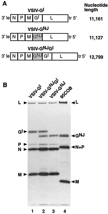
Full-length cDNA clones of the VSIV genome containing an additional heterologous transcriptional unit cloned under XhoI restriction sites at each of the four internal gene junctions and flanked by the conserved gene start signal and 3′ untranslated region and the gene end of the GI gene were previously described (56). The plasmid containing the transcriptional unit (I) at the M-G junction (pMIG) was used to clone the GNJ gene from the VSNJV 95COB strain to generate the VSIV-GNJGI recombinant (Fig. 1A). The coding region of the GNJ gene was amplified by reverse transcription (RT)-PCR from purified viral RNA using two oligodeoxynucleotides containing XhoI restriction sites (5′TGACTCGAGATCAATATGTTGTC3′ and 3′GGGTGAAGGCAATCGAGCTCAGT5′; XhoI recognition sites are underlined, and start and stop codons are in boldface). This RT-PCR product was cloned into the PCR-Blunt II-Topo plasmid (Invitrogen) and used to replace the transcriptional unit in the pMIG plasmid by digestion with XhoI. The same RT-PCR product was cloned into a VSIV plasmid lacking the G gene (pΔG) to generate the VSIV-GNJ recombinant (Fig. 1A). The presence and position of the inserted gene were confirmed by restriction enzyme analysis and sequencing of the insertion sites in the plasmids. To recover infectious virus, BHK-21 cells were infected with the vaccinia virus recombinant that expresses T7 RNA polymerase (vTF7-3) and transfected with the full-length cDNA clones and three support plasmids that expressed the N, P, and L proteins required for RNA encapsidation and replication (3, 58). Infectious viruses were recovered from the supernatant medium, amplified by passage on BHK-21 cells at low multiplicity in the presence of 1-β-d-arabinofuranosylcytosine to inhibit replication of vTF7-3, and filtered through 0.2-μm-pore-size filters. Virus stocks were tested for the presence of vTF7-3 by neutralization with VSIV- or VSNJV-specific antibodies. No vTF7-3 plaques were detected in up to 3 × 10⁶ PFU of recombinant VSIVs.
FIG. 1.
(A) Schematic representation of recombinant VSV genomes. The negative-strand recombinant VSIV genomic RNAs indicating the gene order and expressed proteins are shown. VSIV leader (le), trailer (tr) and viral (N, P, M, GI, and L) genes are represented. GNJ refers to the glycoprotein gene from the VSNJV field isolate 95COB (shaded boxes). The lengths of the genomes are shown on the right. (B) Viral proteins incorporated in virus particles. Virions were labeled with [35S]methionine and purified by centrifugation through 10% sucrose, and the virion proteins were analyzed by sodium dodecyl sulfate-polyacrylamide gel electrophoresis. The infecting viruses are indicated above the lanes, and synthesized proteins are indicated on the left for the recombinant VSIV viruses and on the right for 95COB.
Analysis of virion proteins.
To assess proteins in mature virions, BHK-21 cells were infected at a multiplicity of infection of 5 PFU/cell. At 2 h postadsorption, the cells were washed and incubated in methionine-free medium for 30 min. The cells were labeled with [35S]methionine (50 μCi/ml) overnight. The supernatant fluid was collected, cell debris, was removed by low-speed centrifugation, and the virus was collected by centrifugation through a 10% sucrose cushion. The viral pellet was resuspended in 0.01 M Tris, pH 7.5, and the virion proteins were solubilized in gel loading buffer (27) and separated on a 10% polyacrylamide gel. Viral proteins were quantitated by densitometric analysis of autoradiographs of gels, and molar ratios were calculated after adjustment for the methionine content. Comparisons between lanes were done after normalization against the nucleoprotein.
Neutralization of recombinant viruses with antibodies specific for Indiana or New Jersey serotype.
Serial threefold dilutions of antibodies specific for VSIV (ATCC VR-1238AF) or VSNJV (ATCC VR-1239AF) in Dulbecco's modified Eagle medium plus 2% fetal calf serum (DMEM2) were mixed with an equal volume of the same medium containing 200 PFU of the corresponding virus. After 30 min of incubation at 37°C, the virus-antibody mixture was added to confluent Vero-76 cells growing in six-well dishes. Virus was adsorbed for 1 h, the inoculum was removed, and the cells were washed twice, covered with a nutrient agarose overlay, and incubated for 16 to 18 h at 37°C. The cell monolayers were fixed with formaldehyde and stained with crystal violet, and the virus plaques were counted.
Single-cycle virus replication.
Single-step growth analysis was conducted in BHK-21 cells infected at a multiplicity of infection of 3 PFU/cell. After 1 h of adsorption, the inoculum was removed and the monolayers were washed twice. Fresh medium was added, and the cells were incubated at 37°C. Supernatant fluids were harvested at appropriate intervals, and viral yields were determined by plaque assay on Vero-76 cells.
Lethality in mice.
Three-week old male Swiss Webster mice were purchased from Taconic Farms, Germantown, N.Y., and housed under biosafety level 3 (BL-3) containment conditions. Groups of 10 mice were lightly anesthetized with ketamine-xylazine and inoculated intranasally with 10 μl of 10-fold dilutions of the individual viruses in DMEM2. Control animals were given an equal volume of DMEM2. Following inoculation, the virus preparations were all within three times the original titer as confirmed by plaque assay. The animals were observed every day, and the 50% lethal dose (LD50) for each virus was calculated by the method of Reed and Muench (39).
Swine inoculation and sample collection.
Twenty-four Yorkshire pigs (8 to 10 weeks old and weighing 25 to 30 kg) were obtained from a local breeder and housed under BL-3 isolation conditions at the Plum Island Animal Disease Center. All animals were negative for VSIV and VSNJV, as indicated by serum neutralizing assay. The pigs were divided into two groups of four (for inoculation with VSIV-GI or 95COB) and two groups of eight (for inoculation with VSIV-GNJGI or VSIV-GNJ), and each group was housed in a separate room under BL-3 isolation conditions. The epidermis of the snout in pigs that had been sedated with xylazine-ketamine-telazol was pricked 20 times using a dual-tip skin test applicator (Duotip-Test; Lincoln Diagnostics, Decatur, Ill.), and 3 × 10⁶ PFU of virus was placed on the scarified area in 100 μl of DMEM2. The area under the inoculum was then resensitized by repeating the scarification procedure, and the animals were restrained in a stationary position until the inoculum was adsorbed. Virus preparations were all within three times the original titer following the inoculation procedure. The temperature and appearance of the animals were monitored daily, and samples were collected by individuals wearing protective clothing and portable HEPA respirators, which were changed for entry into each isolation room. Esophageal-pharyngeal fluid (EPF) was obtained before and at various times following virus inoculation by using a modified pharyngeal or probang scraper. Nasal swabs and serum samples were taken before and at various times following inoculation. Swabs of any lesions that appeared were also taken and tested by virus isolation. Swabs and EPF samples were collected in antibiotic-containing basal medium Eagle plus 1% fetal calf serum.
Assessment of clinical disease in swine.
The extents of disease resulting from inoculation with the different viruses were evaluated by assessing lesion formation using a clinical scoring system based on the sizes and locations of the resulting lesions. Scores were assigned as follows: 0 indicated no visible lesions, 1 was given for a lesion that occurred at the site of inoculation and was <2 cm in diameter, 2 indicated a lesion that was >2 cm in diameter or multiple lesions at the site of inoculation, 3 indicated a lesion of <2 cm at a noninoculation site, and 4 indicated a lesion of >2 cm or multiple lesions at noninoculation sites.
Titration of virus isolated from pigs.
Nasal swabs, EPF, and lesion swabs were analyzed for the presence and titer of virus by inoculation onto BHK-21 cell monolayers in 96-well plates after the samples had been clarified by centrifugation. The supernatants were serially diluted and added to the cells in quadruplicate wells, the plates were incubated at 35.5°C for 3 days, and the 50% tissue culture infective dose was calculated.
RESULTS
Generation and recovery of recombinant viruses.
We generated recombinant VSV based on the Indiana serotype by engineering infectious cDNA clones in which the homologous GI open reading frame (ORF) was replaced by the GNJ ORF or in which the GNJ ORF was added to yield a VSIV genome containing both the GI and GNJ ORFs, as described in Materials and Methods and shown schematically in Fig. 1A. These viruses, along with the wild-type Indiana recombinant (VSIV-GI) (58) and the New Jersey field isolate 95COB (45), were used throughout this work. All experiments involving VSNJV 95COB were carried out under appropriate biological containment at the U.S. Department of Agriculture Plum Island Animal Disease Center.
The RNA genomes of viruses recovered from cDNA were characterized by sequence analysis after RT-PCR. The N, P, M, GI, and GNJ genes and the corresponding gene junctions were sequenced to confirm the insertion of the GNJ gene from VSNJV 95COB and to determine whether changes in sequence occurred during the recovery process. The order of genes (Fig. 1A) and the sequence of the intergenic junctions were as in the original cDNAs, and the N, P, M, and GI sequences were identical to those of the parental VSIV-GI. There were two nucleotide differences in the GNJ gene in the recombinant viruses compared to that in 95COB that caused the amino acid changes K113M and P238S. These changes were present in the original GNJ DNA inserted in the two clones. As shown below, these mutations did not alter the antigenicity of the protein and do not map within any reported domain important for structure or functionality, such as glycosylation sites or domains involved in folding, trimerization, intracellular transport, or fusion (13, 16, 17, 32, 50, 59, 60).
Virion proteins.
The synthesis of viral proteins was examined in BHK-21 cells by metabolic labeling with [35S]methionine. Recombinant viruses expressed the expected viral proteins: five in the case of VSIV-GI and VSIV-GNJ and six in the case of VSIV-GNJGI, which expressed both GNJ and GI glycoproteins (data not shown). The protein content of virus particles produced during infection of BHK-21 cells was examined by sodium dodecyl sulfate-polyacrylamide gel electrophoresis of the [35S]methionine-labeled proteins from purified viruses. Each virus contained the appropriate proteins, and VSIV-GNJGI incorporated both GNJ and GI glycoproteins into virions (Fig. 1B). The GNJ protein electrophoretic mobility was slightly faster than that of the GI protein, as described previously (29). VSIV-GI and VSIV-GNJGI virions accumulated similar total amounts of G protein in their envelopes. This amount was ∼20% lower in VSIV-GNJ when normalized to the amounts of N protein in the respective virions. The amount of the GNJ protein incorporated in the virus particles was always slightly lower than the amount of GI protein (Fig. 1B, lanes 1, 2, and 3). In VSIV-GNJGI virions, the amounts of the GNJ and GI proteins accounted for 40 and 60% of total G protein, respectively. The VSNJV (95COB) field isolate incorporated a larger amount of G in its virions than any of the recombinant VSIVs (Fig. 1B, lanes 1 through 4). Interestingly, this virus replicated faster than the recombinant viruses in BHK-21 cells, and it was also the most pathogenic in mice and natural hosts (see below).
Neutralization of recombinant viruses by antibodies against Indiana or New Jersey serotype.
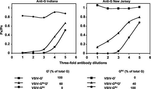
To determine whether the recombinant viruses could be neutralized by antibodies specific to the VSIV or VSNJV serotype, according to the glycoprotein(s) they expressed, the viruses were incubated with neutralizing antibodies against VSIV or VSNJV before inoculation of Vero-76 cells (Fig. 2). As expected, VSIV-GI was completely neutralized by the VSIV-specific antibodies but not by the VSNJV-specific antibodies. Similarly, VSIV-GNJ was neutralized by antibodies against VSNJV but not by antibodies against VSIV (Fig. 2). VSIV-GNJGI was neutralized by antibodies against either VSIV or VSNJV. However, differences were observed in the neutralization curves between VSIV- and VSNJV-specific antibodies. Antibodies against VSIV completely neutralized VSIV-GNJGI at the lower dilutions, indicating that the GNJ and GI glycoproteins were expressed in the same virion. Antibodies against the VSNJV serotype neutralized VSIV-GNJGI, but neutralization was not complete at the lowest dilution tested. This was consistent with the observation that the amount of GI in these virions was higher than that of GNJ (Fig. 2) and suggests that enough GI homo-oligomers may form in some virions to allow infection in the presence of VSNJV-specific antibodies or that some virions may have only GI.
FIG. 2.
Neutralization of recombinant and wild-type viruses by antibodies specific for Indiana or New Jersey serotype. Two hundred PFU of virus were incubated for 30 min at 37°C with serial threefold dilutions (starting at 1:40) of antibody prior to inoculation of Vero-76 cells. After 16 to 18 h of incubation at 37°C, individual plaques were counted. Px and Pn represent the numbers of plaques in the presence and absence of antibody, respectively. The percentages of GI (VSIV) and GNJ (VSNJV) in the total G protein present in the different virions, as determined in Fig. 1B, are indicated at the bottom.
Virus replication in cell culture.
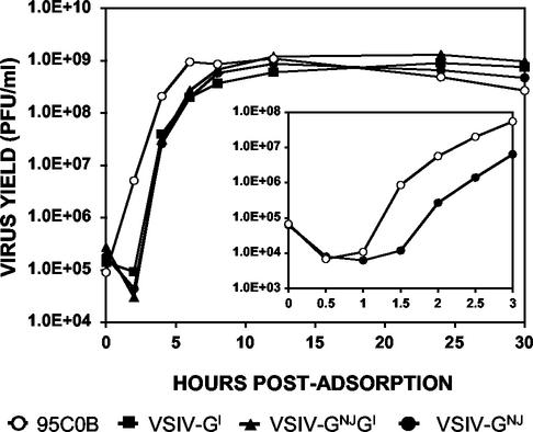
Replication of the recombinant viruses was examined under single-step growth conditions in BHK-21 cells at 37°C. Supernatant fluids were harvested at various times, and the virus yields were measured by plaque assay on Vero-76 cells (Fig. 3). Growth curves from VSIV-GNJGI and VSIV-GNJ were indistinguishable from that of VSIV-GI (Fig. 3), showing that the ability of these viruses to replicate in cell culture was not impaired by the insertion of an additional gene or by the replacement of the GI gene by the GNJ gene. However, replication of recombinant viruses was delayed compared to that of the wild-type VSNJV (95COB) (Fig. 3). This difference was most apparent 90 min after the adsorption period, when the amount of 95COB virus in the supernatant was 100-fold greater than that of VSIV-GNJ, as shown by a second experiment in which samples were taken every 30 min (Fig. 3, inset). After that, both viruses grew with similar kinetics and reached equivalent titers in the supernatant, indicating that the observed differences were most likely due to an early step in the infection cycle.
FIG. 3.
Single-step growth curves of wild-type and recombinant viruses in BHK-21 cells at 37°C. The cells were infected at a multiplicity of infection of 3, and samples of the supernatant medium were harvested at the indicated time points. The samples were titrated in duplicate by plaque assay on Vero-76 cells. Single-step growth curves comparing VSIV-GNJ and 95COB viruses at early times postinfection are shown in the inset.
Lethality in mice.
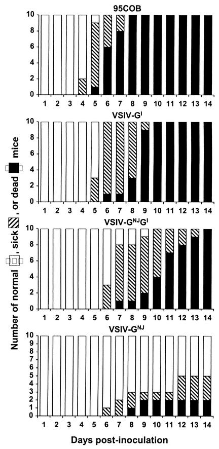
Young mice are susceptible to fatal encephalitis after intranasal inoculation of wild-type VSV and provide a well-studied model for comparing the relative lethalities of VSV and its mutants (36, 48, 55). Inoculation of mice allowed us to compare the pathogenicities of the wild-type and variant viruses in this animal model with those in swine, a natural host for VSV (14) (see below). We inoculated groups of 10 Swiss-Webster mice intranasally with serial 10-fold dilutions ranging from 1 to 106 PFU per mouse. The mice were observed daily for clinical symptoms and death. The LD50s were 11, 123, and >106 PFU/mouse for VSIV-GI, VSIV-GNJGI, and VSIV-GNJ, respectively. The morbidity and mortality of mice inoculated with 1,000 PFU of viruses are shown in Fig. 4. The time to onset of sickness and death increased progressively in VSIV-GNJGI and VSIV-GNJ compared to VSIV-GI. Symptoms of illness started as early as 5 to 6 days postinfection with VSIV-GI and were usually followed by death 7 to 8 days postinfection (Fig. 4). The generalization of symptoms was typically delayed 1 to 2 days in VSIV-GNJGI, and the time between the onset of sickness and death was extended by 3 to 4 days compared to VSIV-GI. Inoculation of VSIV-GNJ into mice rarely resulted in death, so the LD50 could not be determined. In contrast, 95COB virus was highly pathogenic in mice when inoculated intranasally (LD50 = 16 PFU/mouse) (Fig. 4). Although the replacement of GI by GNJ was responsible for the highly attenuated phenotype of VSIV-GNJ, this effect was compensated for in the 95COB virus. Since VSIV-GNJ and 95COB differ in genes other than the G gene, those genes may contribute to the different pathogenicities in the mouse model.
FIG. 4.
Lethalities of wild-type and recombinant viruses in mice. The viruses were inoculated intranasally into groups of 10 mice at a dose of 103 PFU/mouse. The mice were monitored daily for signs of morbidity and mortality. No further changes occurred after day 14.
Pathogenesis in swine.
Previous experiments showed important differences in pathogenicity between VSNJV and VSIV strains when pigs were inoculated experimentally by scarification of the snout, a procedure that leads to development of clinical symptoms and a disease course that closely resembles natural infection (14, 31, 51). VSNJV 95COB is a highly pathogenic field isolate that causes much more severe disease than recombinant or wild-type VSIV (14; L. L. Rodriguez, unpublished results). To test the hypothesis that the G glycoprotein was involved in the differential pathogeneses of VSIV and VSNJV, groups of four (VSIV-GI and 95COB) or eight (VSIV-GNJ and VSIV-GNJGI) Yorkshire pigs (8 to 10 weeks old and weighing 25 to 30 kg), which were negative for VSIV and VSNJV as indicated by serum neutralizing assay, were inoculated intradermally by scarification of the snout with 3 × 10⁶ PFU per animal as previously described (14). Each group was kept in a different room under BL-3 conditions at Plum Island Animal Disease Center and monitored daily for vesicular lesions and for rectal temperature. The extents of disease resulting from inoculation with the different viruses were evaluated by assessing lesion formation using a clinical scoring system based on the size and location of the resulting lesions (see Materials and Methods).
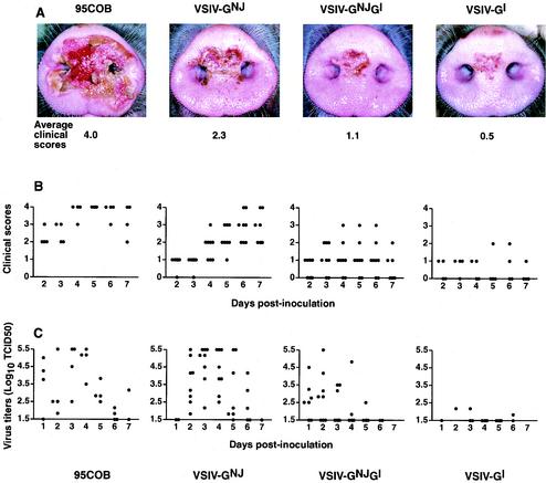
In general, small vesicles (<2 cm in diameter) developed the second day postinoculation at the area of scarification in most of the pigs. In severe cases, these vesicles increased in size to >2 cm and ruptured by days 3 to 5. At 6 to 7 days postinoculation, the lesions started to heal. Important differences, depending on the virus inoculated, were observed in the numbers of pigs that developed vesicles, the sizes and extents of the vesicular lesions, spread to distal areas, and time of appearance and healing of the lesions. The clinical scores from the snout of each pig inoculated with one of the four different viruses are shown for the 7 days following inoculation in Fig. 5B.
FIG. 5.
(A) Snouts of pigs inoculated 5 days before with recombinant and wild-type VSVs. The swine were anesthetized, and 3 × 10⁶ PFU of the indicated virus was applied by scarification of the snout. The snouts of the most severely affected animals from each group are represented. The average clinical scores from the snouts of all animals from each group 5 days postinfection are also shown. (B) Clinical scores after inoculation of pigs with the recombinant and wild-type viruses. The clinical scores refer to the snout and were based on the sizes and locations of viral lesions (see the text). Each symbol represents an individual animal. (C) Titers of virus recovered following inoculation with recombinant and wild-type viruses. Nasal swabs and EPF were taken daily from inoculated swine. Virus was recovered and titrated on BHK-21 cells at 35.5°C for 3 days. Each symbol represents an individual animal and corresponds to the highest titer obtained from the nasal swab or EPF. The virus dilution giving a titer of 5.5 log10 was the highest dilution assayed, and samples to which this value was assigned might actually have higher titers of virus. TCID50, 50% tissue culture infectious dose.
A progressive increase in the severity of the lesions was observed in inoculated pigs in the following order: VSIV-GI < VSIV-GNJGI < VSIV-GNJ < 95COB (Fig. 5A). All four pigs inoculated with 95COB developed snout vesicles >2 cm in diameter as early as 2 days postinoculation, and the lesions reached scores of 4 by days 4 to 5 (Fig. 5B). In addition, two swine developed severe secondary lesions at the coronary bands of the hooves. In contrast, pigs inoculated with VSIV-GI developed only mild lesions. One pig inoculated with VSIV-GI never developed lesions, only one developed snout vesicles >2 cm in diameter 5 days postinfection, and one animal showed a small secondary foot lesion. Six of eight animals inoculated with VSIV-GNJGI developed lesions, two exhibited vesicles >2 cm in diameter, and one developed small lesions on the snout outside the area of inoculation. All pigs inoculated with VSIV-GNJ developed large vesicles at the inoculation site, and five developed lesions on the snout outside the area of scarification. These lesions were especially severe in two pigs, which received clinical scores of 4. Compared to 95COB, a delay of ∼2 days in vesicle development was observed in pigs inoculated with VSIV-GNJ, with vesicles reaching maximum severity by days 6 to 7 postinoculation (Fig. 5B). Neither VSIV-GNJGI nor VSIV-GNJ caused secondary lesions on the feet. The pathological changes primarily involved the epidermis during the early stages but extended down to subcutaneous tissues in 95COB- and VSV-GNJ-infected pigs, causing loss of epithelium and leaving a bleeding surface in the most severe cases (Fig. 5A). This process was less apparent in lesions caused by VSIV-GI. In agreement with previous results (14, 20, 52), we were unable to correlate rectal temperature with the severity of the infection.
As might be expected, none of the recombinant viruses were as virulent as the wild-type VSNJV 95COB. This virus is a field isolate with low passage in tissue culture, and viral products other than G may contribute to its increased pathogenicity. It may also be important that GNJ is expressed at higher levels in 95COB than in the recombinant viruses and/or that the GNJ protein in the engineered viruses might not interact optimally with other VSIV proteins (e.g., M), and this might lead to suboptimal replication and/or assembly of the recombinant viruses containing GNJ. Nevertheless, the results presented above showed that the replacement of the GI protein by the GNJ protein from 95COB alone was able to increase the pathogenicity of VSIV in pigs.
Virus replication in swine.
To investigate the ability of inoculated viruses to replicate in pigs, nasal swabs and EPF samples were taken on days 1 to 7 postinfection and clarified by centrifugation. Viruses were titered on BHK-21 cell monolayers in 96-well plates by serially diluting the supernatants and infecting cells in quadruplicate wells. The cells were incubated at 35.5°C for 3 days, and the 50% tissue culture infective dose was calculated. All animals were negative for virus isolation on the day before inoculation. In all groups, virus was isolated as early as 1 day postinfection from nasal swabs and 2 days postinfection from EPF. Typically, recovery from nasal swabs was more consistent and the titers were higher than those from EPF.
In agreement with the clinical scores shown in Fig. 5B, the virus titers recovered from pigs inoculated with 95COB and VSIV-GNJ were generally highest, followed by VSIV-GNJGI and then by VSIV-GI (Fig. 5C). Within each group, the pigs with higher titers of virus also developed more severe lesions. These results suggest that VSV pathology in swine correlates primarily with virus replication. Differences in virus titers of up to 10,000-fold were detected between pigs inoculated with VSIV-GNJ and those inoculated with VSIV-GI, confirming a role for the GNJ glycoprotein in the replication and pathogenicity of VSV in swine. The increase in the titers of viruses isolated from the VSIV-GNJ group was delayed 1 day compared to those from 95COB and VSIV-GNJGI, as seen from the clinical scores (Fig. 5B and C). There were also differences in the average lengths of time the virus was shed among the groups of pigs inoculated with different viruses. VSIV-GI was shed for the shortest period (2.7 ± 1.7 days), followed by VSIV-GNJGI (3.9 ± 0.9 days), VSIV-GNJ (4.1 ± 0.8 days), and 95COB (5.7 ± 0.9 days) (P < 0.001).
DISCUSSION
The purpose of this work was to begin elucidating the basis of the marked differences in virulence and field occurrence observed between the VSNJV and VSIV serotypes. In this study, we generated recombinant cDNAs from which we recovered VSIVs that expressed one or both of the glycoproteins from the two major VSV serotypes. These recombinant viruses allowed us to explore the possibility of expressing VSIV and VSNJV glycoproteins from a single virus and the roles of these glycoproteins in virus replication, gene expression, and pathogenesis in laboratory animals and natural hosts.
All the engineered viruses were viable and synthesized the appropriate proteins. Neither the insertion of an additional gene (VSIV-GNJGI) nor the replacement of the homologous GI gene by the heterologous GNJ gene (VSIV-GNJ) had any detectable effect on the abilities of the recombinant viruses to replicate in BHK-21 cells, as assayed by single-step growth curves.
Although all the recombinant viruses grew equally well in BHK-21 cells, VSIV-GNJGI and VSIV-GNJ were attenuated in mice. Primary viral infections in animals are often a race, with viral replication and spread versus innate immune response and virus clearance. In this scenario, even small impairments in virus replication and/or spread, not detectable in single-step growth curves in vitro, could become critical for pathogenesis in mice (42, 57). The insertion of an additional heterologous gene and subsequent downregulation of the downstream GI and L genes may account for the moderately attenuated phenotype of VSIV-GNJGI. We do not know the cause(s) of the highly attenuated phenotype of VSIV-GNJ in mice, but several effects, alone or in combination, may have an influence in modulating the pathogenesis of this virus. For example, the lower expression of GNJ and/or the suboptimal interaction of GNJ with other viral protein(s) from the Indiana serotype during VSIV-GNJ infection may be responsible. We are working to test these possibilities. A recombinant VSIV carrying GNJ has been previously reported to grow to lower titers in tissue culture than the parental virus and to be attenuated in mice (28, 47). However, substantial differences exist between the extents of pathogenesis observed in mice for the parental recombinant VSIV used in those studies and the VSIV used here that preclude direct comparison between the VSIV-GNJ viruses (references 15, 41, and 57 and this study).
Although the pathogenesis of VSV New Jersey and Indiana infection in laboratory rodents has been extensively examined, similar studies have not been performed in the natural hosts, and the molecular mechanisms involved remain largely unresolved. We have made use of a domestic-swine model (14) to study the pathogenesis of VSV. Inoculation by scarification of the snout led to the development of clinical symptoms and a disease course that closely resembled that observed in nature (31). Following the experimental inoculation of pigs, we observed that vesicles formed at the site of inoculation and ruptured, leaving reddish denuded erosions with exfoliated tissue adhering to the margins of the lesions. The erosive stage lasted for a week or so, and virus could be isolated from nasal swabs and EPF for 6 to 7 days after inoculation. Although pathological changes were limited to the epithelia of affected areas in most of the pigs, secondary lesions developed in the hooves of some animals. Depression, lameness, and excessive salivation were also seen. In this animal model, we observed that VSIV-GNJ replicated to higher titers and caused more severe lesions than the parental VSIV-GI. These results identified the G glycoprotein as an important determinant for VSV pathogenicity in swine, a natural host for VSV. Data from the recombinant virus expressing glycoproteins from two serotypes (VSIV-GNJGI) further supported this conclusion. VSIV-GNJGI was more pathogenic than VSIV-GI, despite the fact that GNJ represented only 40% of the total G protein incorporated into VSIV-GNJGI virions. These results also illustrate the profound differences between mice and swine in VSV pathogenesis and emphasize the importance of the study of virus-host interactions in the natural hosts whenever possible.
Several studies indicate that the G protein plays an essential role in the pathogenesis of other viruses, including the rhabdoviruses rabies virus and viral hemorrhagic septicemia virus (12, 18, 22, 49, 53). The entry halftime of pathogenic rabies viruses in neuroblastoma cells was seven to eight times shorter than that of nonpathogenic variants (11). In addition, nonpathogenic viruses were unable to spread from cell to cell and displayed a lower rate of spread through the central nervous system. Other studies showed that pathogenic viruses apparently could use receptors or routes of entry different from those used by their nonpathogenic derivatives (10, 26). Fusion of rabies virus-infected cells may also be an important pathogenetic mechanism (37). Changes in the surface envelope glycoprotein of simian-human immunodeficiency virus molecular clones have also been reported to be responsible for increases in fusion capacity, cytopathicity, replication capacity, and neutralization resistance (8).
In order to elucidate the mechanisms responsible for the differences in pathogenicity between VSV-GIN and VSV-GNJ viruses, functional analyses comparing the G proteins from the two serotypes similar to those described above for rabies virus and simian-human immunodeficiency virus are needed. Reverse genetic techniques for manipulating viral genomes and the domestic-swine model for VSV infection described here and elsewhere (14) constitute powerful tools for studies exploring the roles of individual proteins in the life cycle of this pathogen and for vaccine development.
Acknowledgments
This work was supported by NIH grant R37AI12464 to G.W.W. and by USDA/ARS CRIS Project no. 1940-32000-040D (L.L.R.). I. Martinez was the recipient of a fellowship from the Ministerio de Sanidad y Consumo (Spain) (BAE 00/5009 and BAE 01/5006).
We thank S. P. J. Whelan and L. A. Ball for the pMIG and pΔG plasmids and the members of the Wertz, Ball, and Rodriguez laboratories for constructive comments.
REFERENCES
- 1.Abraham, G., and A. K. Banerjee. 1976. Sequential transcription of the genes of vesicular stomatitis virus. Proc. Natl. Acad. Sci. USA 73:1504-1508. [DOI] [PMC free article] [PubMed] [Google Scholar]
- 2.Ball, L. A., and C. N. White. 1976. Order of transcription of genes of vesicular stomatitis virus. Proc. Natl. Acad. Sci. USA 73:442-446. [DOI] [PMC free article] [PubMed] [Google Scholar]
- 3.Ball, L. A., C. R. Pringle, B. Flanagan, V. P. Perepelitsa, and G. W. Wertz. 1999. Phenotypic consequences of rearranging the P, M, and G genes of vesicular stomatitis virus. J. Virol. 73:4705-4712. [DOI] [PMC free article] [PubMed] [Google Scholar]
- 4.Bricker, B. J., R. M. Snyder, J. W. Fox, W. A. Volk, and R. R. Wagner. 1987. Monoclonal antibodies to the glycoprotein of vesicular stomatitis virus (New Jersey serotype): a method for preliminary mapping of epitopes. Virology 161:533-540. [DOI] [PubMed] [Google Scholar]
- 5.Bridges, V., B. J. McCluskey, M. D. Salman, H. S. Hurd, and J. Dick. 1997. Review of the 1995 vesicular stomatitis outbreak in the western United States. J. Am. Vet. Med. Assoc. 211:556-560. [PubMed] [Google Scholar]
- 6.Burkhart, C., G. Freer, R. Castro, L. Adorini, K.-H. Wiesmüller, R. M. Zinkernagel, and H. Hengartner. 1994. Characterization of T-helper epitopes of the glycoprotein of vesicular stomatitis virus. J. Virol. 68:1573-1580. [DOI] [PMC free article] [PubMed] [Google Scholar]
- 7.Cartwright, B., and F. Brown. 1972. Serological relationships between different strains of vesicular stomatitis virus. J. Gen. Virol. 16:391-398. [DOI] [PubMed] [Google Scholar]
- 8.Chakrabarti, L. A., T. Ivanovic, and C. Cheng-Mayer. 2002. Properties of the surface envelope glycoprotein associated with virulence of simian-human immunodeficiency virus SHIVSF33A molecular clones. J. Virol. 76:1588-1599. [DOI] [PMC free article] [PubMed] [Google Scholar]
- 9.Chen, S. S. 1991. Myristylation of the envelope glycoprotein of vesicular stomatitis virus. Intervirology 32:193-197. [DOI] [PubMed] [Google Scholar]
- 10.Coulon, P., J. P. Ternaux, A. Flamand, and C. Tuffereau. 1998. An avirulent mutant of rabies virus is unable to infect motoneurons in vivo and in vitro. J. Virol. 72:273-278. [DOI] [PMC free article] [PubMed] [Google Scholar]
- 11.Dietzschold, B., T. J. Wiktor, J. Q. Trojanovski, R. I. Macfarlan, W. H. Wunner, M. J. Torres-Anjel, and H. Kropowski. 1985. Differences in cell to cell spread of pathogenic and apathogenic rabies virus in vivo and in vitro. J. Virol. 56:12-18. [DOI] [PMC free article] [PubMed] [Google Scholar]
- 12.Dietzschold, B., W. H. Wunner, T. J. Wiktor, A. D. Lopes, M. Lafon, C. L. Smith, and H. Kopowski. 1983. Characterization of an antigenic determinant of the glycoprotein that correlates with pathogenicity of rabies virus. Proc. Natl. Acad. Sci. USA 80:70-74. [DOI] [PMC free article] [PubMed] [Google Scholar]
- 13.Doms, R. W., A. Ruusala, C. Machamer, J. Helenius, A. Helenius, and J. K. Rose. 1988. Differential effects of mutations in three domains on folding, quaternary structure, and intracellular transport of vesicular stomatitis virus G protein. J. Cell Biol. 107:89-99. [DOI] [PMC free article] [PubMed] [Google Scholar]
- 14.Flanagan, E. B., J. M. Zamparo, L. A. Ball, L. L. Rodriguez, and G. W. Wertz. 2001. Rearrangement of the genes of vesicular stomatitis virus eliminates clinical disease in the natural host: new strategy for vaccine development. J. Virol. 75:6107-6114. [DOI] [PMC free article] [PubMed] [Google Scholar]
- 15.Flanagan, E. B., L. A. Ball, and G. W. Wertz. 2000. Moving the glycoprotein gene of vesicular stomatitis virus to promoter-proximal positions accelerates and enhances the protective immune response. J. Virol. 74:7895-7902. [DOI] [PMC free article] [PubMed] [Google Scholar]
- 16.Fredericksen, B. L., and M. A. Whitt. 1995. Vesicular stomatitis virus glycoprotein mutations that affect membrane fusion activity and abolish virus infectivity. J. Virol. 69:1435-1443. [DOI] [PMC free article] [PubMed] [Google Scholar]
- 17.Gallione, C., and J. K. Rose. 1983. Nucleotide sequence of a cDNA clone encoding the entire glycoprotein from the New Jersey serotype of vesicular stomatitis virus. J. Virol. 46:162-169. [DOI] [PMC free article] [PubMed] [Google Scholar]
- 18.Gaudin, Y., P. de Kinkelin, and A. Benmansour. 1999. Mutations in the glycoprotein of viral haemorrhagic septicemia virus that affect virulence for fish and the pH threshold for membrane fusion. J. Gen. Virol. 80:1221-1229. [DOI] [PubMed] [Google Scholar]
- 19.Hale, A. H., M. J. Ruebush, and L. Lefrancois. 1981. Cross-reactive anti-vesicular stomatitis virus (VSV) cytotoxic T lymphocytes recognize the major surface glycoprotein of VSV. Eur. J. Immunol. 11:434-436. [DOI] [PubMed] [Google Scholar]
- 20.Howerth, E. W., D. E. Stallknecht, M. Dorminy, T. Pisell, and G. R. Clarke. 1997. Experimental vesicular stomatitis in swine: effects of route of inoculation and steroid treatment. J. Vet. Diagn. Investig. 9:136-142. [DOI] [PubMed] [Google Scholar]
- 21.Huneycutt, B. S., I. V. Plakhov, Z. Shusterman, S. M. Bartido, A. Huang, C. S. Reiss, and C. Aoki. 1994. Distribution of vesicular stomatitis virus proteins in the brains of BALB/c mice following intranasal inoculation: an immunohistochemical analysis. Brain Res. 635:81-95. [DOI] [PubMed] [Google Scholar]
- 22.Ito, N., M. Takayama, K. Yamada, M. Sugiyama, and N. Minamoto. 2001. Rescue of rabies virus from cloned cDNA and identification of the pathogenicity-related gene: glycoprotein gene is associated with virulence for adult mice. J. Virol. 75:9121-9128. [DOI] [PMC free article] [PubMed] [Google Scholar]
- 23.Iverson, L. E., and J. K. Rose. 1981. Localized attenuation and discontinuous synthesis during vesicular stomatitis virus transcription. Cell 23:477-484. [DOI] [PubMed] [Google Scholar]
- 24.Keil, W., and R. R. Wagner. 1989. Epitope mapping by deletion mutants and chimeras of two vesicular stomatitis virus glycoprotein genes expressed by a vaccinia virus vector. Virology 170:392-407. [DOI] [PubMed] [Google Scholar]
- 25.Kelley, J. R., S. U. Emerson, and R. R. Wagner. 1972. The glycoprotein of vesicular stomatitis virus is the antigen that gives rise to and reacts with neutralizing antibody. J. Virol. 10:1231-1235. [DOI] [PMC free article] [PubMed] [Google Scholar]
- 26.Kucera, P., M. Dolivo, P. Coulon, and A. Flamand. 1985. Pathways of the early propagation of virulent and avirulent rabies strains from the eye to the brain. J. Virol. 55:158-162. [DOI] [PMC free article] [PubMed] [Google Scholar]
- 27.Laemmli, U. K. 1970. Cleavage of structural proteins during the assembly of the head of bacteriophage T4. Nature 227:680-685. [DOI] [PubMed] [Google Scholar]
- 28.Lawson, N. D., E. A. Stillman, M. A. Whitt, and J. K. Rose. 1995. Recombinant vesicular stomatitis viruses from DNA. Proc. Natl. Acad. Sci. USA 92:4477-4481. [DOI] [PMC free article] [PubMed] [Google Scholar]
- 29.Lefrancois, L., and D. Lyles. 1982. The interaction of antibody with the major surface glycoprotein of vesicular stomatitis virus. I. Analysis of neutralizing epitopes with monoclonal antibodies. Virology 121:157-167. [PubMed] [Google Scholar]
- 30.Lefrancois, L., and D. Lyles. 1982. The interaction of antibody with the major surface glycoprotein of vesicular stomatitis virus. II. Monoclonal antibodies to nonneutralizing and cross-reactive epitopes. Virology 121:168-174. [DOI] [PubMed] [Google Scholar]
- 31.Letchworth, G. J., L. L. Rodriguez, and J. del C. Barrera. 1999. Vesicular stomatitis. Vet. J. 157:239-260. [DOI] [PubMed] [Google Scholar]
- 32.Li, Y., C. Drone, E. Sat, and H. P. Ghosh. 1993. Mutational analysis of the vesicular stomatitis virus glycoprotein G for membrane fusion domains. J. Virol. 67:4070-4077. [DOI] [PMC free article] [PubMed] [Google Scholar]
- 33.Machamer, C. E., R. W. Doms, D. G. Bole, A. Helenius, and J. K. Rose. 1990. Heavy chain binding protein recognizes incompletely disulfide-bonded forms of vesicular stomatitis virus G protein. J. Biol. Chem. 265:6879-6883. [PubMed] [Google Scholar]
- 34.Mathieu, M. E., P. R. Grigera, A. Helenius, and R. R. Wagner. 1996. Folding, unfolding, and refolding of the vesicular stomatitis virus glycoprotein. Biochemistry 35:4084-4093. [DOI] [PubMed] [Google Scholar]
- 35.McCluskey, B. J., H. S. Hurd, and E. L. Mumford. 1999. Review of the 1997 outbreak of vesicular stomatitis in the western United States. J. Am. Vet. Med. Assoc. 215:1259-1262. [PubMed] [Google Scholar]
- 36.Miyoshi, K., D. H. Harter, and K. C. Hsu. 1971. Neuropathological and immunofluorescence studies of experimental vesicular stomatitis encephalitis in mice. J. Neuropathol. Exp. Neurol. 30:266-277. [DOI] [PubMed] [Google Scholar]
- 37.Morimoto, K., Y. J. Ni, and A. Kawai. 1992. Syncytium formation is induced in the murine neuroblastoma cell cultures which produce pathogenic type G proteins of the rabies virus. Virology 189:203-216. [DOI] [PMC free article] [PubMed] [Google Scholar]
- 38.Plakhov, I. V., E. E. Arlund, C. Aoki, and C. S. Reiss. 1995. The earliest events in vesicular stomatitis virus infection of the murine olfactory neuroepithelium and entry of the central nervous system. Virology 209:257-262. [DOI] [PubMed] [Google Scholar]
- 39.Reed, E. J., and H. Muench. 1938. A simple method of estimating fifty percent endpoints. Am. J. Hyg. 27:493-497. [Google Scholar]
- 40.Reiss, C. S., I. V. Plakhov, and T. Komatsu. 1998. Viral replication in olfactory receptor neurons and entry into the olfactory bulb and brain. Ann. N. Y. Acad. Sci. 855:751-761. [DOI] [PubMed] [Google Scholar]
- 41.Roberts, A., E. Kretzschmar, A. S. Perkins, J. Forman, R. Price, L. Buonocore, Y. Kawaoka, and J. K. Rose. 1998. Vaccination with a recombinant vesicular stomatitis virus expressing an influenza virus hemagglutinin provides complete protection from influenza virus challenge. J. Virol. 72:4704-4711. [DOI] [PMC free article] [PubMed] [Google Scholar]
- 42.Roberts, A., L. Buonocore, R. Price, J. Forman, and J. K. Rose. 1999. Attenuated vesicular stomatitis virus as vaccine vectors. J. Virol. 73:3723-3732. [DOI] [PMC free article] [PubMed] [Google Scholar]
- 43.Rodriguez, L. L. 2002. Emergence and re-emergence of vesicular stomatitis in the United States. Virus Res. 85:211-219. [DOI] [PubMed] [Google Scholar]
- 44.Rodriguez, L. L., and S. T. Nichol. 1999. Vesicular stomatitis viruses, p. 1910-1919. In R. G. Webster and A. Granoff (ed.), Encyclopedia of virology. Academic Press, London, United Kingdom.
- 45.Rodriguez, L. L., T. A. Bunch, M. Fraire, and Z. N. Llewellyn. 2000. Re-emergence of vesicular stomatitis in the western United States is associated with distinct viral genetic lineages. Virology 271:171-181. [DOI] [PubMed] [Google Scholar]
- 46.Rose, J. K., and C. J. Gallione. 1981. Nucleotide sequence of the mRNA encoding the vesicular stomatitis virus G and M proteins determined from cDNA clones containing the complete coding regions. J. Virol. 39:519-528. [DOI] [PMC free article] [PubMed] [Google Scholar]
- 47.Rose, N. F., A. Roberts, L. Buonocore, and J. K. Rose. 2000. Glycoprotein exchange vectors based on vesicular stomatitis virus allow effective boosting and generation of neutralizing antibodies to a primary isolate of human immunodeficiency virus type 1. J. Virol. 74:10903-10910. [DOI] [PMC free article] [PubMed] [Google Scholar]
- 48.Sabin, A., and P. Olitsky. 1938. Influence of host factors on neuroinvasiveness of vesicular stomatitis virus. J. Exp. Med. 67:201-227. [DOI] [PMC free article] [PubMed] [Google Scholar]
- 49.Seif, I. P., P. Coulon, P. E. Rollin, and A. Flamand. 1985. Rabies virulence: effect on pathogenicity and sequence characterization of rabies virus mutations affecting antigenic site III of the glycoprotein. J. Virol. 53:926-934. [DOI] [PMC free article] [PubMed] [Google Scholar]
- 50.Shokralla, S., Y. He, E. Wanas, and H. P. Ghosh. 1998. Mutations in a carboxy-terminal region of vesicular stomatitis virus glycoprotein G that affect membrane fusion activity. Virology 242:39-50. [DOI] [PubMed] [Google Scholar]
- 51.Stallknecht, D. E., D. E. Perzak, L. D. Bauer, M. D. Murphy, and E. W. Howerth. 2001. Contact transmission of vesicular stomatitis virus New Jersey in pigs. Am. J. Vet. Res. 62:516-520. [DOI] [PubMed] [Google Scholar]
- 52.Stallknecht, D. E., E. W. Howerth, C. L. Reeves, and B. S. Seal. 1999. Potential for contact and mechanical vector transmission of vesicular stomatitis virus New Jersey in pigs. Am. J. Vet. Res. 60:43-48. [PubMed] [Google Scholar]
- 53.Tuffereau, C., H. Leblois, J. Benejean, P. Coulon, F. Lafay, and A. Flamand. 1989. Arginine or lysine in position 333 of ERA and CVS glycoprotein is necessary for rabies virulence in adult mice. Virology 172:206-212. [DOI] [PubMed] [Google Scholar]
- 54.Villarreal, L. P., M. Breindl, and J. J. Holland. 1976. Determination of molar ratios of vesicular stomatitis virus induced RNA species in BHK21 cells. Biochemistry 15:1663-1667. [DOI] [PubMed] [Google Scholar]
- 55.Wagner, R. R. 1974. Pathogenicity and immunogenicity for mice of temperature-sensitive mutants of vesicular stomatitis virus. Infect. Immun. 10:309-315. [DOI] [PMC free article] [PubMed] [Google Scholar]
- 56.Wertz, G. W., R. Moudy, and L. A. Ball. 2002. Adding genes to the RNA genome of vesicular stomatitis virus: positional effects on stability of expression. J. Virol 76:7642-7650. [DOI] [PMC free article] [PubMed] [Google Scholar]
- 57.Wertz, G. W., V. P. Perepelitsa, and L. A. Ball. 1998. Gene rearrangement attenuates expression and lethality of a nonsegmented negative strand RNA virus. Proc. Natl. Acad. Sci. USA 95:3501-3506. [DOI] [PMC free article] [PubMed] [Google Scholar]
- 58.Whelan, S. P. J., L. A. Ball, J. N. Barr, and G. W. Wertz. 1995. Recovery of infectious vesicular stomatitis virus entirely from cDNA clones. Proc. Natl. Acad. Sci. USA 92:8388-8392. [DOI] [PMC free article] [PubMed] [Google Scholar]
- 59.Whitt, M. A., P. Zagouras, B. Crise, and J. K. Rose. 1990. A fusion-defective mutant of the vesicular stomatitis virus glycoprotein. J. Virol. 64:4907-4913. [DOI] [PMC free article] [PubMed] [Google Scholar]
- 60.Zhang, L., and H. P. Ghosh. 1994. Characterization of the putative fusogenic domain in vesicular stomatitis virus glycoprotein G. J. Virol. 68:2186-2193. [DOI] [PMC free article] [PubMed] [Google Scholar]


