Abstract
Many transcription factors, particularly those involved in the control of cell growth, are unstable proteins destroyed by ubiquitin-mediated proteolysis. In a previous study of sequences targeting the transcription factor Myc for destruction, we observed that the region in Myc signaling ubiquitin-mediated proteolysis overlaps closely with the region in Myc that activates transcription. Here, we present evidence that the overlap of these two activities is not unique to Myc, but reflects a more general phenomenon. We show that a similar overlap of activation domains and destruction elements occurs in other unstable transcription factors and report a close correlation between the ability of an acidic activation domain to activate transcription and to signal proteolysis. We also show that destruction elements from yeast cyclins, when tethered to a DNA-binding domain, activate transcription. The intimate overlap of activation domains and destruction elements reveals an unexpected convergence of two very different processes and suggests that transcription factors may be destroyed because of their ability to activate transcription.
Cells exploit a variety of mechanisms to keep the function of transcriptional activators tightly in check. Processes that limit the activity, location, and abundance of transcription factors play an important role in regulating gene expression and maintaining cellular homeostasis. One prominent mechanism regulating transcription factor function is proteolysis; the rapid and controlled destruction of transcription factors like Myc (1), Jun (2), p53 (3), and E2F-1 (4) keeps the intracellular levels of these proteins low and responsive to environmental stimuli. Although several proteolytic processes have been implicated in transcription factor destruction—such as cleavage by calpains (5) and lysosomal proteases (6)—the most widespread pathway of transcription factor turnover is ubiquitin (Ub)-mediated proteolysis.
Ub-mediated proteolysis is a process in which proteins to be destroyed are tagged by linkage to a small, highly conserved protein known as Ub (7). Covalent attachment of Ub to proteins signals their destruction by the 26S proteasome, a large complex with multiple proteolytic activities (8). Ub-mediated proteolysis features in many cellular events, including the cell cycle, antigen presentation, and DNA repair (7). The versatility of this system stems from both its diversity and its specificity. Because cells possess a diverse collection of ubiquitylating enzymes, Ub-mediated proteolysis targets a wide range of proteins for destruction. Because destruction by the proteasome generally depends on prior substrate ubiquitylation, Ub-mediated proteolysis destroys its substrates with extreme precision.
Central to the control of Ub-mediated proteolysis, therefore, are the interactions between target proteins and components of the ubiquitylation machinery. Despite the importance of this process, however, the mechanisms governing substrate recognition in this pathway are poorly understood. It is known that substrate recognition is usually mediated by the action of Ub-protein ligases, and that substrate proteins contain an element—sometimes referred to as a degron (7)—that signals ubiquitylation. With the notable exceptions of N-end rule degrons (9) and cyclin destruction boxes (10), however, most degrons are poorly defined and are characterized, at best, by a preponderance of certain types of amino acids (e.g., PEST sequences; ref. 11). The crude characterization of degrons has hampered the understanding of how proteins are targeted for Ub-mediated destruction.
During a previous analysis of sequences targeting the transcription factor Myc for destruction, we found that the Myc degron overlaps closely with the Myc transcriptional activation domain (TAD; ref. 1). The Myc TAD and degron both reside within the first 143 aa of Myc, they both are diffuse elements—made up of a number of smaller subelements—and they both are able to function in yeast as well as in mammalian cells. Moreover, the ability of these sequences to activate transcription accurately reflects their ability to signal proteolysis. Based on these similarities, we concluded that it is the Myc TAD per se that signals Myc destruction.
Here, we have asked whether the overlap of transcriptional activation and protein destruction elements is peculiar to Myc or reflects a more general phenomenon. We report that this overlap is observed in other unstable transcription factors, that an intimate relationship can exist between these two activities, and that, just as activation domains can signal proteolysis, so too can degrons activate transcription. The surprising duality of these sequences implies a functional link between transcription and Ub-mediated proteolysis, and suggests that activation domain-directed protein destruction is a common mechanism regulating transcription factor stability.
Materials and Methods
Plasmid DNA Manipulations.
Mammalian expression constructs encoding GAL4-fusion activators were created by subcloning activation domain-encoding sequences into the XbaI and BamHI sites of pCG-GAL(HA) (1). The relevant details for activation domains used are: (i) CCAAT-box transcription factor (CTF), one copy of the proline-rich CTF TAD (residues 399–499) (12); (ii) Q18, four copies of an 18-aa glutamine-rich TAD from Oct-2 (13); (iii) Q19, four copies of a 19-aa TAD from Oct-1 (13); (v) Sp1, one copy of the glutamine-rich Sp1 B domain (residues 263–391; ref. 14); E2F, one copy of the acidic E2F-1 TAD (residues 389–437; ref. 15); (vi) VP16, one copy of the acidic VP16 TAD (residues 413–490; ref. 16); and (vii) VN8, one, two, three, or six copies of a wild-type 8-aa sequence from the VP16 TAD, DFDLDMLG, or three copies of the mutant sequence DADADMLG (M. Tanaka, Tokai University; ref. 17). Yeast expression constructs encoding hemagglutinin (HA)-tagged GAL4-fusion activators were created by excising XhoI/BamHI fragments from the relevant pCG-GAL(HA) constructs and transferring these into the XhoI and BamHI sites of pGBT9 (CLONTECH). To construct pGBT9–Cln2 and pGBT9–Cln3, sequences encoding the Cln2 PEST (residues 368–545; ref. 18) and Cln3 PEST (residues 380–580; ref. 19) regions, respectively, were first cloned into the XbaI/BamHI sites of pCG-GAL(HA). These constructs then were cleaved with XhoI and BamHI, and the appropriate fragments were cloned into the XhoI and BamHI sites of pGBT9.
Human Cell Experiments.
All human cell experiments were performed by transient transfection of human HeLa cells using calcium phosphate coprecipitation (1). To determine steady-state protein levels, 5 × 10⁵ HeLa cells, growing on a 6-cm dish, were transfected with 400 ng of the indicated pCG-GAL(HA) expression construct, along with 10 μg of pUC119 carrier DNA. Twenty hours after transfection, cells were treated with proteasome inhibitors—either 25 μM proteasome inhibitor 1 (PS1; Calbiochem) or 25 mM N-acetyl-Leu-Leu-norleucinal (LLnL; Calbiochem), as indicated—or an equivalent amount of DMSO. Twelve hours later, cells were harvested (1), and the steady-state level of GAL4-fusion proteins was determined by immunoblotting, probing with the anti-HA antibody 12CA5 (C. Bautista, Cold Spring Harbor Laboratory). Protein stability was determined by pulse–chase analysis using the method of Hofmann et al. (20) as previously modified (1). Transcriptional activation was determined by transfecting 10-cm dishes of HeLa cells with: (i) 1 μg of the reporter plasmid p4xGal.c-fos.TAT.luc, (ii) 1 μg of the β-galactosidase expression plasmid pSVβgal, (iii) 400 ng of the appropriate pCG–GAL(HA) expression plasmid, and (iv) 18 μg pUC119, as described (1). Forty hours later, cells were harvested and luciferase and β-galactosidase activities were determined as described. Protein ubiquitylation was measured by using the polyhistidine-tagged Ub method of Treier et al. (2).
Yeast Experiments.
Saccharomyces cerevisiae yeast strain HF7c (21) (G. Hannon, Cold Spring Harbor Laboratory) carries two integrated, GAL4-dependent, reporter constructs, one driving synthesis of HIS3p, the other driving synthesis of β-galactosidase: GAL4 activity thus can be measured as both growth in the presence of 3-amino-triazole (3AT), a competitive inhibitor of the His3 product, and as β-galactosidase activity. HF7c cells were transformed with the indicated pGBT9 constructs and transformants were selected by growth on complete synthetic media (CSM) lacking tryptophan. Growth in the presence of 3AT was scored on CSM plates lacking tryptophan and histidine and including 2 mM 3AT (Sigma). β-galactosidase activity was measured in solution according to the protocol of Herskowitz (www.sacs.ucsf.edu/home/HerskowitzLab/protocols/bgal2.html).
Results and Discussion
Activation Domains and Degrons Overlap in Many Unstable Transcription Factors.
Our previous analysis of Myc destruction revealed that the Myc degron overlaps closely with the Myc TAD (1). As a first approach to understanding the significance of this overlap, we asked whether a similar overlap occurs in other transcription factors destroyed by Ub-mediated proteolysis. We reviewed the literature and identified 10 transcription factors in which both TADs and degrons have been mapped; we then compared the positions of the TADs and degrons within these factors. The results of this comparison are presented in Table 1. In two instances—β-catenin and Myb—TADs and degrons are clearly separate. In eight instances, however—E2F-1, Fos, GCN4, HIF-1α, Jun, Myc, p53, and Rel—TADs and degrons overlap. The overlap of TADs and degrons in these proteins is extensive, and striking considering that, for each protein, different groups have mapped the different elements by using different strategies. The frequency with which these two types of elements overlap suggests that, rather than being peculiar to Myc, the colocalization of activation domains and degrons is common among unstable transcriptional factors.
Table 1.
Relationship of TADs and degrons in unstable transcription factors
| Factor | Position of TAD* | Position of degron* | References† |
|---|---|---|---|
| E2F-1 | 368–437 | 418–437 | (15) |
| (4) | |||
| Fos | 308–380 | 359–380 | (22) |
| (23) | |||
| GCN4 | 87–152 | 99–106 | (24) |
| (25) | |||
| HIF-1α | 531–575 | 401–603 | (26) |
| (27) | |||
| Jun | 5–196 | 1–67 | (28) |
| (2) | |||
| Myc | 1–146 | 1–143 | (29) |
| (1) | |||
| p53 | 1–73 | 1–40 | (30) |
| (31) | |||
| Rel | 416–521 | 427–480 | (32) |
| (33) | |||
| β-catenin | 695–781 | 32–37 | (34) |
| (35) | |||
| Myb | 198–356 | 549–536 | (36) |
| (37) |
In cases where two or more TADs or degrons have been mapped within a single transcription factor, only the overlapping TADs and degrons are listed.
†References are listed for the activation domain first, degron second.
Activation Domain Function Is Not Sufficient to Direct Protein Instability.
To account for the widespread overlap of TADs and degrons we observed, we considered two explanations. First, we considered that any sequence that activates transcription also signals proteolysis. Although the separate TADs and degrons of β-catenin and Myb (Table 1) argue against this possibility, it is conceivable that TADs and degrons do overlap in these proteins, but that this overlap has not yet been reported. Second, we considered that degron function occurs in a subset of activation domains, and that there is some unique aspect of the E2F-1, Fos, GCN4, HIF-1α, Jun, Myc, p53, and Rel TADs that gives them degron function. To distinguish between these two possibilities, and to see whether we could identify other sequences with TAD/degron function, we surveyed several activation domains for their ability to signal protein instability. Results of this analysis are presented in Fig. 1.
Figure 1.
The VP16 activation domain, but not other domains that activate transcription, confers protein instability. (A) Steady-state levels of GAL4-fusion activators. Human HeLa cells were transiently transfected with expression constructs encoding the indicated GAL4–fusion activators (lanes 2–9) or with pUC119 carrier DNA (lane 1). After transfection, cells were treated with either DMSO solvent (−PS1) or PS1 (+PS1). Cells then were harvested, and equal volumes of total cell lysates were resolved by SDS/PAGE. GAL4-fusion proteins were detected by immunoblotting. (B) Stability of GAL4-fusion activators. The indicated GAL4-fusion proteins were transiently expressed in HeLa cells and pulse–chase analysis was performed as described (1). Labeled GAL4–fusion proteins were recovered by denaturing immunoprecipitation, and visualized by SDS/PAGE followed by autoradiography.
We selected a number of activation domains, classified as either proline-rich (CTF), glutamine-rich (Q18; Q19; Sp1), or acidic (VP16; E2F-1; Myc), and expressed these domains in human HeLa cells as fusions to the DNA-binding domain (DBD) of the yeast transcription factor GAL4. We first examined the steady-state levels of these proteins, either in the absence or presence of PS1, an inhibitor of the proteasome, as shown in Fig. 1A. In the absence of proteasome inhibitor (−PS1), the GAL4 DBD alone (Δ; lane 2), as well as GAL4 fused to the CTF, Q18, Q19, and Sp1 activation domains (lanes 3–6), accumulated to levels readily detected by immunoblotting. GAL4-fused to the VP16, E2F, and Myc TADs, in contrast, did not accumulate to detectable levels (lanes 7–9), consistent with the possibility that these TADs destabilize the GAL4 DBD. This possibility was further reinforced by the observation that treatment of transfected cells with PS1 (+PS1) caused the GAL4–VP16, –E2F, and –Myc activators to accumulate to detectable levels (lanes 7–9), without appreciably altering expression of the other GAL4-fusion proteins (lanes 2–6).
To ask whether these differences in protein accumulation reflect differences in protein stability, we performed pulse–chase analysis, a representative sample of which is shown in Fig. 1B. Consistent with our previous observations, the GAL4 DBD alone (Δ) had a half-life of approximately 90 min. Fusion of GAL4 DBD to the CTF activation domain (CTF), or the Q18, Q19, and Sp1 activation domains (data not shown) did not decrease GAL4 DBD stability. Fusion of GAL4 to the VP16 activation domain, in contrast, resulted in a significant decrease in protein stability, yielding a protein with a half-life of around 60 min. Consistent with previous reports (4), the E2F activation domain also destabilized the GAL4 DBD. Both GAL4-VP16 and GAL4-E2F were stabilized by treatment with PS1 (data not shown), demonstrating that proteasome function is required for the rapid turnover of these proteins.
Taken together, these data demonstrate that an activation domain per se does not signal proteolysis. Out of the activation domains we compared, only the VP16, E2F-1, and Myc TADs could signal protein destruction, suggesting there is something unique to these domains that gives them degron function. When we compared the sequences of these activation domains with the other TAD/degrons listed in Table 1, we were unable to identify any convincing regions of sequence homology that would distinguish a TAD with degron function from an activation domain without. We do note, however, that most of the TAD/degrons listed in Table 1, together with the VP16 activation domain, are characterized by a preponderance of acidic amino acid residues. We therefore conclude that degron function is restricted to a subset of TADs enriched in acidic residues, and that, like TAD function itself, the degron function of these elements cannot be related to a clearly identifiable sequence motif.
Close Correlation Between Activation Domain and Degron Function in a Family of Synthetic Transcriptional Activators.
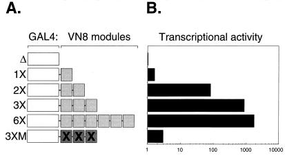
Our previous analysis of Myc (1) revealed a close correlation between activation domain and degron function. To determine whether this is true in other cases, we asked whether a series of TADs, derived by reiteration of an 8-aa sequence (DFDLDMLG; referred to as VN8) from the VP16 TAD (17), also could signal proteolysis. We chose these TADs because they are well defined and potent, and because their transcriptional potency can be varied predictably by changing their copy number and sequence. Fig. 2 shows the structure of GAL4–VN8 activators, together with their relative transcriptional activities in human cells. Each activator was constructed by fusing the GAL4 DBD to either one, two, three, or six tandem copies of VN8, or three copies of a mutant VN8 sequence—DADADMLG (17). Transcriptional activity then was measured by the ability of each activator to drive expression of a GAL4-binding site-containing reporter in human HeLa cells. Consistent with their activities in yeast (17), increasing the copy number of VN8 modules increased transcriptional potency. One copy of VN8 fused to the GAL4 DBD activated transcription 2-fold more than the GAL4 DBD alone, two copies activated transcription more than 100-fold, and three and six copies resulted in approximately 1,000- and 2,000-fold increases in transcriptional activation, respectively. As in yeast (17), the mutant VN8 module was much less potent (compare the activity of the 3× and 3×M activators).
Figure 2.
Tandem reiteration of the VN8 module generates transcriptional activators of widely differing potencies. (A) Structure of synthetic activators used in this study. Each activator carried the GAL4 DBD fused to 1×, 2×, 3×, or 6× copies of the wild-type VN8 sequence, or three copies of mutant VN8 sequence (3×M). (B) Relative transcriptional potency of each activator. GAL4-fusion proteins were assayed for transcriptional activation in human HeLa cells as described in Materials and Methods. Relative transcriptional activity is shown on a logarithmic scale, setting the transcriptional activity of the GAL4 DBD alone (Δ) at 1.
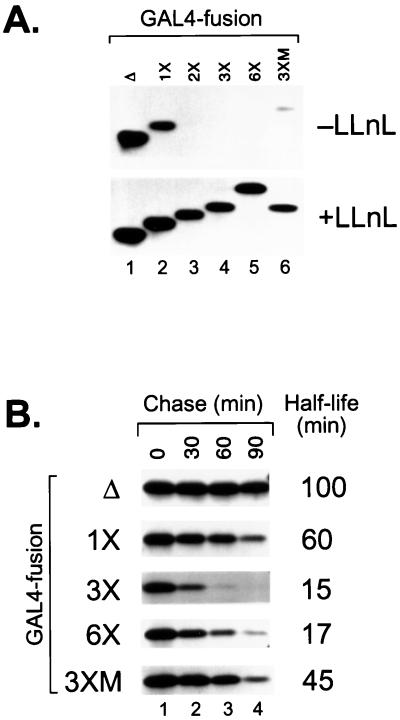
To address whether these TADs also function as degrons, we first compared the steady-state levels of the different GAL4–VN8 fusion proteins in the absence (−LLnL) or presence (+LLnL) of the proteasome inhibitor LLnL. The results of these studies are shown in Fig. 3A. The GAL4 DBD alone accumulated to the highest level (lane 1). Addition of one copy of VN8 reduced protein accumulation by approximately 50% (lane 2), whereas addition of two (lane 3), three (lane 4), or six (lane 5) copies of VN8 reduced protein accumulation to beneath detectable levels. Strikingly, however, the mutant 3×VN8 activator (lane 6) did accumulate to low, but detectable, levels under these conditions. Treatment of transfected cells with the proteasome inhibitor LLnL significantly altered the accumulation of these proteins; indeed, in the presence of LLnL (+LLnL), all GAL4-fusion proteins accumulated to comparable levels (compare lanes 1–6). Together, these data demonstrate: (i) that the accumulation of these transcriptional activators in human cells is inversely related to their transcriptional potency, and (ii) that a functioning proteasome is required for the low steady-state levels of the potent transcriptional activators.
Figure 3.
The ability of VN8 modules to signal proteolysis correlates with their ability to activate transcription. (A) Steady-state levels of the GAL4-VN8 activators. Human HeLa cells were transiently transfected with expression constructs encoding the indicated GAL4-fusion proteins. After transfection, cells were either untreated (−LLnL) or treated with the proteasome inhibitor LLnL (+LLnL). Total proteins were prepared and HA-tagged GAL4-fusion proteins revealed by SDS/PAGE and immunoblotting analysis. (B) Stability of GAL4–VN8 activators. GAL4-VN8 proteins were transiently expressed in HeLa cells and their stabilities were determined by pulse–chase analysis as described in Materials and Methods.
We next used pulse–chase analysis to measure the stability of the GAL4-VN8 fusion proteins, as shown in Fig. 3B. As before, the GAL4 DBD had a half-life of around 100 min. Addition of one VN8 module reduced the protein half-life to around 60 min, whereas addition of three and six copies of VN8 further reduced the protein half-life to 15 and 17 min, respectively. The mutant 3× activation domain, in contrast, had less effect on protein stability, reducing the half-life to 45 min. These data demonstrate that the VN8 TADs cause protein destabilization to an extent that is strikingly similar to their relative abilities to activate transcription. Importantly, the increased stability of the mutant activator (3×M), relative to its wild-type counterpart (3×), also demonstrates that tandem reiteration of sequences per se is not responsible for this effect.
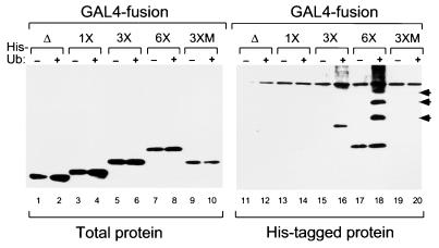
Lastly, we asked whether the VN8 TADs direct protein ubiquitylation. To do this, we asked whether GAL4–VN8 derivatives could become covalently linked to polyhistidine-tagged human Ub (His–Ub; ref. 2), as shown in Fig. 4. Under these conditions, the GAL4 DBD alone displayed little if any ubiquitylation (lane 12). Addition of one copy of VN8 did not result in any increase in ubiquitylation (lane 14). Addition of three and six copies of the VN8 module did, however, result in a significant increase in ubiquitylation: GAL4–3×VN8 (lane 16) displayed a significant level of monoubiquitylation, as well as a smaller level of higher molecular weight species, and GAL4–6×VN8 (lane 18) displayed at least three prominent high molecular weight Ub conjugates (arrowed; compare lanes 17 and 18), as well as a smear of larger Ub conjugates. In contrast to the wild-type 3×VN8 protein (lane 16), the mutant 3×M derivative (lane 20) failed to show any detectable level of ubiquitylation in this assay. Thus, as with steady-state protein accumulation and protein stability, there appears to be a direct relationship between transcriptional potency and ubiquitylation status, with potent transcriptional activators being more heavily ubiquitylated than weaker activators.
Figure 4.
The ubiquitylation status of GAL4-VN8 proteins is directly related to transcriptional potency. HeLa cells were transfected with expression constructs encoding the indicated GAL4-fusion proteins, either alone (odd-numbered lanes) or with an expression construct encoding polyhistidine-tagged human Ub (His-Ub; even-numbered lanes). After transfection, cells were treated with LLnL to allow the unstable GAL4-derivatives to accumulate. Cellular proteins were harvested as described in Materials and Methods, and HA-tagged GAL4-fusion proteins present in the total lysate (lanes 1–10) or the nickel-affinity-purified material (lanes 11–20) were revealed by SDS/PAGE and immunoblotting. The arrows indicate the position of His-Ub-GAL4–6×VN8 conjugates (lane 18).
These results clearly demonstrate that VN8 modules signal Ub-mediated proteolysis in a manner that reflects their ability to activate transcription. The tight overlap of these two activities within a set of small, well-defined protein sequences reveals that the relationship between activation domain and degron function is intimate. Together with the results of our previous studies of Myc, these data suggest that, in some cases, activation domains and degrons are identical elements. We refer to sequences with dual degron/activation domain function by the acronym DAD (destruction and activation domain).
Degrons from Yeast Cyclins Cln2 and Cln3 Activate Transcription.
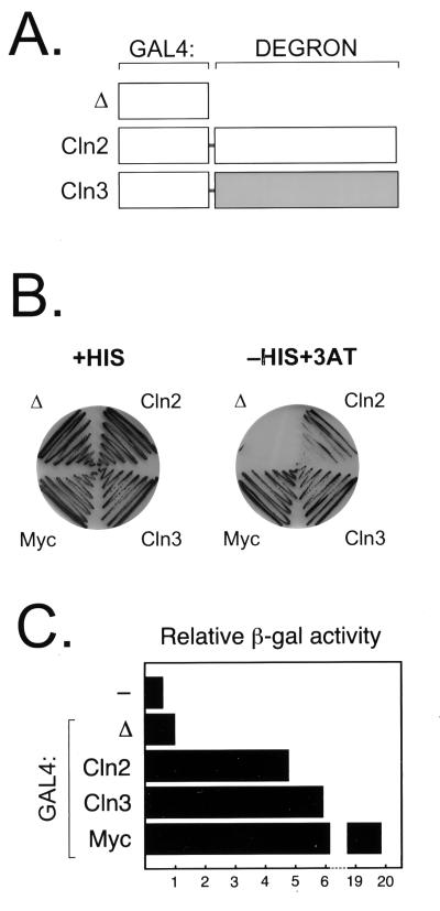
The realization that activation domains can signal proteolysis prompted us to ask the complementary question: Can degrons activate transcription? For this purpose, we chose degrons from the yeast cyclins Cln2 (18) and Cln3 (19). We fused the Cln2 and Cln3 degrons to the GAL4 DBD (as illustrated in Fig. 5A) and examined their ability to activate an appropriate reporter construct. We originally examined transcriptional activation by these proteins in human HeLa cells and found that GAL4-Cln2 and GAL4-Cln3 did not activate transcription (data not shown). We also found, however, that the Cln2 and Cln3 degrons did not destabilize the GAL4 DBD in HeLa cells (data not shown), suggesting that these elements could not function in a human cell environment. We therefore tested the activity of these GAL4-fusion proteins in yeast cells, where the Cln2 and Cln3 degrons can signal protein instability (18, 19). Results of this analysis are presented in Fig. 5 B and C.
Figure 5.
The Cln2 and Cln3 degrons activate transcription in yeast. (A) Structure of the GAL4–Cln2 and –Cln3 activators. (B) Activation of His3p expression. The indicated GAL4-fusion proteins were expressed in yeast strain HF7c, and their ability to activate transcription was scored by yeast growth in the presence of 3AT. Yeast were grown either in media containing histidine (nonselective; Left) or media lacking histidine and containing 2 mM 3AT (selective media; Right). (C) Activation of β-galactosidase (β-gal) expression. Liquid cultures of HF7c cells carrying an empty expression vector (−) or vectors expressing the indicated GAL4–fusion proteins were assayed for β-gal expression as described in Materials and Methods. Relative β-gal activity is expressed relative to β-gal activity directed by the GAL4 DBD alone (Δ), which is set at 1.
The yeast reporter strain we used in these experiments (HF7c; ref. 21) carries two GAL-responsive reporters that allow GAL4-fusion protein activity to be measured as either growth of yeast in the presence of 3AT or as β-galactosidase activity. By both criteria, the Cln2 and Cln3 degrons activate transcription. Although the GAL4 DBD alone did not support yeast growth in the presence of 2 mM 3AT, the GAL4–Cln2 and GAL4–Cln3 fusion proteins supported yeast growth under these conditions (Fig. 5B). In addition, the GAL4–Cln2 and –Cln3 proteins activated β-galactosidase synthesis to levels 5–6 times higher than that of the GAL4 DBD alone (Fig. 5C). This level of activation was considerably less than that seen with GAL4–Myc; this difference is consistent with the fact that the Cln2 and Cln3 degrons are also less effective at destabilizing GAL4 than the Myc DAD (data not shown).
The normal role of Cln3 is to activate the transcription factors SBF and MBF in late G1 phase of the cell cycle (38). Because of our finding that the GAL4–Cln3 fusion activated transcription, we were concerned that this could reflect a natural role of Cln3; that is, perhaps Cln3 becomes part of the SBF/MBF DNA complex and activates transcription directly. If this were so, it would invalidate our assumption that the C-terminal domain of Cln3 has evolved as a degron, rather than as a TAD. However, we were able to identify mutant alleles of CLN3 that lacked the ability to activate transcription as part of a GAL4 fusion, and yet complemented a cln1 cln2 cln3 deletion (data not shown). Thus, it appears that the normal role of Cln3 does not involve direct transcriptional activation.
The experiments presented in Fig. 5 demonstrate that the Cln2 and Cln3 degrons can activate transcription. The finding that these degrons can activate transcription provides further, compelling evidence for an intimate functional relationship between TADs and degrons. It is curious to note that, unlike the other DADs we have discussed, the Cln2 and Cln3 degrons are not enriched in acidic amino acid residues. These sequences do, however, contain a large number of serine and threonine residues and have been shown to be phosphorylated (39, 40). In the case of Cln2, this phosphorylation is a prerequisite for degron function (41), and this is likely true for Cln3 as well (42). Perhaps the negative charge provided by phosphorylation of these sequences mimics the environment provided by an acidic activation domain. Similarly, perhaps some acidic activation domains have degron function because they mimic the environment provided by a phosphorylated degron.
What Is the Significance of Overlapping Activation Domains and Degrons?
Our data reveal that two very different events—transcriptional activation and Ub-mediated proteolysis—have converged on a set of sequences capable of functioning in both processes. The dual function of these DADs raises the question of why transcriptional activation and proteolysis can be signaled by a common element. One possibility is that cells have evolved a machinery to destroy potent transcriptional activators; in this case, recognition of TADs by the ubiquitylation machinery would provide an efficient means to target these proteins for destruction. Such regulation could be important for rapid reprogramming of transcriptional patterns, to limit activation by any one single activator, or to prevent an accumulation of excess activator that could lead to “squelching” of the basal machinery.
Considering our observation that degrons can activate transcription, however, another possibility is that the overlap of TADs and degrons reflects a functional link between transcriptional activation and Ub-mediated proteolysis. Perhaps there is a common cellular machinery that participates in both processes. Indeed, there are many observations that support such a scenario. For example, two subunits of the proteasome, Sug1 and Sug2, interact genetically (43, 44) and biochemically (45) with transcriptional activation domains. Sug1 also interacts with a subunit of the basal transcription factor TFIIH (46). In addition, the yeast Ub-protein ligase RSP5, and its human homolog hPRF1, have coactivator function in vivo (47), directly interact with TADs in vitro (48), and can ubiquitylate the largest subunit of RNA polymerase II (49). Finally, we note that histones are ubiquitylated and that their ubiquitylation status correlates with transcriptional activity (e.g., ref. 50). Perhaps, therefore, some activators can stimulate transcription by recruiting components of the Ub-proteasome pathway that subsequently modify histones, basal factors, and ultimately the activators themselves.
In conclusion, our data demonstrate that some activation domains have a second function—the ability to signal Ub-mediated proteolysis. The surprising overlap of two very different processes suggests that many transcriptional activators are destroyed because of their ability to activate transcription and implies that a common targeting mechanism underlies the destruction of transcriptional activators.
Acknowledgments
For reagents we thank C. Bautista, D. Bohmann, G. Hannon, and M. Tanaka. We thank D. Conklin, S. Kim, K. Tworkowski, and W. Herr for critical comments and suggestions. M.M. thanks the Atsushi Kitaueda Memorial Fund for supporting his travel to New York. W.P.T. is a Kimmel Scholar. This work was supported by Cold Spring Harbor Laboratory Cancer Center Support Grant CA45508, The Robertson Research Fund, The Cold Spring Harbor Laboratory Association, National Institutes of Health Grant GM 39978 and U.S. Public Health Service Grant CA-13106 from the National Cancer Institute.
Abbreviations
- DAD
destruction and activation domain
- DBD
DNA-binding domain
- TAD
transcriptional activation domain
- Ub
ubiquitin
- 3AT
3-amino-triazole
- HA
hemagglutinin
- PS1
proteasome inhibitor 1
- LLnL
N-acetyl-Leu-Leu-norleucinal
Footnotes
Article published online before print: Proc. Natl. Acad. Sci. USA, 10.1073/pnas.050007597.
Article and publication date are at www.pnas.org/cgi/doi/10.1073/pnas.050007597
References
- 1.Salghetti S E, Kim S Y, Tansey W P. EMBO J. 1999;18:717–726. doi: 10.1093/emboj/18.3.717. [DOI] [PMC free article] [PubMed] [Google Scholar]
- 2.Treier M, Staszewski L M, Bohmann D. Cell. 1994;78:787–798. doi: 10.1016/s0092-8674(94)90502-9. [DOI] [PubMed] [Google Scholar]
- 3.Chowdary D R, Dermody J J, Jha K K, Ozer H L. Mol Cell Biol. 1994;14:1997–2003. doi: 10.1128/mcb.14.3.1997. [DOI] [PMC free article] [PubMed] [Google Scholar]
- 4.Campanero M R, Flemington E K. Proc Natl Acad Sci USA. 1997;94:2221–2226. doi: 10.1073/pnas.94.6.2221. [DOI] [PMC free article] [PubMed] [Google Scholar]
- 5.Pariat M, Carillo S, Molinari M, Salvat C, Debussche L, Bracco L, Milner J, Piechaczyk M. Mol Cell Biol. 1997;17:2806–2815. doi: 10.1128/mcb.17.5.2806. [DOI] [PMC free article] [PubMed] [Google Scholar]
- 6.Cuervo A M, Hu W, Lim B, Dice J F. Mol Biol Cell. 1998;9:1995–2010. doi: 10.1091/mbc.9.8.1995. [DOI] [PMC free article] [PubMed] [Google Scholar]
- 7.Varshavsky A. Trends Biochem Sci. 1997;22:383–387. doi: 10.1016/s0968-0004(97)01122-5. [DOI] [PubMed] [Google Scholar]
- 8.DeMartino G N, Slaughter C A. J Biol Chem. 1999;274:22123–22126. doi: 10.1074/jbc.274.32.22123. [DOI] [PubMed] [Google Scholar]
- 9.Varshavsky A. Proc Natl Acad Sci USA. 1996;93:12142–12149. doi: 10.1073/pnas.93.22.12142. [DOI] [PMC free article] [PubMed] [Google Scholar]
- 10.Glotzer M, Murray A W, Kirschner M W. Nature (London) 1991;349:132–138. doi: 10.1038/349132a0. [DOI] [PubMed] [Google Scholar]
- 11.Rogers S, Wells R, Rechsteiner M. Science. 1986;234:364–368. doi: 10.1126/science.2876518. [DOI] [PubMed] [Google Scholar]
- 12.Mermod N, O'Neill E A, Kelly T J, Tjian R. Cell. 1989;58:741–753. doi: 10.1016/0092-8674(89)90108-6. [DOI] [PubMed] [Google Scholar]
- 13.Tanaka M, Herr W. Mol Cell Biol. 1994;14:6056–6067. doi: 10.1128/mcb.14.9.6056. [DOI] [PMC free article] [PubMed] [Google Scholar]
- 14.Courey A J, Tjian R. Cell. 1988;55:887–898. doi: 10.1016/0092-8674(88)90144-4. [DOI] [PubMed] [Google Scholar]
- 15.Flemington E K, Speck S H, Kaelin W G., Jr Proc Natl Acad Sci USA. 1993;90:6914–6918. doi: 10.1073/pnas.90.15.6914. [DOI] [PMC free article] [PubMed] [Google Scholar]
- 16.Triezenberg S J, Kingsbury R C, McKnight S L. Genes Dev. 1988;2:718–729. doi: 10.1101/gad.2.6.718. [DOI] [PubMed] [Google Scholar]
- 17.Tanaka M. Proc Natl Acad Sci USA. 1996;93:4311–4315. doi: 10.1073/pnas.93.9.4311. [DOI] [PMC free article] [PubMed] [Google Scholar]
- 18.Salama S R, Hendricks K B, Thorner J. Mol Cell Biol. 1994;14:7953–7966. doi: 10.1128/mcb.14.12.7953. [DOI] [PMC free article] [PubMed] [Google Scholar]
- 19.Tyers M, Tokiwa G, Nash R, Futcher B. EMBO J. 1992;11:1773–1784. doi: 10.1002/j.1460-2075.1992.tb05229.x. [DOI] [PMC free article] [PubMed] [Google Scholar]
- 20.Hofmann F, Martelli F, Livingston D M, Wang Z. Genes Dev. 1996;10:2949–2959. doi: 10.1101/gad.10.23.2949. [DOI] [PubMed] [Google Scholar]
- 21.Feilotter H E, Hannon G J, Ruddell C J, Beach D. Nucleic Acids Res. 1994;22:1502–1503. doi: 10.1093/nar/22.8.1502. [DOI] [PMC free article] [PubMed] [Google Scholar]
- 22.Brown H J, Sutherland J A, Cook A, Bannister A J, Kouzarides T. EMBO J. 1995;14:124–131. doi: 10.1002/j.1460-2075.1995.tb06982.x. [DOI] [PMC free article] [PubMed] [Google Scholar]
- 23.Tsurumi C, Ishida N, Tamura T, Kakizuka A, Nishida E, Okumura E, Kishimoto T, Inagaki M, Okazaki K, Sagata N, et al. Mol Cell Biol. 1995;15:5682–5687. doi: 10.1128/mcb.15.10.5682. [DOI] [PMC free article] [PubMed] [Google Scholar]
- 24.Hope I A, Mahadevan S, Struhl K. Nature (London) 1988;333:635–640. doi: 10.1038/333635a0. [DOI] [PubMed] [Google Scholar]
- 25.Kornitzer D, Raboy B, Kulka R G, Fink G R. EMBO J. 1994;13:6021–6030. doi: 10.1002/j.1460-2075.1994.tb06948.x. [DOI] [PMC free article] [PubMed] [Google Scholar]
- 26.Jiang B H, Zheng J Z, Leung S W, Roe R, Semenza G L. J Biol Chem. 1997;272:19253–19260. doi: 10.1074/jbc.272.31.19253. [DOI] [PubMed] [Google Scholar]
- 27.Huang L E, Gu J, Schau M, Bunn H F. Proc Natl Acad Sci USA. 1998;95:7987–7992. doi: 10.1073/pnas.95.14.7987. [DOI] [PMC free article] [PubMed] [Google Scholar]
- 28.Baichwal V R, Tjian R. Cell. 1990;63:815–825. doi: 10.1016/0092-8674(90)90147-7. [DOI] [PubMed] [Google Scholar]
- 29.Kato G J, Barrett J, Villa-Garcia M, Dang C V. Mol Cell Biol. 1990;10:5914–5920. doi: 10.1128/mcb.10.11.5914. [DOI] [PMC free article] [PubMed] [Google Scholar]
- 30.Fields S, Jang S K. Science. 1990;249:1046–1049. doi: 10.1126/science.2144363. [DOI] [PubMed] [Google Scholar]
- 31.Haupt Y, Maya R, Kazaz A, Oren M. Nature (London) 1997;387:296–299. doi: 10.1038/387296a0. [DOI] [PubMed] [Google Scholar]
- 32.Moore P A, Ruben S M, Rosen C A. Mol Cell Biol. 1993;13:1666–1674. doi: 10.1128/mcb.13.3.1666. [DOI] [PMC free article] [PubMed] [Google Scholar]
- 33.Chen E, Hrdlickova R, Nehyba J, Longo D L, Bose H R, Jr, Li C C. J Biol Chem. 1998;273:35201–35207. doi: 10.1074/jbc.273.52.35201. [DOI] [PubMed] [Google Scholar]
- 34.Simcha I, Shtutman M, Salomon D, Zhurinsky J, Sadot E, Geiger B, Ben-Ze'ev A. J Cell Biol. 1998;141:1433–1448. doi: 10.1083/jcb.141.6.1433. [DOI] [PMC free article] [PubMed] [Google Scholar]
- 35.Orford K, Crockett C, Jensen J P, Weissman A M, Byers S W. J Biol Chem. 1997;272:24735–24738. doi: 10.1074/jbc.272.40.24735. [DOI] [PubMed] [Google Scholar]
- 36.Chen R H, Fields S, Lipsick J S. Oncogene. 1995;11:1771–1779. [PubMed] [Google Scholar]
- 37.Bies J, Nazarov V, Wolff L. J Virol. 1999;73:2038–2044. doi: 10.1128/jvi.73.3.2038-2044.1999. [DOI] [PMC free article] [PubMed] [Google Scholar]
- 38.Tyers M, Tokiwa G, Futcher B. EMBO J. 1993;12:1955–1968. doi: 10.1002/j.1460-2075.1993.tb05845.x. [DOI] [PMC free article] [PubMed] [Google Scholar]
- 39.Willems A R, Lanker S, Patton E E, Craig K L, Nason T F, Mathias N, Kobayashi R, Wittenberg C, Tyers M. Cell. 1996;86:453–463. doi: 10.1016/s0092-8674(00)80118-x. [DOI] [PubMed] [Google Scholar]
- 40.Yaglom J A, Goldberg A L, Finley D, Sherman M Y. Mol Cell Biol. 1996;16:3679–3684. doi: 10.1128/mcb.16.7.3679. [DOI] [PMC free article] [PubMed] [Google Scholar]
- 41.Skowyra D, Craig K L, Tyers M, Elledge S J, Harper J W. Cell. 1997;91:209–219. doi: 10.1016/s0092-8674(00)80403-1. [DOI] [PubMed] [Google Scholar]
- 42.Yaglom J, Linskens M H, Sadis S, Rubin D M, Futcher B, Finley D. Mol Cell Biol. 1995;15:731–741. doi: 10.1128/mcb.15.2.731. [DOI] [PMC free article] [PubMed] [Google Scholar]
- 43.Rubin D M, Coux O, Wefes I, Hengartner C, Young R A, Goldberg A L, Finley D. Nature (London) 1996;379:655–657. doi: 10.1038/379655a0. [DOI] [PubMed] [Google Scholar]
- 44.Russell S J, Sathyanarayana U G, Johnston S A. J Biol Chem. 1996;271:32810–32817. doi: 10.1074/jbc.271.51.32810. [DOI] [PubMed] [Google Scholar]
- 45.Swaffield J C, Melcher K, Johnston S A. Nature (London) 1995;374:88–91. doi: 10.1038/374088a0. [DOI] [PubMed] [Google Scholar]
- 46.Weeda G, Rossignol M, Fraser R A, Winkler G S, Vermeulen W, van't Veer L J, Ma L, Hoeijmakers J H, Egly J M. Nucleic Acids Res. 1997;25:2274–2283. doi: 10.1093/nar/25.12.2274. [DOI] [PMC free article] [PubMed] [Google Scholar]
- 47.Imhof M O, McDonnell D P. Mol Cell Biol. 1996;16:2594–2605. doi: 10.1128/mcb.16.6.2594. [DOI] [PMC free article] [PubMed] [Google Scholar]
- 48.Gavva N R, Gavva R, Ermekova K, Sudol M, Shen C J. J Biol Chem. 1997;272:24105–24108. doi: 10.1074/jbc.272.39.24105. [DOI] [PubMed] [Google Scholar]
- 49.Huibregtse J M, Yang J C, Beaudenon S L. Proc Natl Acad Sci USA. 1997;94:3656–3661. doi: 10.1073/pnas.94.8.3656. [DOI] [PMC free article] [PubMed] [Google Scholar]
- 50.Davie J R, Murphy L C. Biochemistry. 1990;29:4752–4757. doi: 10.1021/bi00472a002. [DOI] [PubMed] [Google Scholar]


