Abstract
Gene delivery to skeletal muscle is a promising strategy for the treatment of muscle disorders and for the systemic secretion of therapeutic proteins. However, present DNA delivery technologies have to be improved with regard to both the level of expression and interindividual variability. We report very efficient plasmid DNA transfer in muscle fibers by using square-wave electric pulses of low field strength (less than 300 V/cm) and of long duration (more than 1 ms). Contrary to the electropermeabilization-induced uptake of small molecules into muscle fibers, plasmid DNA has to be present in the tissue during the electric pulses, suggesting a direct effect of the electric field on DNA during electrotransfer. This i.m. electrotransfer method increases reporter and therapeutic gene expression by several orders of magnitude in various muscles in mouse, rat, rabbit, and monkey. Moreover, i.m. electrotransfer strongly decreases variability. Stability of expression was observed for at least 9 months. With a pCMV-FGF1 plasmid coding for fibroblast growth factor 1, this protein was immunodetected in the majority of muscle fibers subjected to the electric pulses. DNA electrotransfer in muscle may have broad applications in gene therapy and in physiological, pharmacological, and developmental studies.
Nonviral gene transfer for gene therapy is a rapidly expanding field (1–3). Tissues for which highly efficient gene transfer is sought include tumors, various epithelia or endothelia, and organs such as the liver, heart, or brain. Special interest has been devoted to gene delivery to skeletal muscle fibers, e.g., for the correction of myopathies such as Duchenne’s muscular dystrophy (4), for the local secretion of angiogenic or neurotrophic factors, and also for vaccination (5). Another exciting application is the use of highly vascularized muscle as an endocrine organ for the systemic secretion of therapeutic proteins, such as erythropoietin, α1-antitrypsin, clotting factors, antiinflammatory cytokines, etc. (6–10).
The transfer of a foreign gene into muscle fibers by injection of “naked” plasmid DNA has been demonstrated (11–13), leading to transgene expression and physiological or therapeutic responses, such as vaccinal and antiinflammatory response or hematocrit increase (8, 9, 14–16). However, there are several major limitations to the transfer of naked DNA. First, there is a very high interindividual variability of the level of foreign gene expression. The origin of this lack of reproducibility is not yet understood, and it might strongly impair the clinical development of therapeutic use of i.m. DNA administration. Second, the level of therapeutic protein expression is still too low for efficient treatment of diseases such as hemophilia or muscular dystrophy. Thus, alternative strategies aimed at improving the level and reliability of gene expression after i.m. DNA injection are needed. The use of ballistic technology (a “gene gun”) resulted in a moderate enhancement of gene transfer into skin (17) or into muscle after surgical limb opening (18); the principal limitation was a poor penetration across tissues. Until now, no enhancement of gene transfer in muscle, as compared with naked DNA, has been achieved by using cationic lipids or polymers (19, 20; C. Dubertret, B. Pitard, and D.S., unpublished data). It is surprising that this enhancement has not been observed, considering that cationic lipids lead to efficient transfection in vitro (1, 2, 21–25) and in vivo in tissues such as brain or tumor (26–29). Neutral polymers, such as polyvinyl derivatives, have been reported to enhance plasmid delivery to rat muscle by 2- to 6-fold (30).
Since the 1982 publication by Neumann et al. (31), the use of electric pulses for cell electropermeabilization (also called cell electroporation) has been used to introduce foreign DNA into prokaryotic and eukaryotic cells in vitro (32) and has been used in other applications in cell biology and pharmacology (33). By using optimal electrical parameters, it is possible to achieve excellent levels of cell permeabilization that are compatible with cell survival (34). Cell electropermeabilization also can be achieved locally in vivo by means of simple electrodes (35, 36). A relevant clinical application, electrochemotherapy, is being pursued in oncology, by using cell electropermeabilization to favor the cellular entry of hydrophilic anticancer agents like bleomycin (35, 37–39). This approach results in a drastically increased antitumor effect of bleomycin. The short (100-μs) and intense (800- to 1,500-V/cm) electric pulses used in current electrochemotherapy protocols are safe and well tolerated (35, 37–39).
Electric pulses also have been used to transfer DNA in vivo to skin, liver, and tumor cells (40–44). Initial experiments were performed by using runs of short (100- to 300-μs) and intense (400- to 1,200-V/cm) electric pulses (40, 41). Long electric pulses of lower voltage-to-distance ratios have been reported to increase DNA transfer in vivo to tumor cells (10 pulses; 800 V/cm; 4 ms per pulse; ref. 42), to liver cells (8 pulses; 250 V/cm; 50 ms per pulse; ref. 43), and, in a study published during the completion of the present article, to muscle cells (6 pulses; 200 V/cm; 50 ms per pulse; ref. 44). We report here the characteristics and mechanisms of highly efficient and reproducible plasmid transfer to muscle fibers achieved in vivo by the local application of electric pulses, and we discuss the features of this i.m. DNA electrotransfer method that open possibilities for nonviral gene therapy.
MATERIALS AND METHODS
Plasmids and DNA Preparation.
The plasmids pXL2774 (pCMV-Luc), pXL3031 (pCMV-Luc+), pXL3004 (pCMV-βgal), and pXL3096 (pCMV-FGF1) contain the cytomegalovirus (CMV) promoter (nucleotides 229–890 of pcDNA3; Invitrogen) inserted upstream of the coding sequence, respectively, of the Photinus pyralis peroxisomal luciferase (pGL2 basic; Promega), of the modified cytosolic luc+ luciferase, of the Escherichia coli LacZ gene coding for the β-galactosidase, and of the gene coding for a fusion peptide between the signal secretion peptide of human fibroblast interferon and fibroblast growth factor 1 (FGF1; ref. 45). Plasmids were prepared by using standard procedures (46). All plasmid preparations contained a high percentage of supercoiled DNA (70–80%), and no RNA was detectable by gel electrophoresis.
DNA Injection and Electric-Pulse Delivery.
Unless otherwise stated, 15 μg of DNA in 30 μl of 0.9% NaCl was injected into the tibial cranial muscle of anesthetized C57Bl/6 mice (Iffa Credo) with a Hamilton syringe. There were 10 tibial cranial muscles included in each experimental group. At defined times after DNA injection (25 s or 1 min, depending on the experiment), transcutaneous electric pulses were applied by two stainless steel plate electrodes placed 4.2–5.3 mm apart, at each side of the leg. The 1-cm plates encompassed the whole leg of each mouse. Electrical contact with the leg skin was ensured by shaving each leg and applying a conductive gel. Square-wave electric pulses were generated by a PS-15 electropulsator (Jouan, St. Herblain, France), except in the analysis of pulse-length influence, for which we used a T820 electroporator (BTX, San Diego), which allowed the delivery of pulses for 1–99 ms. Electric-field strengths (in V/cm) are reported in terms of the ratio of the applied voltage to the distance between the electrodes.
For pulse delivery to rat and rabbit muscles, two implanted stainless steel needles (Terumo, Leuven, Belgium; 0.08 mm in diameter × 50 mm in length) (the depth of insertion was 2 cm; the gap between the two needles was 0.9 cm) were used as electrodes and connected to the generator. The efficiency of needle electrodes for DNA electrotransfer to rat muscle is similar to that of external plate electrodes (data not shown). pXL3031 (150 μg; 0.5 μg/μl) was injected into rat muscle, and pXL2774 (200 μg; 1 μg/μl) was injected into rabbit muscle. The needle electrodes also were used in the monkey trial. All animals were anesthetized during the whole procedure. Experiments were conducted following the recommendations of the National Institutes of Health for animal experimentation and those of the Rhône-Poulenc-Rorer local Ethics Committee on Animal Care and Experimentation.
Measurement of Luciferase Activity.
Unless otherwise stated, mice were killed 7 days after DNA transfer. Muscles were removed and homogenized in 1.5 ml of Cell Culture Lysis reagent (Promega) supplemented with aprotinin (Sigma) at a final concentration of 2 μg/ml. After centrifugation at 5,000 × g for 15 min, luciferase activity was assessed on 10 μl of supernatant with a Berthold Lumat LB 9501 luminometer (EG & G, Evry, France). This assessment was made by measuring the integration of the light produced during 10 s, starting 1 s after the addition of 50 μl of Luciferase Assay Substrate (Promega) to the muscle-fiber lysate. Calibration performed with purified firefly luciferase protein (Sigma and Boehringer Mannheim) established that 106 relative light units was approximately equivalent to 30 pg of luciferase. Results are expressed as picograms of luciferase per muscle ±SEM (see the figures) or nanograms per muscle ±SEM (see Table 1). Because electric fields have been shown to induce production of free radicals, which could interfere with the luciferase detection assay, we verified, by using a plasmid coding for β-galactosidase, that electrotransfer did not result in false-positive detection of luciferase activity (data not shown).
Table 1.
Increase of foreign gene expression after electric-pulse delivery in various mouse, rat, and rabbit muscles
| Species | Electric pulses | Amount of luciferase expressed, ng
|
|||
|---|---|---|---|---|---|
| Gastrocnemius | Rectus femoris | Triceps brachii | Tibialis cranialis | ||
| Mouse | − | 0.3 ± 0.1 | 0.2 ± 0.1 | 0.03 ± 0.03 | — |
| + | 64.6 ± 16.4 | 82.2 ± 26.1 | 33.6 ± 17.3 | — | |
| Rat | − | — | 0.8 ± 0.4 | 13.3 ± 7.5 | 17.4 ± 5.9 |
| + | — | 130.2 ± 65.1 | 175.6 ± 23.8 | 3249.1 ± 617.4 | |
| Rabbit | − | — | 0.01 ± 0.01 | 0.31 ± 0.1 | 0.02 ± 0.01 |
| + | — | 43.1 ± 24.5 | 223.8 ± 157.7 | 640.5 ± 159.4 | |
Chromium-51-EDTA (51Cr-EDTA) Uptake.
A small molecule, 51Cr-EDTA (Amersham Pharmacia), was used as a permeabilization indicator. In parallel experiments, either 51Cr-EDTA (0.01 MBq in 30 μl saline) or pXL2774 plasmid (15 μg in 30 μl saline) was injected i.m. into mouse tibial cranial muscle at different times either before or after electric-pulse delivery, by using the same procedure as described above for DNA (eight pulses; 200 V/cm; 20 ms per pulse; 1 Hz). For the experiments on the influence of voltage and the number of pulses on 51Cr-EDTA entrapment, 50 μl of 51Cr-EDTA (0.2 MBq) was i.v. injected 2.5 min before pulsation. In all cases, mice were killed 1 h after 51Cr-EDTA injection. The treated muscle was excised and weighed, and radioactivity was measured in a Packard γ counter. Taking into account the specific radioactivity, the decay, and the period from the reference date, calculations were performed to determine the uptake in nanomoles of 51Cr-EDTA per gram of treated muscle.
Immunohistochemical Procedures.
Muscles were removed, fixed, and sliced 7 days after DNA electrotransfer. To show β-galactosidase expression, staining with 5-bromo-4-chloro-3-indolyl β-d-galactoside was performed in toto according to described procedures (47). For detection of FGF1 expression, sections were subjected to indirect immunohistochemistry on an automat (Ventana Medical Systems, Tucson, AZ), by first using a polyclonal rabbit anti-human FGF1 antibody dilvent, Ventana Medical Systems; 1/200) and then a biotinylated donkey polyclonal anti-rabbit antibody (The Jackson Laboratory; 1/800) and then an avidin-biotin-alkaline phosphatase revelation kit (Jackson ImmunoResearch).
RESULTS
Influence of the Electric-Pulse Parameters on Reporter-Gene Expression.
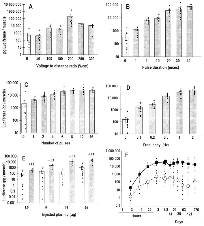
A luciferase gene-encoding plasmid was injected i.m. into mouse-leg tibial cranial muscle, and electric pulses were delivered through external plate electrodes placed at each side of the leg. When we applied electric pulses similar to those used for electrochemotherapy and for liver DNA transfer (eight pulses; 100 μs per pulse; 1,200 V/cm; 1 Hz), we observed a 50-fold decrease of muscle luciferase expression, as compared with the values observed in the absence of an electric field. In contrast, we found that eight long (20-ms) and less intense (200-V/cm) electric pulses applied at a 1-Hz frequency resulted in a marked enhancement of luciferase expression and in a drastic reduction in interindividual variability (Fig. 1A). This effect was observed constantly in 149 separate muscles (data not shown). We refer to this procedure as electrotransfer.
Figure 1.
Characteristics of DNA electrotransfer in muscle fibers. Bars represent the mean ± SEM of individual values (triangles), observed in the absence (white bars) or the presence (gray bars) of electric-pulse delivery. The ordinate scale is logarithmic. (A) Influence of the applied voltage-to-distance ratio (eight pulses; 20 ms per pulse; 1 Hz). (B) Influence of pulse length (eight pulses; 200 V/cm; 1 Hz). (C) Influence of pulse number (200 V/cm; 20 ms per pulse; 1 Hz). (D) Influence of the frequency of pulse delivery (eight pulses; 200 V/cm; 20 ms per pulse). (E) Dependence of luciferase gene expression on the amount of injected DNA in a constant volume of 30 μl (eight pulses; 200 V/cm; 20 ms per pulse; 1 Hz); (F) Kinetics of luciferase gene expression in the absence (open circles) or presence (filled squares) of electrotransfer (eight pulses; 200 V/cm; 20 ms per pulse; 2 Hz). Abscissa: time after plasmid injection. Mice were killed 7 days (A–E) or at the indicated times (F) after DNA transfer.
Electrotransfer efficiency depended strongly on the characteristics of the electric pulses. Enhancement of expression was detected from a threshold field strength of 100 V/cm and was optimal at 200 V/cm (eight pulses; 20 ms per pulse; 1 Hz; Fig. 1A). Under these conditions, luciferase expression was enhanced approximately 100-fold. Transgene expression was reduced at 300 V/cm or more (Fig. 1A and data not shown). At 200 V/cm (eight pulses; 1 Hz), the electrotransfer efficiency increased with the duration of each pulse up to 40 ms (Fig. 1B). In other experiments, a plateau value was obtained at 20 ms per pulse, and luciferase expression clearly leveled off for pulses lasting >40 ms per pulse (data not shown). At 200 V/cm and 20 ms per pulse and 1 Hz, 8–16 pulses led to the highest enhancement (Fig. 1C). In other experiments that used similar pulse characteristics, 8 pulses yielded the best results, and a clear decrease of the electrotransfer effect was observed with 32 pulses (data not shown). Subsequent experiments showed that at the optimal conditions of eight pulses, 200 V/cm, and 20 ms per pulse, an additional gain could be obtained by increasing the frequency of pulse administration (Fig. 1D).
Characteristics of the Expression of the Electrotransferred DNA.
i.m. gene electrotransfer increased as a function of the amount of DNA injected, whereas no clear dose–effect relationship was observed in the absence of the electric pulses (Fig. 1E).
Note that, after electrotransfer, a clear reduction in the interindividual variability of gene expression, as indicated by the relative SEM, was observed constantly, in contrast to the classical high variability observed with DNA administered alone (Fig. 1 A–E). For example, in Fig. 1E, the relative SEM (in percentage of the mean value) decreased from 57.2–64.3% for controls without electric pulses to 16.2–16.9% when electric pulses were applied.
The time course of expression of electrotransferred luciferase gene expression was studied (Fig. 1F). Luciferase expression was already detectable 3 h after DNA injection in the absence or presence of electric pulses. It increased progressively and, at day 3, reached a plateau value that persisted for at least 270 days. At all time points of the reported experiment, expression of the electrotransferred DNA was much higher than that of DNA injected without electric pulses (from 25-fold at 3 h to 734-fold at 121 days).
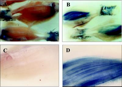
Under optimal conditions (eight pulses; 200 V/cm; 20 ms per pulse; 1 Hz), histological analysis with the β-galactosidase reporter gene showed that electrotransfer increased global β-galactosidase expression, the number of muscle fibers positive for β-galactosidase, and the volume of tissue expressing the foreign gene (Fig. 2). i.m. gene electrotransfer was restricted to the muscle volume exposed to the electric pulses (data not shown).
Figure 2.
β-Galactosidase expression in mouse tibial cranial muscle after DNA injection with or without the application of electric pulses. (A and B) Legs from mice injected with DNA without (A) or with (B) exposure to the electric pulses (eight pulses; 200 V/cm; 20 ms per pulse; 1 Hz). (C and D) Macroscopic photographs of the whole tibial cranial muscle of mice injected with DNA without (C) or with (D) exposure to electric pulses.
The in vivo electropermeabilization of muscle fibers, which results from membrane perturbation caused by the electric field, was determined by assaying the uptake of low molecular mass 51Cr-EDTA, a small complex used as a permeabilization indicator. At eight pulses, 20 ms per pulse, 1 Hz, the same field-threshold value of 100 V/cm was observed for muscle-fiber electropermeabilization as for electrotransfer.
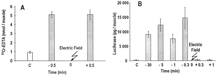
The uptake of 51Cr-EDTA was increased whether its i.m. injection was performed before or after the electric pulses (Fig. 3A). In contrast, DNA had to be present in the tissue at the time of the electric-pulse application to observe electrotransfer. Indeed, when DNA injection was performed 20 s (or later) after the delivery of electric pulses, no increase in foreign gene expression was detected (Fig. 3B). Interestingly, the electrotransfer effect was detected when DNA was injected as long as 30–60 min before the delivery of electric pulses but was lost when DNA was injected 2 h before the delivery (Fig. 3A and data not shown).
Figure 3.
Comparative effect of the time of injection vs. time of electric-pulse delivery on the in vivo 51Cr-EDTA uptake and on luciferase gene expression by muscle fibers. Bars represent the mean of the individuals values; error bars represent SEM. Gray bars represent the uptake of 51Cr-EDTA (A) or the uptake of plasmid DNA (B) as assessed by luciferase gene expression 7 days after the electric pulses (eight pulses; 200 V/cm; 20 ms per pulse; 1 Hz). White bars represent control values observed in the absence of electric pulses.
Finally, for 20-ms electric pulses, in contrast to plasmid electrotransfer, 51Cr-EDTA uptake increased up to a voltage-to-distance ratio of 400 V/cm before leveling off and increased with the number of pulses up to 32 pulses (data not shown). Note that significant 51Cr-EDTA uptake was also observed with eight 100-μs pulses at 1,200 V/cm (data not shown), conditions that led to strong decrease of electrotransferred plasmid expression, as reported above.
Electrotransfer to Rat, Rabbit, and Primate Muscles of Reporter or Therapeutic Genes.
All preceding experiments were performed on mouse tibialis cranialis muscle. A 2- to 4-log enhancement of luciferase gene transfer by electric pulses also was observed in various mouse, rat, and rabbit muscles (Table 1). Either external plate electrodes or needle electrodes directly implanted in the muscle were used. A similar electrotransfer efficacy was found for both types of electrodes in rat muscles.
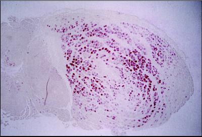
An electrotransfer-induced increase of gene expression also was obtained with DNA coding for FGF1, which, when secreted locally, is a candidate for therapeutic angiogenesis in peripheral artery disease and in ischemic heart disease (48, 49). After electrotransfer of plasmid pXL3096, immunohistology identified FGF1 expression in the majority of the fibers of the injected mouse leg muscle (Fig. 4). Interestingly, histology shows that injected plasmid DNA diffuses extensively in muscle tissue, because fibers positive for FGF1 were detected in the whole muscle section. In contrast, in the absence of electric pulses, FGF1 immunodetection was restricted to the vicinity of the path of the DNA-injecting needle. At days 3 and 7 after DNA injection, an average of 0 and 4 ± 2 fibers immunopositive for FGF1 per muscle (n = 5) were observed in the absence of electric pulses, and an average of 320 ± 45 and 710 ± 122 fibers immunopositive for FGF1 were observed with electrotransfer, respectively (n = 5; mean ± SEM).
Figure 4.
FGF1 expression in mouse tibial cranial muscle. A run of eight pulses of 200 V/cm and 20 ms per pulse at 2 Hz was used, and muscle was removed 7 days after electrotransfer of the pXL3096 plasmid.
We also tested the injection of 500 μg of plasmid pCMV-FGF1 (pXL3096) in the tibialis cranialis, rectus femoris, triceps, and biceps of a Maccaca fascicularis monkey. One side was injected with plasmid DNA alone, and, for the other side, DNA injection was followed by application of electric pulses through implanted electrodes. Fibers positive for FGF1 were detected in three of the four different muscles treated with the electric field, whereas no fibers positive for FGF1 were immunodetected in the four contralateral muscles.
DISCUSSION
We report here an efficient method for DNA transfer in muscle fibers, which consists of plasmid DNA i.m. injection, followed by the delivery of low-field-strength, long-duration, square-wave electric pulses through external or invasive electrodes. During completion of this paper, two papers were published describing the use of low-field-strength, long-duration electric pulses for gene transfer to muscle (44, 50). Harrison et al. (50) reported ex vivo plasmid electrotransfer into a newborn mouse heart maintained in organotypic culture for several days, conditions quite different from the in vivo gene transfer to skeletal muscle. Aihara and Miyazaki (44) reported an increase in interleukin 5 and β-galactosidase expression in mouse muscle after a similar electrotransfer procedure. The electric-field strength used was similar to the one we used, but the pulse duration was 50 ms, a condition that we found to be suboptimal. This condition and/or the fact that a different promoter was used might explain why these authors observed transgene expression peaking at day 7 and a duration of only 2 weeks. With respect to this concomitant work (44), we have shown the electrotransfer effect for two new genes (luciferase and FGF1), and we have explored the influence of electric-pulse parameters on gene expression thoroughly. In addition, we have illustrated the potentiality of i.m. electrotransfer on four species, including monkey, and shown that the use of external plate electrodes leads to results similar to those obtained with invasive needle electrodes. We also show a significant decrease in interindividual variability by using optimal electrotransfer parameters, as well as very long-term foreign gene expression (at least 9 months); both of these observations are highly relevant for further fundamental or clinical applications. Finally, we also have investigated the mechanisms by which electric pulses increase i.m. plasmid DNA transfer.
The mechanism of DNA electrotransfer was investigated by comparison with the uptake of a radioactive marker, 51Cr-EDTA. The uptake of 51Cr-EDTA can be considered evidence of muscle-fiber permeabilization. The fact that both 51Cr-EDTA uptake and DNA expression started at a threshold of 100 V/cm indicates that electropermeabilization is a prerequisite for DNA electrotransfer. For 20-ms pulses, transfection was optimal at 200 V/cm, whereas 51Cr-EDTA uptake continued to increase up to 400 V/cm, indicating that minimizing damage to electropermeabilized cells is important for transfection efficiency. We also found that adding 51Cr-EDTA shortly after the delivery of electric pulses resulted in uptake, whereas gene transfer did not occur when DNA was added after pulse delivery. This result suggests that the electric pulses increase gene transfer not only by muscle-fiber permeabilization but also by a direct active effect on the DNA molecule, promoting DNA migration and cellular uptake. This hypothesis agrees with in vitro studies (51) and with the beneficial effects of long-duration pulses in vivo into tumor and liver cells (42, 43).
Experiments showing that DNA has to be injected in muscles before the delivery of electric pulses also argue against a direct transcriptional or translational effect of the electric fields in muscle fibers; such an effect would have occurred similarly whether the pulses were applied shortly before or after DNA injection. A hypothetical transcriptional or translational effect also is ruled out by the fact that an identical electric-field-induced enhancement of expression was observed with three different promoters upstream of the luciferase gene (data not shown).
The following conclusions are relevant to the clinical development of i.m. electrotransfer.
(i) Electrotransfer drastically increases the efficiency of i.m. gene transfer, resulting in a 2- to 4-log enhancement of gene expression. Both the number of muscle fibers positive for FGF1 and the volume of transfected tissue were increased significantly. Therefore, i.m. electrotransfer may be crucial in obtaining a therapeutic response.
(ii) Electrotransfer strongly decreases interindividual variability, which is one of the largest restrictions to the use of i.m. naked DNA.
(iii) Electrotransfer efficacy does not depend on the period between DNA preinjection and electric-pulse delivery (up to 1 h). This result, which indicates that no fast extracellular degradation of plasmid DNA occurs in muscle tissue (at least for the electrotransferred DNA fraction), contributes to the homogenization and predictability of foreign gene expression, and it makes the overall procedure easier to perform.
(iv) Electrotransfer gives long-lasting expression, which is important for long-term clinical applications.
(v) Electrotransfer is achievable in different muscles of various species, including primates, indicating wide applicability.
(vi) Electrotransfer allows modulation of foreign gene expression by varying either the amount of DNA injected, electric-pulse parameters, and/or the volume of tissue exposed to the electric pulses. Only local effects were obtained, in agreement with the known local permeabilizing effects of appropriate electric pulses (35, 37–39). These various control levels permit adjustment of foreign gene expression to achieve the desired investigative or therapeutic goal.
DNA electrotransfer in muscle may have broad applications in the therapeutic field and in physiological, pharmacological, and developmental studies.
Acknowledgments
We thank J. Crouzet and J. F. Mayaux for discussion and support and F. Soubrier, B. Cameron, and M. Ollivier for plasmid construction and production. We acknowledge the French Centre National de la Recherche Scientifique, Institut Gustave Roussy, Rhône-Poulenc-Rorer, the Rhône-Poulenc Group Scientific Direction, Det Medicinske Selskab i København (Tode-legatet), and the SAS Institute (Denmark) for support.
ABBREVIATIONS
- CMV
cytomegalovirus
- FGF
fibroblast growth factor
References
- 1.Ledley F D. Hum Gene Ther. 1995;6:1129–1144. doi: 10.1089/hum.1995.6.9-1129. [DOI] [PubMed] [Google Scholar]
- 2.Lee R J, Huang L. Crit Rev Ther Drug Carrier Syst. 1997;14:173–206. [PubMed] [Google Scholar]
- 3.Smith J, Zhang Y, Niven R. Adv Drug Delivery Rev. 1997;26:135–150. doi: 10.1016/s0169-409x(97)00031-8. [DOI] [PubMed] [Google Scholar]
- 4.Ragot T, Vincent N, Chafey P, Vigne E, Gilgenkrantz H, Couton D, Cartaud J, Briand P, Kaplan J C, Perricaudet M. Nature (London) 1993;361:647–650. doi: 10.1038/361647a0. [DOI] [PubMed] [Google Scholar]
- 5.Montgomery D L, Ulmer J B, Donnelly J J, Liu M A. Pharmacol Ther. 1997;74:195–205. doi: 10.1016/s0163-7258(97)82003-7. [DOI] [PubMed] [Google Scholar]
- 6.Tripathy S K, Goldwasser E, Min-Min L, Barr E, Leiden J F. Proc Natl Acad Sci USA. 1994;91:11557–11561. doi: 10.1073/pnas.91.24.11557. [DOI] [PMC free article] [PubMed] [Google Scholar]
- 7.Naffakh N, Pinset C, Montarras D, Li Z, Paulin D, Danos O, Heard J M. Hum Gene Ther. 1996;7:11–21. doi: 10.1089/hum.1996.7.1-11. [DOI] [PubMed] [Google Scholar]
- 8.Miller G, Steinbrecher R A, Murdock P J, Tuddenham E G D, Lee C A, Pasi K J, Goldspink G. Gene Ther. 1995;2:736–742. [PubMed] [Google Scholar]
- 9.Levy M Y, Barron L G, Meyer K B, Szocka F. Gene Ther. 1996;3:201–211. [PubMed] [Google Scholar]
- 10.Kessler P D, Podsakoff G M, Chen X, McQuiston S, Colosi P C, Matelis L A, Kurtzman G J, Byrne B J. Proc Natl Acad Sci USA. 1996;93:14082–14087. doi: 10.1073/pnas.93.24.14082. [DOI] [PMC free article] [PubMed] [Google Scholar]
- 11.Wolff J A, Malone R W, Williams P, Chong W, Acsadi G, Jani A, Felgner P L. Science. 1990;247:1465–1468. doi: 10.1126/science.1690918. [DOI] [PubMed] [Google Scholar]
- 12.Davis H L, Demeneix B A, Quantin B, Coulombe J, Whalen R. Hum Gene Ther. 1993;4:733–740. doi: 10.1089/hum.1993.4.6-733. [DOI] [PubMed] [Google Scholar]
- 13.Manthorpe M, Cornefert-Jensen F, Hartikka J, Felgner J, Rundell A, Margalith M, Dwarki V. Hum Gene Ther. 1993;4:419–431. doi: 10.1089/hum.1993.4.4-419. [DOI] [PubMed] [Google Scholar]
- 14.Davis H L, McCluskie M J, Gerin J L, Purcell R H. Proc Natl Acad Sci USA. 1996;93:7213–7218. doi: 10.1073/pnas.93.14.7213. [DOI] [PMC free article] [PubMed] [Google Scholar]
- 15.Tripathy S K, Svensson E C, Black H B, Goldwasser E, Margalith M, Hobart P M, Leiden J M. Proc Natl Acad Sci USA. 1996;93:10876–10880. doi: 10.1073/pnas.93.20.10876. [DOI] [PMC free article] [PubMed] [Google Scholar]
- 16.Song X Y, Gu M L, Klinman D M, Wahl S M. J Clin Invest. 1998;101:2615–2621. doi: 10.1172/JCI2480. [DOI] [PMC free article] [PubMed] [Google Scholar]
- 17.Fynan E F, Webster R G, Fuller D H, Haynes J R, Santoro J C, Robinson H L. Proc Natl Acad Sci USA. 1993;90:11478–11482. doi: 10.1073/pnas.90.24.11478. [DOI] [PMC free article] [PubMed] [Google Scholar]
- 18.Baranov V, Zelenin A, Tarasenko O, Kolesnikov V, Mikhailov V, Ivaschenko T, Kiselev A, Artemyeva O, Evgrafov O, Zelina I, et al. In: Gene Therapy, North Atlantic Treaty Organization Advanced Science Institute series. Xanthopoulos K G, editor. H105. Berlin: Springer; 1998. pp. 219–223. [Google Scholar]
- 19.Wolff J A, Dowty M E, Jiao S, Repetto G, Berg R K, Ludtke J J, Williams P, Slautterback D B. J Cell Sci. 1992;103:1249–1259. doi: 10.1242/jcs.103.4.1249. [DOI] [PubMed] [Google Scholar]
- 20.Schwartz B, Benoist C, Abdallah B, Rangara R, Hassan A, Scherman D, Demeinex B. Gene Ther. 1996;3:405–411. [PubMed] [Google Scholar]
- 21.Felgner P L, Gadek T R, Holm M, Roman R, Chan H W, Wenz M, Northrop J P, Ringold G M, Danielsen M. Proc Natl Acad Sci USA. 1987;84:7413–7417. doi: 10.1073/pnas.84.21.7413. [DOI] [PMC free article] [PubMed] [Google Scholar]
- 22.Boussif O, Lezoualc’h F, Zanta M A, Mergny M, Scherman D, Demeinex B, Behr J P. Proc Natl Acad Sci USA. 1995;92:7297–7301. doi: 10.1073/pnas.92.16.7297. [DOI] [PMC free article] [PubMed] [Google Scholar]
- 23.Pitard B, Aguerre O, Airiau M, Lachages A-M, Bouknikachvilli T, Byk G, Dubertret C, Daniel J-C, Herviou C, Scherman D, et al. Proc Natl Acad Sci USA. 1997;94:14412–14417. doi: 10.1073/pnas.94.26.14412. [DOI] [PMC free article] [PubMed] [Google Scholar]
- 24.Escriou V, Ciolina C, Lacroix F, Byk G, Scherman D, Wils P. Biochim Biophys Acta. 1998;1368:276–288. doi: 10.1016/s0005-2736(97)00194-6. [DOI] [PubMed] [Google Scholar]
- 25.Byk G, Dubertret C, Escriou V, Frederic M, Jaslin G, Rangara R, Pitard B, Crouzet J, Wils P, Schwartz B, et al. J Med Chem. 1998;41:224–225. doi: 10.1021/jm9704964. [DOI] [PubMed] [Google Scholar]
- 26.Nabel E G, Plautz G, Nabel G J. Science. 1990;249:1285–1288. doi: 10.1126/science.2119055. [DOI] [PubMed] [Google Scholar]
- 27.Plautz G E, Yang Z Y, Wu B W, Gao G, Huang L, Nabel G. Proc Natl Acad Sci USA. 1993;90:4645–4649. doi: 10.1073/pnas.90.10.4645. [DOI] [PMC free article] [PubMed] [Google Scholar]
- 28.Logan J J, Bebok Z, Walker L C, Peng S, Felgner P L, Siegal G P, Frizzell R A, Dong J, Howard M, Matalon S, et al. Gene Ther. 1995;2:38–49. [PubMed] [Google Scholar]
- 29.Schwartz B, Benoist C, Abdallah B, Scherman D, Behr J P, Demeinex B A. Hum Gene Ther. 1995;6:1515–1524. doi: 10.1089/hum.1995.6.12-1515. [DOI] [PubMed] [Google Scholar]
- 30.Mumper R J, Duguid J G, Anwer K, Barron M K, Nitta H, Rolland A P. Pharm Res. 1996;13:701–709. doi: 10.1023/a:1016039330870. [DOI] [PubMed] [Google Scholar]
- 31.Neumann E, Schaefer-Ridder M, Wang Y, Hofschneider P H. EMBO J. 1982;1:841–845. doi: 10.1002/j.1460-2075.1982.tb01257.x. [DOI] [PMC free article] [PubMed] [Google Scholar]
- 32.Potter H. Anal Biochem. 1988;174:361–373. doi: 10.1016/0003-2697(88)90035-8. [DOI] [PubMed] [Google Scholar]
- 33.Orlowski S, Mir L M. Biochim Biophys Acta. 1993;1154:51–63. doi: 10.1016/0304-4157(93)90016-h. [DOI] [PubMed] [Google Scholar]
- 34.Gehl J, Skovsgaard T, Mir L M. Anticancer Drugs. 1998;9:319–325. doi: 10.1097/00001813-199804000-00005. [DOI] [PubMed] [Google Scholar]
- 35.Mir L M, Belehradek M, Domenge C, Orlowski S, Poddevin B, Belehradek J, Jr, Schwaab G, Luboinski B, Paoletti C. C R Acad Sci Ser III. 1991;313:613–618. [PubMed] [Google Scholar]
- 36.Gilbert R, Jaroszeski M J, Heller R. Biochim Biophys Acta. 1997;1334:9–14. doi: 10.1016/s0304-4165(96)00119-5. [DOI] [PubMed] [Google Scholar]
- 37.Mir L M, Glass F L, Seröa G, Teissié J, Domenge C, Miklavcic D, Jaroszeski M J, Orlowski S, Reintgen D S, Rudolf Z R, et al. Br J Cancer. 1998;77:2336–2342. doi: 10.1038/bjc.1998.388. [DOI] [PMC free article] [PubMed] [Google Scholar]
- 38.Heller R, Jaroszeski M J, Reintgen D S, Puleo C A, DeConti R C, Gilbert R A, Glass L F. Cancer. 1998;83:148–157. doi: 10.1002/(sici)1097-0142(19980701)83:1<148::aid-cncr20>3.0.co;2-w. [DOI] [PubMed] [Google Scholar]
- 39.Seröa G, Stabuc B, Cemazar M, Jancar B, Miklavcic D, Rudolf Z. Eur J Cancer. 1998;34:1213–1218. doi: 10.1016/s0959-8049(98)00025-2. [DOI] [PubMed] [Google Scholar]
- 40.Titomirov A V, Sukharev S, Kristanova E. Biochim Biophys Acta. 1991;1088:131–134. doi: 10.1016/0167-4781(91)90162-f. [DOI] [PubMed] [Google Scholar]
- 41.Heller R, Jaroszeski M, Atkin A, Moradpour D, Gilbert R, Wands J, Nicolau C. FEBS Lett. 1996;389:225–228. doi: 10.1016/0014-5793(96)00590-x. [DOI] [PubMed] [Google Scholar]
- 42.Rols M-P, Delteil C, Golzio M, Dumond P, Cros S, Teissié J. Nat Biotechnol. 1998;16:168–171. doi: 10.1038/nbt0298-168. [DOI] [PubMed] [Google Scholar]
- 43.Suzuki T, Shin B C, Fujikura K, Matsuzaki T, Takata K. FEBS Lett. 1998;425:436–440. doi: 10.1016/s0014-5793(98)00284-1. [DOI] [PubMed] [Google Scholar]
- 44.Aihara H, Miyazaki J I. Nat Biotechnol. 1998;16:867–870. doi: 10.1038/nbt0998-867. [DOI] [PubMed] [Google Scholar]
- 45.Jouanneau J, Gavrilovic J, Caruelle D, Jaye M, Moens G, Caruelle J P, Thiery J P. Proc Natl Acad Sci USA. 1991;88:2893–2897. doi: 10.1073/pnas.88.7.2893. [DOI] [PMC free article] [PubMed] [Google Scholar]
- 46.Ausubel F M, Brent R, Kingston R E, Moore D D, Seidman J G, Smith J A, Struhl K, editors. Current Protocols in Molecular Biology. New York: Wiley; 1994. pp. 1.7.9–1.7.10. [Google Scholar]
- 47.Demeneix B A, Abdel-Taweb H, Benoist C, Seugnet I, Behr J P. BioTechniques. 1994;16:496–501. [PubMed] [Google Scholar]
- 48.Tabata H, Silver M, Isner J M. Cardiovasc Res. 1997;35:470–479. doi: 10.1016/s0008-6363(97)00152-1. [DOI] [PubMed] [Google Scholar]
- 49.Schumacher B, Pecher P, von Spech B U, Stegman T. Circulation. 1998;97:645–650. doi: 10.1161/01.cir.97.7.645. [DOI] [PubMed] [Google Scholar]
- 50.Harrison R L, Byrne B J, Tung L. FEBS Lett. 1998;435:1–5. doi: 10.1016/s0014-5793(98)00987-9. [DOI] [PubMed] [Google Scholar]
- 51.Sukharev S I, Klenchin V A, Serov S M, Chernomordik L V, Chizmadzhev Y A. Biophys J. 1992;63:1320–1327. doi: 10.1016/S0006-3495(92)81709-5. [DOI] [PMC free article] [PubMed] [Google Scholar]


