

Abstract
Heart diameter, lung width, and lung length have been measured on serial chest radiographs taken at annual intervals on 84 boys and 78 girls who were tuberculosis contacts but free of disease. Most were followed from age 5 or 6 to age 15; some till age 20. Supplementary data were available on 46 boys and 40 girls from the Harpenden Growth Study.
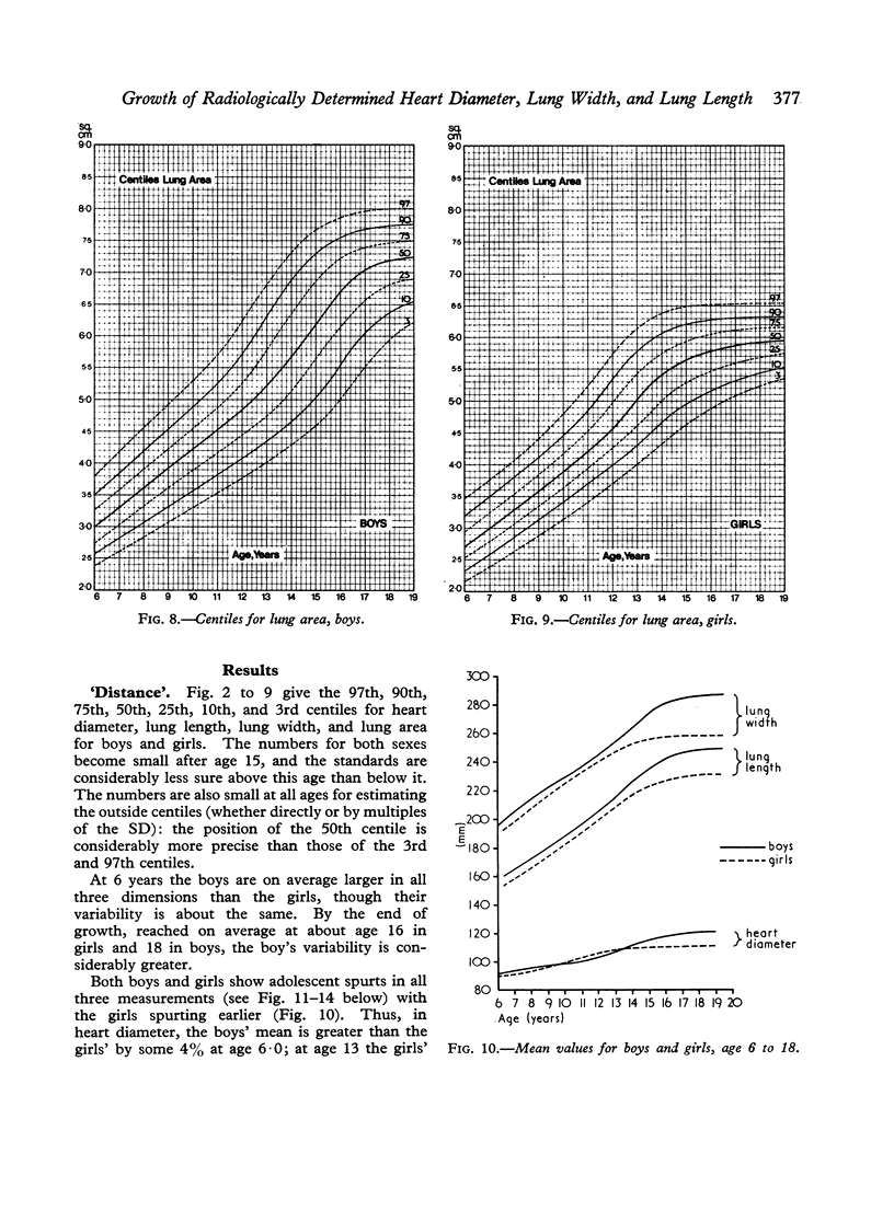
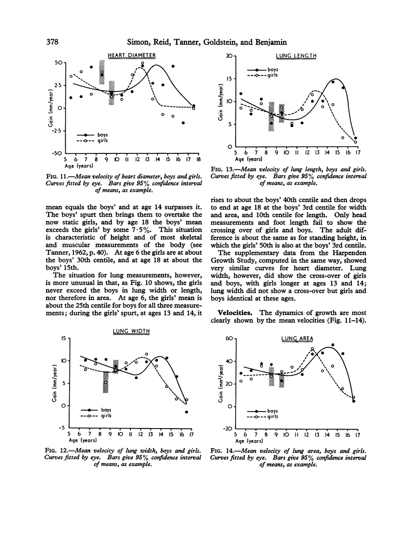
Centile standards for these measurements and for lung area (width × length) at ages from 6 to 19 years are presented for clinical use. Mean velocity curves are given over the same age range.
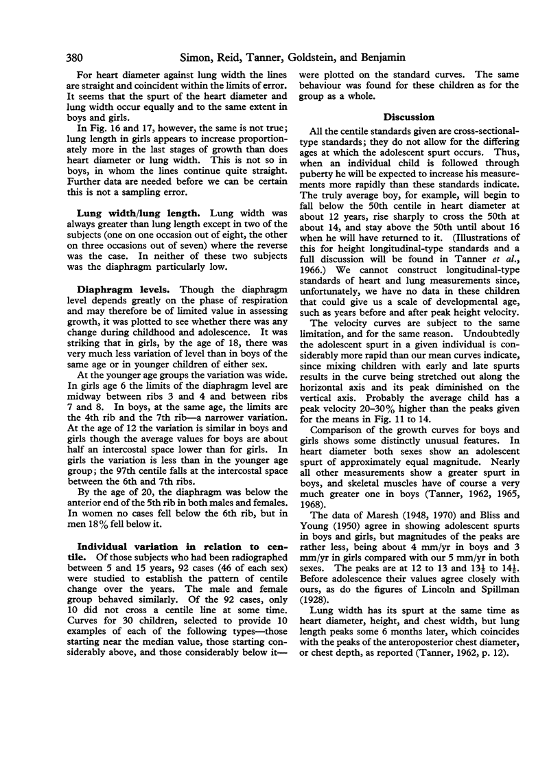
The lungs are exceptional in that the adolescent growth spurt is of similar magnitude in both boys and girls, and the girls' mean value does not exceed the boys' mean value in lung width even at the peak of the girls' spurt, as it does in all other body measurements except those of head and feet.
The age at peak velocity for lung width and heart diameter coincides with the age for peak height velocity; the peak for lung length occurs about 6 months later than that for lung width.
The heart diameter has an adolescent spurt of about equal magnitude in both sexes in these data. At age 6, heart diameter is 78% and 81% of its adult value, respectively, in boys and girls, compared with figures of 66 to 67% for lung width, 62 to 63% for lung length, and 66 to 67% for height.
A wide variation in the level of the diaphragm was found, but by the age of 20 the diaphragm was below the anterior end of the 5th rib in both males and females. In women no case fell below the 6th rib, but in men some 18% fell below it.
Full text
PDF


Images in this article

Selected References
These references are in PubMed. This may not be the complete list of references from this article.
- BLISS C. I., YOUNG M. S. An analysis of heart measurements of growing boys. Hum Biol. 1950 Dec;22(4):271–280. [PubMed] [Google Scholar]
- Hiernaux J. Bodily shape differentiation of ethnic groups and of the sexes through growth. Hum Biol. 1968 Feb;40(1):44–62. [PubMed] [Google Scholar]
- JEURISSEN A. [Technic for measuring the volume of the thoracic cavity in living subjects]. Acta Tuberc Pneumol Belg. 1959 Sep-Oct;50:346–351. [PubMed] [Google Scholar]
- JEURISSEN A. [The development of the thoracic cavity during the growth of young and adolescent girls]. Acta Tuberc Pneumol Belg. 1959 Sep-Oct;50:352–370. [PubMed] [Google Scholar]
- Tanner J. M., Whitehouse R. H., Takaishi M. Standards from birth to maturity for height, weight, height velocity, and weight velocity: British children, 1965. I. Arch Dis Child. 1966 Oct;41(219):454–471. doi: 10.1136/adc.41.219.454. [DOI] [PMC free article] [PubMed] [Google Scholar]