

Abstract
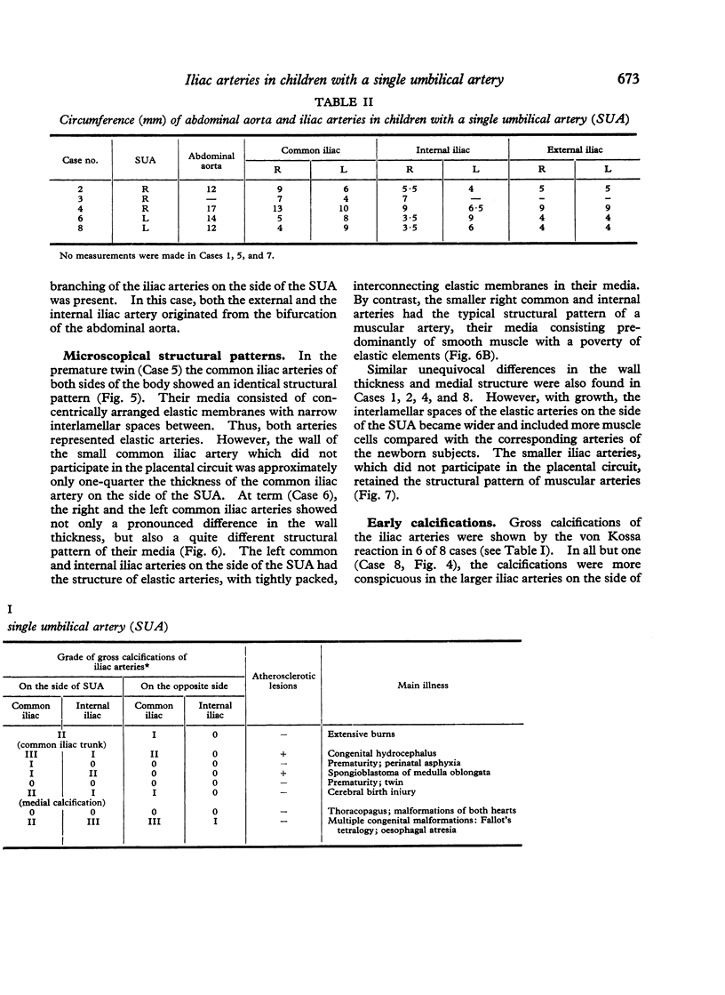
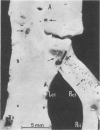
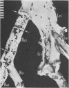
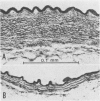
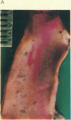
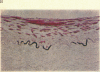
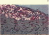
With a single umbilical artery a unique haemodynamic situation arises during fetal development, resulting in a different calibre and structure of the iliac arteries on the two sides of the body. On the side of the single umbilical artery, the enlarged iliac arteries have the structure of elastic arteries, whereas the smaller and thin-walled iliac arteries, which do not participate in the placental circuit, show the typical structure of muscular arteries. These differences of the arterial structure determine the morphological pattern of early calcifications which are regularly present in the iliac arteries on both sides of the body, but are usually more conspicuous in the large iliac arteries on the side of the single umbilical artery. In 2 children, aged 18 months and 4 years, atherosclerotic lesions were present in the wide common iliac artery on the side of the obliterated single umbilical artery. These lesions represent the earliest atherosclerotic changes to be found in the human. They are probably related to the remodelling of these arteries, beginning after birth as an adaptation to decreased blood flow.
Full text
PDF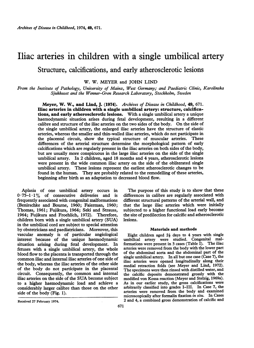

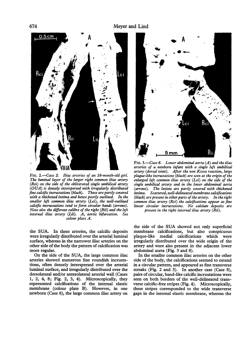
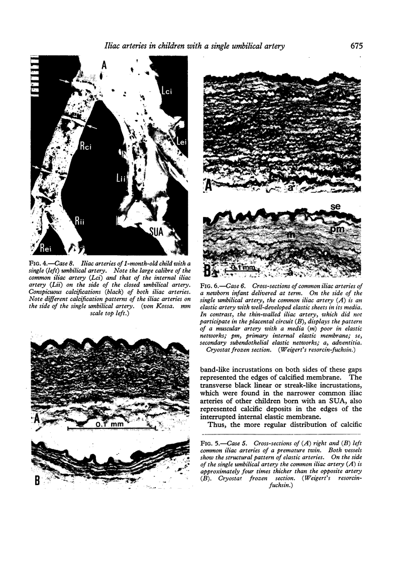

Images in this article


Selected References
These references are in PubMed. This may not be the complete list of references from this article.
- BENIRSCHKE K., BOURNE G. L. The incidence and prognostic implication of congenital absence of one umbilical artery. Am J Obstet Gynecol. 1960 Feb;79:251–254. doi: 10.1016/0002-9378(60)90183-6. [DOI] [PubMed] [Google Scholar]
- COX G. E., TRUEHEART R. E., KAPLAN J., TAYLOR C. B. ATHEROSCLEROSIS IN RHESUS MONKEYS. IV. REPAIR OF ARTERIAL INJURY--AN IMPORTANT SECONDARY ATHEROGENIC FACTOR. Arch Pathol. 1963 Aug;76:166–176. [PubMed] [Google Scholar]
- FAIERMAN E. The significance of one umbilical artery. Arch Dis Child. 1960 Jun;35:285–288. doi: 10.1136/adc.35.181.285. [DOI] [PMC free article] [PubMed] [Google Scholar]
- HASS G. M., TRUEHEART R. E., HEMMENS A. Experimental athero-arteriosclerosis due to calcific medial degeneration and hypercholesteremia. Am J Pathol. 1961 Mar;38:289–323. [PMC free article] [PubMed] [Google Scholar]
- Meyer W. W., Lind J. Calcifications of iliac arteries in newborns and infants. Arch Dis Child. 1972 Jun;47(253):364–372. doi: 10.1136/adc.47.253.364. [DOI] [PMC free article] [PubMed] [Google Scholar]
- Meyer W. W., Stelzig H. H. A simple method for gross demonstration of calcific deposits in the arteries. Angiology. 1969 Sep;20(8):423–427. doi: 10.1177/000331976902000801. [DOI] [PubMed] [Google Scholar]
- Meyer W. W., Stelzig H. H. Calcification patterns of the internal elastic membrane. Calcif Tissue Res. 1969;3(3):266–273. doi: 10.1007/BF02058668. [DOI] [PubMed] [Google Scholar]
- SEKI M., STRAUSS L. ABSENCE OF ONE UMBILICAL ARTERY. ANALYSIS OF 60 CASES WITH EMPHASIS ON ASSOCIATED DEVELOPMENTAL ABERRATIONS. Arch Pathol. 1964 Oct;78:446–453. [PubMed] [Google Scholar]