Abstract
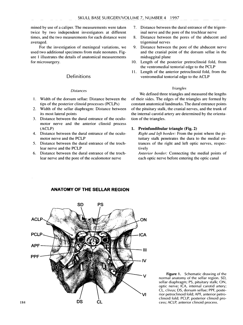
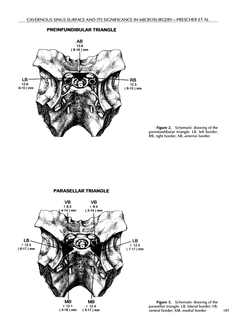
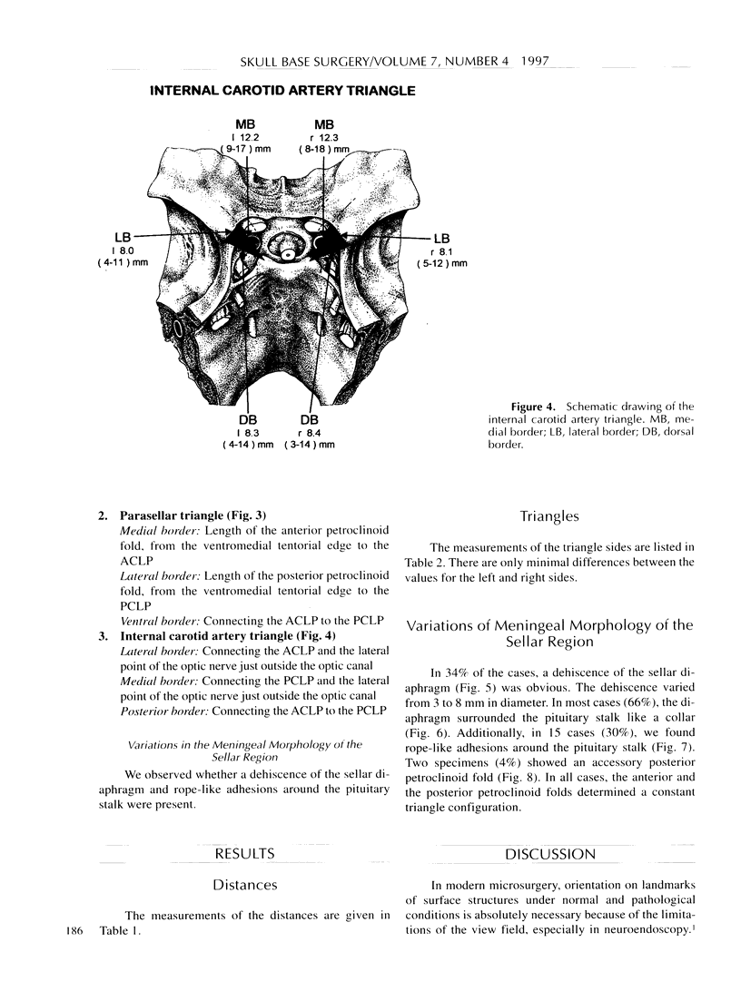
The anatomical and surgical approach to the sella region is of special interest for microsurgeons involved in ear, nose, and throat surgery, neurosurgery, ophthalmology, maxillofacial surgery, and skull base surgery. We investigated the surface morphology of the cavernous sinus and the sella turcica in 48 adult and 2 neonate specimens. To simplify the morphometric recording, distances between anatomical landmarks were defined. In addition, three triangles—the preinfundibular, the parasellar, and the internal carotid artery triangle—are introduced. These triangles are defined in order to determine the location where cranial nerves III, IV, V, and VI penetrate the dura with respect to the anterior and posterior clinoid processes and the tuberculum and dorsum sellae. The triangles were found to be symmetrical, with identical bilateral measurements, and the entry points of the cranial nerves were found to be constant. In 17 cases (34%), we found a dehiscence of the sellar diaphragm, and in 15 cases (30%), rope-like adhesions at the pituitary stalk.
Full text
PDF
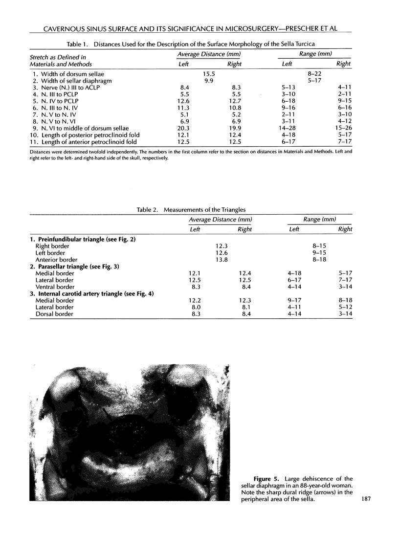
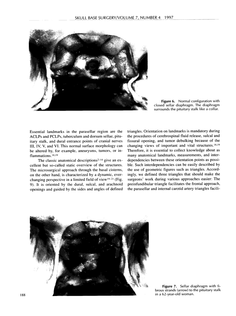
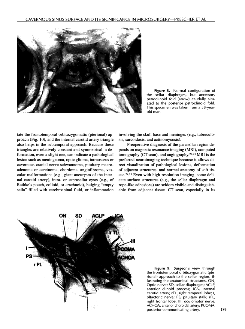
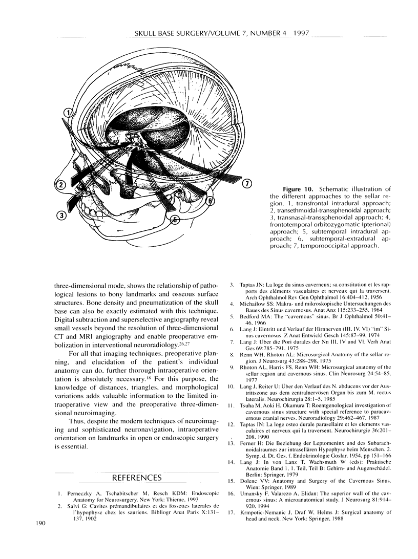
Images in this article
Selected References
These references are in PubMed. This may not be the complete list of references from this article.
- Bedford M. A. The "cavernous" sinus. Br J Ophthalmol. 1966 Jan;50(1):41–46. doi: 10.1136/bjo.50.1.41. [DOI] [PMC free article] [PubMed] [Google Scholar]
- Bergland R. M., Ray B. S., Torack R. M. Anatomical variations in the pituitary gland and adjacent structures in 225 human autopsy cases. J Neurosurg. 1968 Feb;28(2):93–99. doi: 10.3171/jns.1968.28.2.0093. [DOI] [PubMed] [Google Scholar]
- Lang J. Eintritt und Verlauf der Hirnnerven (III, IV, VI) "im" Sinus cavernosus. Z Anat Entwicklungsgesch. 1974;145(1):87–99. [PubMed] [Google Scholar]
- Lang J., Reiter U. Uber den Verlauf des N. abducens vor der Austrittszone aus dem zentralnervösen Organ bis zum M. rectus lateralis. Neurochirurgia (Stuttg) 1985 Jan;28(1):1–5. doi: 10.1055/s-2008-1054170. [DOI] [PubMed] [Google Scholar]
- Lang J. Uber die Pori durales der Nn. III, IV und VI. Verh Anat Ges. 1975;69:785–791. [PubMed] [Google Scholar]
- MICHAILOW S. S. MAKRO- UND MIKROSKOPISCHE UNTERSUCHUNGEN DES BAUES DES SINUS CAVERNOSUS. Anat Anz. 1964 Oct 13;115:233–255. [PubMed] [Google Scholar]
- Renn W. H., Rhoton A. L., Jr Microsurgical anatomy of the sellar region. J Neurosurg. 1975 Sep;43(3):288–298. doi: 10.3171/jns.1975.43.3.0288. [DOI] [PubMed] [Google Scholar]
- Reul J., Spetzger U., Fricke C., Konert T., Bertalanffy H., Thron A. Endovaskuläre Behandlung zerebraler arterieller Aneurysmen mit selektiv ablösbaren Platinspiralen. Dtsch Med Wochenschr. 1995 May 12;120(19):669–675. doi: 10.1055/s-2008-1055394. [DOI] [PubMed] [Google Scholar]
- Rhoton A. L., Jr, Harris F. S., Renn W. H. Microsurgical anatomy of the sellar region and cavernous sinus. Clin Neurosurg. 1977;24:54–85. doi: 10.1093/neurosurgery/24.cn_suppl_1.54. [DOI] [PubMed] [Google Scholar]
- Sekhar L. N., Burgess J., Akin O. Anatomical study of the cavernous sinus emphasizing operative approaches and related vascular and neural reconstruction. Neurosurgery. 1987 Dec;21(6):806–816. doi: 10.1227/00006123-198712000-00005. [DOI] [PubMed] [Google Scholar]
- TAPTAS J. N. La loge du sinus caverneux; sa constitution et les rapports des éléments vasculaires et nerveux qui la traversent. Arch Ophtalmol Rev Gen Ophtalmol. 1956 Jun;16(4):404–412. [PubMed] [Google Scholar]
- Taptas J. N. La loge ostéo-durale parasellaire et les éléments vasculaires et nerveux qui la traversent. Une conception anatomique qui doit remplacer celle du sinus caverneux des classiques. Neurochirurgie. 1990;36(4):201–208. [PubMed] [Google Scholar]
- Tsuha M., Aoki H., Okamura T. Roentgenological investigation of cavernous sinus structure with special reference to paracavernous cranial nerves. Neuroradiology. 1987;29(5):462–467. doi: 10.1007/BF00341744. [DOI] [PubMed] [Google Scholar]
- Umansky F., Valarezo A., Elidan J. The superior wall of the cavernous sinus: a microanatomical study. J Neurosurg. 1994 Dec;81(6):914–920. doi: 10.3171/jns.1994.81.6.0914. [DOI] [PubMed] [Google Scholar]


