Abstract
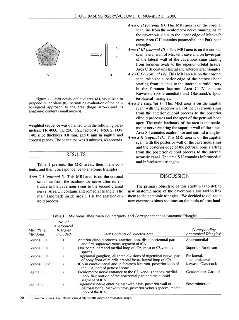
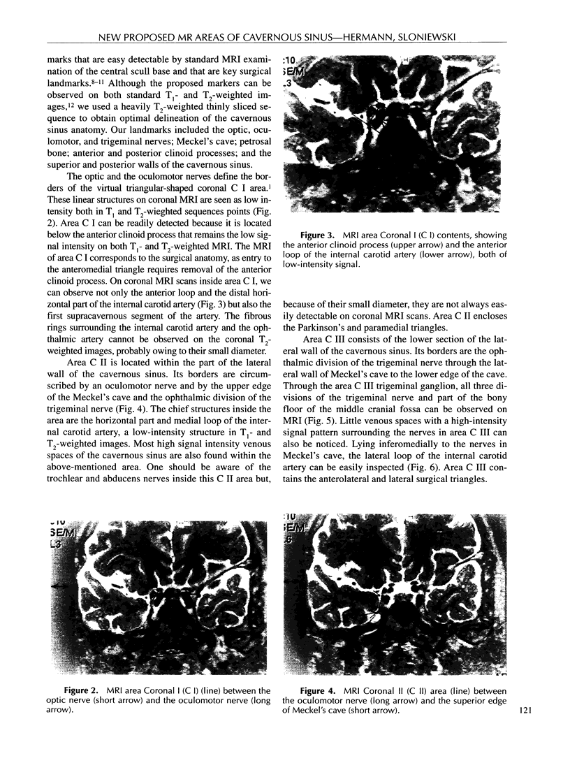
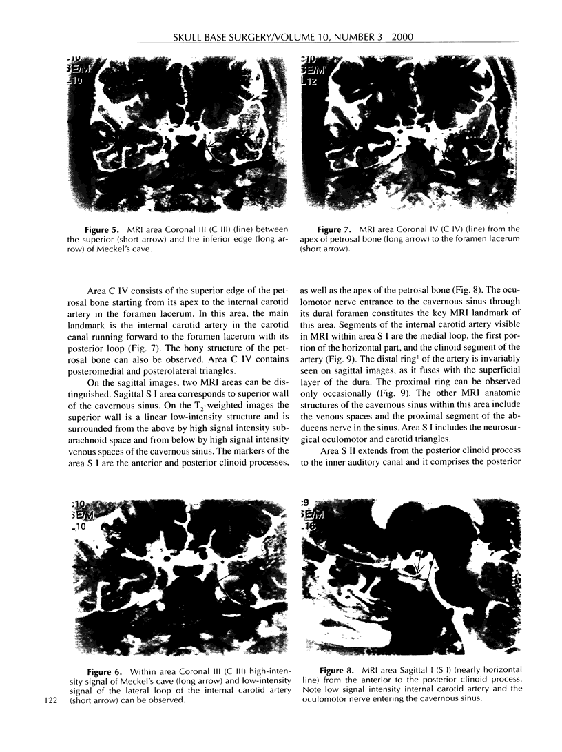
MRI appears to be the best contemporary method with which to evaluate cavernous sinus anatomy. Based on anatomic landmarks that are easily detectable in the standard MRI examination, the cavernous sinus is divided into six areas. Each of the newly defined areas corresponds to the previously described neuroanatomic triangles. Evaluation of the newly defined areas in a plane perpendicular to these triangles on a MRI scan permits expansion of these two-dimensional areas into three-dimensional spaces; thus, the neurosurgical approach can be observed and their anatomic and pathologic content examined. Radiologic and surgical evaluation of the cavernous sinus is presented.
Full text
PDF
Images in this article
Selected References
These references are in PubMed. This may not be the complete list of references from this article.
- Bradac G. B., Riva A., Schörner W., Stura G. Cavernous sinus meningiomas: an MRI study. Neuroradiology. 1987;29(6):578–581. doi: 10.1007/BF00350447. [DOI] [PubMed] [Google Scholar]
- Daniels D. L., Pech P., Mark L., Pojunas K., Williams A. L., Haughton V. M. Magnetic resonance imaging of the cavernous sinus. AJR Am J Roentgenol. 1985 May;144(5):1009–1014. doi: 10.2214/ajr.144.5.1009. [DOI] [PubMed] [Google Scholar]
- Demaerel P., Marchal G., Casteels I., Wilms G., Bosmans H., Dralands G., Baert A. L. Intracavernous aneurysm. Superior demonstration by magnetic resonance angiography. Neuroradiology. 1990;32(4):322–324. doi: 10.1007/BF00593054. [DOI] [PubMed] [Google Scholar]
- Guy R. L., Benn J. J., Ayers A. B., Bingham J. B., Lowy C., Cox T. C., Sonksen P. H. A comparison of CT and MRI in the assessment of the pituitary and parasellar region. Clin Radiol. 1991 Mar;43(3):156–161. doi: 10.1016/s0009-9260(05)80470-2. [DOI] [PubMed] [Google Scholar]
- Kawase T., van Loveren H., Keller J. T., Tew J. M. Meningeal architecture of the cavernous sinus: clinical and surgical implications. Neurosurgery. 1996 Sep;39(3):527–536. doi: 10.1097/00006123-199609000-00019. [DOI] [PubMed] [Google Scholar]
- Oikawa S., Kyoshima K., Kobayashi S. Surgical anatomy of the juxta-dural ring area. J Neurosurg. 1998 Aug;89(2):250–254. doi: 10.3171/jns.1998.89.2.0250. [DOI] [PubMed] [Google Scholar]
- Seoane E., Rhoton A. L., Jr, de Oliveira E. Microsurgical anatomy of the dural collar (carotid collar) and rings around the clinoid segment of the internal carotid artery. Neurosurgery. 1998 Apr;42(4):869–886. doi: 10.1097/00006123-199804000-00108. [DOI] [PubMed] [Google Scholar]


