Abstract
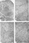
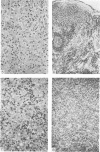
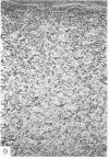
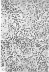
Seventy-seven lymph nodes were examined histologically from sixty-two leprosy patients representing the whole range of the disease spectrum from the high resistance form (tuberculoid) to the specific immunity deficiency form (lepromatous). At the lepromatous end of the spectrum paracortical areas were infiltrated with undifferentiated cells of the histiocyte–macrophage series which failed to eliminate mycobacteria. As resistance to infection increased across the leprosy spectrum, histiocytes became more differentiated eventually appearing epithelioid. This was paralleled by increasing numbers of small lymphocytes in the paracortical areas. In the borderline tuberculoid form of the disease an appearance was seen similar to that found in sarcoidosis. In polar tuberculoid leprosy where there is a high degree of cellular immunity, paracortical areas were well developed and populated with lymphocytes and immunoblasts. The immunological significance of these findings are discussed, especially the relation of the changes in morphological appearance of cells of the histiocyte series to the ability of the patient to develop cell-mediated immune reactions.
Full text
PDF
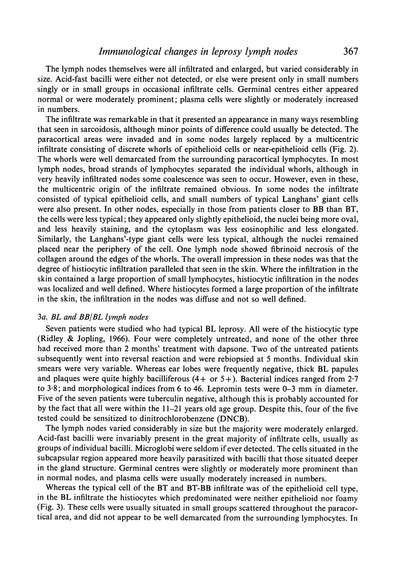
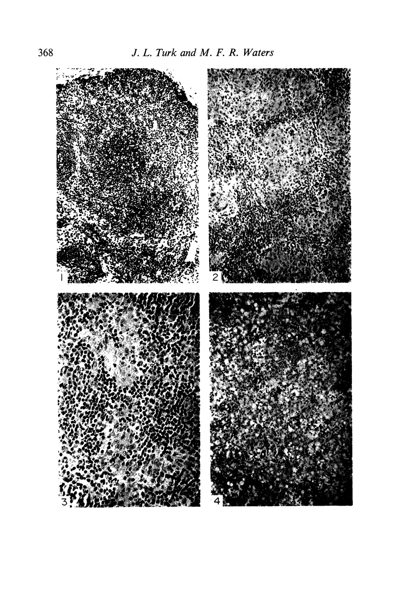
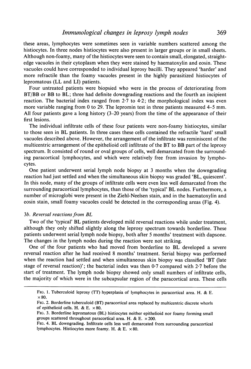
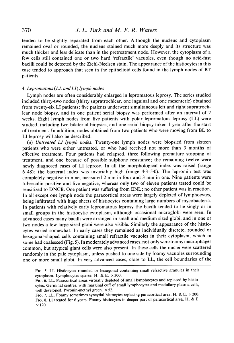
Images in this article
Selected References
These references are in PubMed. This may not be the complete list of references from this article.
- Blanden R. V. Modification of macrophage function. J Reticuloendothel Soc. 1968 Jun;5(3):179–202. [PubMed] [Google Scholar]
- Desikan K. V., Job C. K. Leprous lymphadenitis. Demonstration of tuberculoid lesions. Int J Lepr Other Mycobact Dis. 1966 Apr-Jun;34(2):147–154. [PubMed] [Google Scholar]
- Gaafar S. M., Turk J. L. Granuloma formation in lymph-nodes. J Pathol. 1970 Jan;100(1):9–20. doi: 10.1002/path.1711000103. [DOI] [PubMed] [Google Scholar]
- Guinto R. S. Biology of the mycobacterioses. Skin tests in leprosy. Ann N Y Acad Sci. 1968 Sep 5;154(1):149–156. doi: 10.1111/j.1749-6632.1968.tb16705.x. [DOI] [PubMed] [Google Scholar]
- McDevitt H. O., Benacerraf B. Genetic control of specific immune responses. Adv Immunol. 1969;11:31–74. doi: 10.1016/s0065-2776(08)60477-0. [DOI] [PubMed] [Google Scholar]
- RIDLEY D. S. Therapeutic trials in leprosy using serial biopsies. Lepr Rev. 1958 Jan;29(1):45–52. doi: 10.5935/0305-7518.19580004. [DOI] [PubMed] [Google Scholar]
- Ridley D. S., Jopling W. H. Classification of leprosy according to immunity. A five-group system. Int J Lepr Other Mycobact Dis. 1966 Jul-Sep;34(3):255–273. [PubMed] [Google Scholar]
- Ridley D. S. Reactions in leprosy. Lepr Rev. 1969 Apr;40(2):77–81. doi: 10.5935/0305-7518.19690016. [DOI] [PubMed] [Google Scholar]
- Ridley D. S., Waters M. F. Significance of variations within the lepromatous group. Lepr Rev. 1969 Jul;40(3):143–152. doi: 10.5935/0305-7518.19690026. [DOI] [PubMed] [Google Scholar]
- Turk J. L. Cell-mediated immunological processes in leprosy. Bull World Health Organ. 1969;41(6):779–792. doi: 10.5935/0305-7518.19700030. [DOI] [PMC free article] [PubMed] [Google Scholar]
- Turk J. L., Waters M. F. Cell-mediated immunity in patients with leprosy. Lancet. 1969 Aug 2;2(7614):243–246. doi: 10.1016/s0140-6736(69)90009-9. [DOI] [PubMed] [Google Scholar]
- Turk J. L., Waters M. F. Immunological basis for depression of cellular immunity and the delayed allergic response in patients with lepromatous leprosy. Lancet. 1968 Aug 24;2(7565):436–438. doi: 10.1016/s0140-6736(68)90472-8. [DOI] [PubMed] [Google Scholar]
- Wemambu S. N., Turk J. L., Waters M. F., Rees R. J. Erythema nodosum leprosum: a clinical manifestation of the arthus phenomenon. Lancet. 1969 Nov 1;2(7627):933–935. doi: 10.1016/s0140-6736(69)90592-3. [DOI] [PubMed] [Google Scholar]


