Full Text
The Full Text of this article is available as a PDF (114.6 KB).
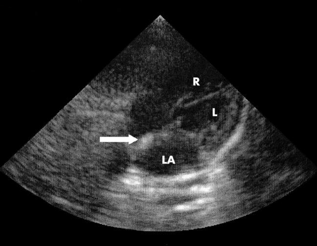
Figure 1 .

Echocardiographic subcostal four chamber view of the heart depicting the left atrium (LA) with the arrow pointing to the echodensity in the atrial septum. R, Right ventricle; L, left ventricle.
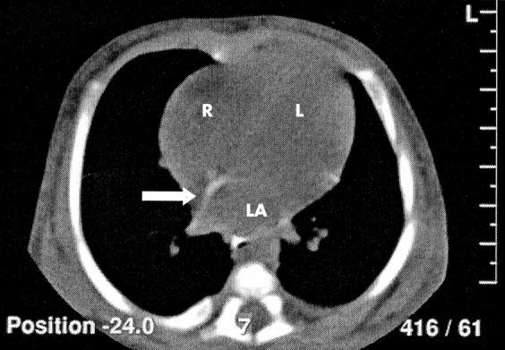
Figure 2 .

Computed tomographic image of an axial plane through the heart depicting the left atrium (LA) with the arrow pointing to the atrial septum. Note that the image intensity of the atrial septum approaches that of the ribs and is more intense than the ventricular walls and ventricular septum, which cannot be clearly distinguished. R, Right ventricle; L, left ventricle.
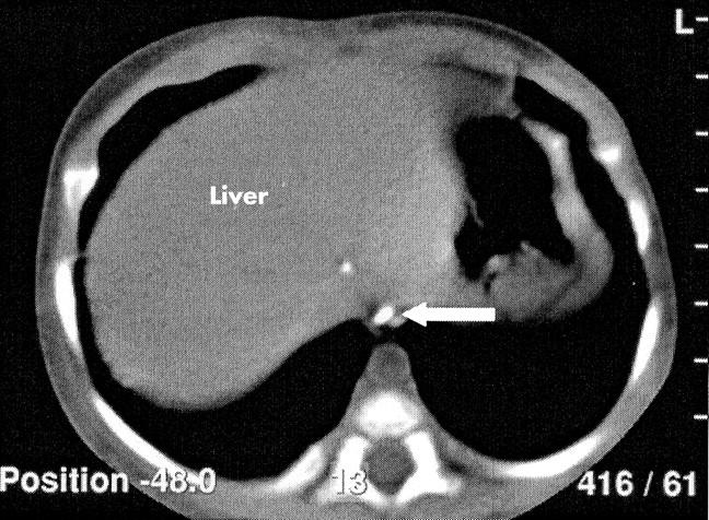
Figure 3 .

Computed tomographic image of an axial section through the upper abdomen depicting the liver with one spot in the parenchyma. The arrow points to a white spot in the inferior vena cava.


