Abstract
A woman presented during two pregnancies (at 25 and 23 weeks' gestation, respectively) because the fetuses had rapid, irregular tachycardia and hydrops. After maternal drug treatment and achievement of slower fetal heart rates, the hydrops gradually resolved. Both babies were born full term with continuing atrial fibrillation. In the first, an ectopic atrial rhythm was temporarily achieved during high dose flecainide treatment but, in the younger sibling, all medications and repeated cardioversions failed even temporarily to convert the atrial fibrillation with an almost isoelectric baseline in ECG to sinus rhythm. Good rate control has been achieved with digoxin in both patients. No infective, immunological, or structural cause was found in either case, and thus an inherited aetiology is probable. Keywords: atrial fibrillation; arrhythmias; fetal atrial fibrillation; familial arrhythmias
Full Text
The Full Text of this article is available as a PDF (168.5 KB).
Figure 1 .
m mode echocardiographic recording of the fetal heart during the first pregnancy at 27 weeks of gestation. Fetal heart rate was 206 beats/min at the time of recording. A, left atrial wall; M, mitral valve movement.
Figure 2 .
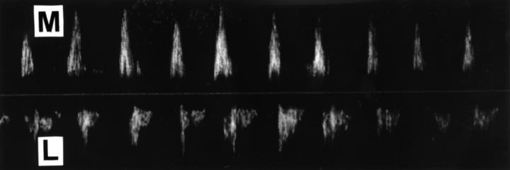
Doppler echocardiographic recording of mitral inflow and left ventricular outflow at 27 weeks of gestation during the first pregnancy. The fetal heart rate was 210 beats/min at the time of recording, with beat intervals varying between 250 and 320 ms. M, mitral inflow; L, left ventricular outflow.
Figure 3 .
Postnatal ECG recordings of the first baby: (A) on the day of birth after intravenous flecainide loading (mean QT time corrected according to Bazett's formula (QTc) 0.35 seconds); (B) at five months old (mean QTc 0.42 seconds); (C) at two years old (mean QTc 0.41 seconds). (Paper speed 50 mm/s.)
Figure 4 .
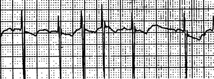
Oesophageal ECG recording of the first baby at one day old. Note the varying polarity of the small P waves. (Paper speed 25 mm/s.)
Figure 5 .
Doppler echocardiographic recording of mitral inflow and left ventricular outflow of the second baby at 23 weeks of gestation. Fetal heart rate was 218 beats/min at the time of recording, with beat intervals varying between 210 and 340 ms. M, mitral inflow; L, left ventricular outflow.
Figure 6 .
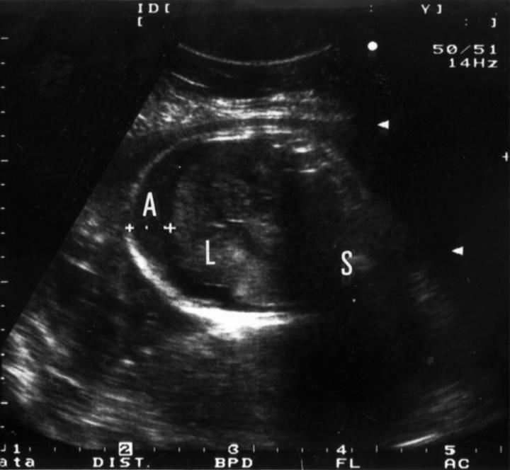
Ultrasonographic cross sectional image of the fetal abdomen of the younger sibling during the 28th gestational week. A, ascites; S, spine; L, liver.
Figure 7 .
Postnatal ECG recordings of the second baby: (A) on the day of birth (mean QTc 0.42 seconds); (B) after intravenous adenosine bolus; (C) at three months old (mean QTc 0.42 seconds). (Paper speed 50 mm/s.)


