Abstract
OBJECTIVE—To identify any potential relations between the size of an emptied plaque cavity and the remodelling pattern, plaque or vessel dimensions, lumen narrowing, and other ultrasonic lesion characteristics. DESIGN—Intravascular ultrasound was used to examine prospectively 51 ruptured ulcerated coronary plaques. Cross sectional area measurements comprised lumen, vessel, plaque, and emptied plaque cavity. Lumen narrowing was calculated as 1 − (lesion lumen area/reference lumen area) × 100%. A remodelling index was calculated as lesion vessel area/reference vessel area, and plaques were divided into those with values > 1.05 (group A) and ⩽ 1.05 (group B). RESULTS—Of the total of 51 plaques, 36 (71%) were assigned to group A and 15 (29%) to group B. In neither group was there a significant difference in reference dimensions and lumen narrowing. However, lesion vessel (mean (SD): 22.6 (8.1) mm2 v 17.5 (4.3) mm2; p = 0.006) and plaque areas (15.8 (6.2) mm2 v 12.8 (3.2) mm2; p = 0.03) were greater in group A than in group B. The cavity inside the plaque was larger in group A than in group B (2.8 (1.6) mm2 v 1.8 (0.9) mm2; p = 0.007) and showed a positive linear relation with lesion and reference vessel size (r = 0.58 and 0.56, respectively; p < 0.001), but not with lumen narrowing. CONCLUSIONS—The size of the emptied cavity inside ruptured plaques is on average larger in lesions with adaptive vascular remodelling, and shows a linear relation with lesion plaque and vessel size and with the reference dimensions, but not with the degree of lumen narrowing. Keywords: intravascular ultrasound; ultrasonic scanning; plaque rupture; remodelling
Full Text
The Full Text of this article is available as a PDF (223.6 KB).
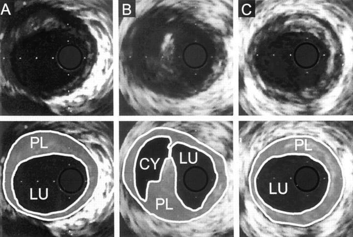
Figure 1 .
Intravascular ultrasound planimetry in ruptured right coronary plaque of a patient in group A (remodelling index > 1.05). Lumen (LU), plaque (PL), plaque cavity (CY), and vessel area (within external contour) were measured in the proximal (A) and distal reference segment (C), and at the lesion site (B). Lesion and mean reference vessel cross sectional area measured 24.4 mm2 and 22.25 mm2, respectively; the remodelling index was 1.09. Plaque cavity cross sectional area measured 4.6 mm2. Lumen area narrowing was 37%.
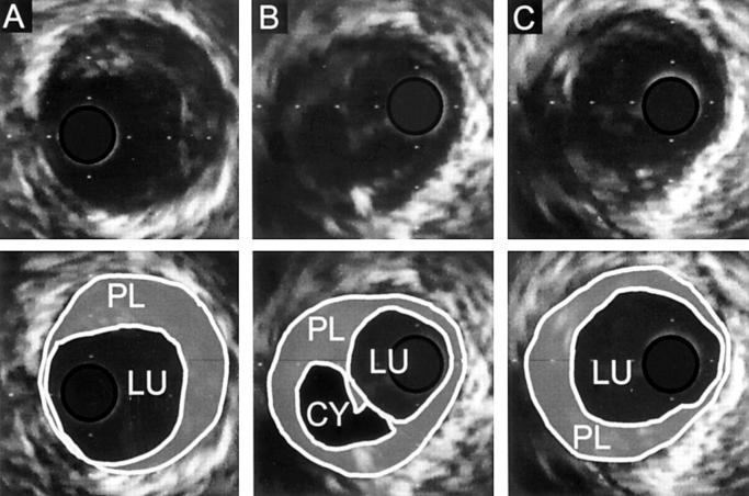
Figure 2 .
Example of ruptured right coronary plaque from a patient in group B (remodelling index ⩽ 1.05). Lumen (LU), plaque (PL), plaque cavity (CY), and vessel area (within external contour) were measured in the proximal (A) and distal reference segment (C), and at lesion site (B). Remodelling index was 0.97. Lesion and mean reference vessel area measured 15.2 mm2 and 15.7 mm2, respectively. Plaque cavity area was 2.6 mm2 and lumen area narrowing measured 40%. Despite comparable lumen area narrowing in the two examples in figs 1 and 2, plaque cavity area was significantly smaller in the present example, which showed smaller vascular dimensions than the vessel segment of fig 1 (note the difference in the 1 mm calibration grid in figs 1 and 2).
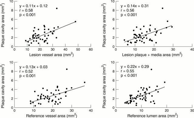
Figure 3 .
Linear regression analyses comparing vascular dimensions with plaque cavity size. The emptied cavity inside the ruptured plaque showed a significant linear relation with lesion plaque and vessel size (upper panels), and with the reference dimensions (lower panels).
Figure 4 .
Comparison of remodelling index and lumen narrowing with plaque cavity size. The emptied cavity inside the ruptured plaque showed a significant linear relation with lesion plaque and vessel size, and with reference dimensions. There was no significant relation between plaque cavity size and remodelling index (left panel) or the per cent lumen area narrowing (right panel, note scattered distribution of data points).
Selected References
These references are in PubMed. This may not be the complete list of references from this article.
- Arnold A. E., Simoons M. L., Van de Werf F., de Bono D. P., Lubsen J., Tijssen J. G., Serruys P. W., Verstraete M. Recombinant tissue-type plasminogen activator and immediate angioplasty in acute myocardial infarction. One-year follow-up. The European Cooperative Study Group. Circulation. 1992 Jul;86(1):111–120. doi: 10.1161/01.cir.86.1.111. [DOI] [PubMed] [Google Scholar]
- Baroldi G., Silver M. D., Mariani F., Giuliano G. Correlation of morphological variables in the coronary atherosclerotic plaque with clinical patterns of ischemic heart disease. Am J Cardiovasc Pathol. 1988;2(2):159–172. [PubMed] [Google Scholar]
- Baumgart D., Liu F., Haude M., Görge G., Ge J., Erbel R. Acute plaque rupture and myocardial stunning in patient with normal coronary arteriography. Lancet. 1995 Jul 15;346(8968):193–194. [PubMed] [Google Scholar]
- Bocksch W. G., Schartl M., Beckmann S. H., Dreysse S., Paeprer H. Intravascular ultrasound imaging in patients with acute myocardial infarction: comparison with chronic stable angina pectoris. Coron Artery Dis. 1994 Sep;5(9):727–735. [PubMed] [Google Scholar]
- Davies M. J., Thomas A. Thrombosis and acute coronary-artery lesions in sudden cardiac ischemic death. N Engl J Med. 1984 May 3;310(18):1137–1140. doi: 10.1056/NEJM198405033101801. [DOI] [PubMed] [Google Scholar]
- Di Mario C., Görge G., Peters R., Kearney P., Pinto F., Hausmann D., von Birgelen C., Colombo A., Mudra H., Roelandt J. Clinical application and image interpretation in intracoronary ultrasound. Study Group on Intracoronary Imaging of the Working Group of Coronary Circulation and of the Subgroup on Intravascular Ultrasound of the Working Group of Echocardiography of the European Society of Cardiology. Eur Heart J. 1998 Feb;19(2):207–229. doi: 10.1053/euhj.1996.0433. [DOI] [PubMed] [Google Scholar]
- Erbel R., Ge J., Görge G., Baumgart D., Haude M., Jeremias A., von Birgelen C., Jollet N., Schwedtmann J. Intravascular ultrasound classification of atherosclerotic lesions according to American Heart Association recommendation. Coron Artery Dis. 1999 Oct;10(7):489–499. doi: 10.1097/00019501-199910000-00009. [DOI] [PubMed] [Google Scholar]
- Falk E. Plaque rupture with severe pre-existing stenosis precipitating coronary thrombosis. Characteristics of coronary atherosclerotic plaques underlying fatal occlusive thrombi. Br Heart J. 1983 Aug;50(2):127–134. doi: 10.1136/hrt.50.2.127. [DOI] [PMC free article] [PubMed] [Google Scholar]
- Fishbein M. C., Siegel R. J. How big are coronary atherosclerotic plaques that rupture? Circulation. 1996 Nov 15;94(10):2662–2666. doi: 10.1161/01.cir.94.10.2662. [DOI] [PubMed] [Google Scholar]
- Fuster V., Badimon L., Badimon J. J., Chesebro J. H. The pathogenesis of coronary artery disease and the acute coronary syndromes (1). N Engl J Med. 1992 Jan 23;326(4):242–250. doi: 10.1056/NEJM199201233260406. [DOI] [PubMed] [Google Scholar]
- Ge J., Chirillo F., Schwedtmann J., Görge G., Haude M., Baumgart D., Shah V., von Birgelen C., Sack S., Boudoulas H. Screening of ruptured plaques in patients with coronary artery disease by intravascular ultrasound. Heart. 1999 Jun;81(6):621–627. doi: 10.1136/hrt.81.6.621. [DOI] [PMC free article] [PubMed] [Google Scholar]
- Ge J., Erbel R., Zamorano J., Koch L., Kearney P., Görge G., Gerber T., Meyer J. Coronary artery remodeling in atherosclerotic disease: an intravascular ultrasonic study in vivo. Coron Artery Dis. 1993 Nov;4(11):981–986. doi: 10.1097/00019501-199311000-00005. [DOI] [PubMed] [Google Scholar]
- Ge J., Haude M., Görge G., Liu F., Erbel R. Silent healing of spontaneous plaque disruption demonstrated by intracoronary ultrasound. Eur Heart J. 1995 Aug;16(8):1149–1151. doi: 10.1093/oxfordjournals.eurheartj.a061061. [DOI] [PubMed] [Google Scholar]
- Glagov S., Weisenberg E., Zarins C. K., Stankunavicius R., Kolettis G. J. Compensatory enlargement of human atherosclerotic coronary arteries. N Engl J Med. 1987 May 28;316(22):1371–1375. doi: 10.1056/NEJM198705283162204. [DOI] [PubMed] [Google Scholar]
- Gussenhoven E. J., Geselschap J. H., van Lankeren W., Posthuma D. J., van der Lugt A. Remodeling of atherosclerotic coronary arteries assessed with intravascular ultrasound in vitro. Am J Cardiol. 1997 Mar 1;79(5):699–702. doi: 10.1016/s0002-9149(96)00849-1. [DOI] [PubMed] [Google Scholar]
- Gyöngyösi M., Yang P., Hassan A., Weidinger F., Domanovits H., Laggner A., Glogar D. Arterial remodelling of native human coronary arteries in patients with unstable angina pectoris: a prospective intravascular ultrasound study. Heart. 1999 Jul;82(1):68–74. doi: 10.1136/hrt.82.1.68. [DOI] [PMC free article] [PubMed] [Google Scholar]
- Hangartner J. R., Charleston A. J., Davies M. J., Thomas A. C. Morphological characteristics of clinically significant coronary artery stenosis in stable angina. Br Heart J. 1986 Dec;56(6):501–508. doi: 10.1136/hrt.56.6.501. [DOI] [PMC free article] [PubMed] [Google Scholar]
- Kearney P., Erbel R., Rupprecht H. J., Ge J., Koch L., Voigtländer T., Stähr P., Görge G., Meyer J. Differences in the morphology of unstable and stable coronary lesions and their impact on the mechanisms of angioplasty. An in vivo study with intravascular ultrasound. Eur Heart J. 1996 May;17(5):721–730. doi: 10.1093/oxfordjournals.eurheartj.a014939. [DOI] [PubMed] [Google Scholar]
- Lee R. T., Libby P. The unstable atheroma. Arterioscler Thromb Vasc Biol. 1997 Oct;17(10):1859–1867. doi: 10.1161/01.atv.17.10.1859. [DOI] [PubMed] [Google Scholar]
- Little W. C., Constantinescu M., Applegate R. J., Kutcher M. A., Burrows M. T., Kahl F. R., Santamore W. P. Can coronary angiography predict the site of a subsequent myocardial infarction in patients with mild-to-moderate coronary artery disease? Circulation. 1988 Nov;78(5 Pt 1):1157–1166. doi: 10.1161/01.cir.78.5.1157. [DOI] [PubMed] [Google Scholar]
- Mann J. M., Davies M. J. Assessment of the severity of coronary artery disease at postmortem examination. Are the measurements clinically valid? Br Heart J. 1995 Nov;74(5):528–530. doi: 10.1136/hrt.74.5.528. [DOI] [PMC free article] [PubMed] [Google Scholar]
- Mann J. M., Davies M. J. Vulnerable plaque. Relation of characteristics to degree of stenosis in human coronary arteries. Circulation. 1996 Sep 1;94(5):928–931. doi: 10.1161/01.cir.94.5.928. [DOI] [PubMed] [Google Scholar]
- McPherson D. D., Sirna S. J., Hiratzka L. F., Thorpe L., Armstrong M. L., Marcus M. L., Kerber R. E. Coronary arterial remodeling studied by high-frequency epicardial echocardiography: an early compensatory mechanism in patients with obstructive coronary atherosclerosis. J Am Coll Cardiol. 1991 Jan;17(1):79–86. doi: 10.1016/0735-1097(91)90707-g. [DOI] [PubMed] [Google Scholar]
- Mintz G. S., Kent K. M., Pichard A. D., Satler L. F., Popma J. J., Leon M. B. Contribution of inadequate arterial remodeling to the development of focal coronary artery stenoses. An intravascular ultrasound study. Circulation. 1997 Apr 1;95(7):1791–1798. doi: 10.1161/01.cir.95.7.1791. [DOI] [PubMed] [Google Scholar]
- Moriuchi M., Saito S., Takaiwa Y., Honye J., Fukui T., Horiuchi K., Takayama T., Yajima J., Shimizu T., Chiku M. Assessment of plaque rupture by intravascular ultrasound. Heart Vessels. 1997;Suppl 12:178–181. [PubMed] [Google Scholar]
- Nishioka T., Luo H., Eigler N. L., Berglund H., Kim C. J., Siegel R. J. Contribution of inadequate compensatory enlargement to development of human coronary artery stenosis: an in vivo intravascular ultrasound study. J Am Coll Cardiol. 1996 Jun;27(7):1571–1576. doi: 10.1016/0735-1097(96)00071-x. [DOI] [PubMed] [Google Scholar]
- Pasterkamp G., Schoneveld A. H., van der Wal A. C., Haudenschild C. C., Clarijs R. J., Becker A. E., Hillen B., Borst C. Relation of arterial geometry to luminal narrowing and histologic markers for plaque vulnerability: the remodeling paradox. J Am Coll Cardiol. 1998 Sep;32(3):655–662. doi: 10.1016/s0735-1097(98)00304-0. [DOI] [PubMed] [Google Scholar]
- Pasterkamp G., Wensing P. J., Post M. J., Hillen B., Mali W. P., Borst C. Paradoxical arterial wall shrinkage may contribute to luminal narrowing of human atherosclerotic femoral arteries. Circulation. 1995 Mar 1;91(5):1444–1449. doi: 10.1161/01.cir.91.5.1444. [DOI] [PubMed] [Google Scholar]
- Qiao J. H., Fishbein M. C. The severity of coronary atherosclerosis at sites of plaque rupture with occlusive thrombosis. J Am Coll Cardiol. 1991 Apr;17(5):1138–1142. doi: 10.1016/0735-1097(91)90844-y. [DOI] [PubMed] [Google Scholar]
- Sabaté M., Kay I. P., de Feyter P. J., van Domburg R. T., Deshpande N. V., Ligthart J. M., Gijzel A. L., Wardeh A. J., Boersma E., Serruys P. W. Remodeling of atherosclerotic coronary arteries varies in relation to location and composition of plaque. Am J Cardiol. 1999 Jul 15;84(2):135–140. doi: 10.1016/s0002-9149(99)00222-2. [DOI] [PubMed] [Google Scholar]
- Smits P. C., Bos L., Quarles van Ufford M. A., Eefting F. D., Pasterkamp G., Borst C. Shrinkage of human coronary arteries is an important determinant of de novo atherosclerotic luminal stenosis: an in vivo intravascular ultrasound study. Heart. 1998 Feb;79(2):143–147. doi: 10.1136/hrt.79.2.143. [DOI] [PMC free article] [PubMed] [Google Scholar]
- Smits P. C., Pasterkamp G., de Jaegere P. P., de Feyter P. J., Borst C. Angioscopic complex lesions are predominantly compensatory enlarged: an angioscopy and intracoronary ultrasound study. Cardiovasc Res. 1999 Feb;41(2):458–464. doi: 10.1016/s0008-6363(98)00320-4. [DOI] [PubMed] [Google Scholar]
- Varnava A. Coronary artery remodelling. Heart. 1998 Feb;79(2):109–110. doi: 10.1136/hrt.79.2.109. [DOI] [PMC free article] [PubMed] [Google Scholar]
- Zamorano J., Erbel R., Ge J., Görge G., Kearney P., Koch L., Scholte A., Meyer J. Spontaneous plaque rupture visualized by intravascular ultrasound. Eur Heart J. 1994 Jan;15(1):131–133. doi: 10.1093/oxfordjournals.eurheartj.a060365. [DOI] [PubMed] [Google Scholar]
- von Birgelen C., Airiian S. G., Mintz G. S., van der Giessen W. J., Foley D. P., Roelandt J. R., Serruys P. W., de Feyter P. J. Variations of remodeling in response to left main atherosclerosis assessed with intravascular ultrasound in vivo. Am J Cardiol. 1997 Dec 1;80(11):1408–1413. doi: 10.1016/s0002-9149(97)00700-5. [DOI] [PubMed] [Google Scholar]
- von Birgelen C., Mintz G. S., de Vrey E. A., Kimura T., Popma J. J., Airiian S. G., Leon M. B., Nobuyoshi M., Serruys P. W., de Feyter P. J. Atherosclerotic coronary lesions with inadequate compensatory enlargement have smaller plaque and vessel volumes: observations with three dimensional intravascular ultrasound in vivo. Heart. 1998 Feb;79(2):137–142. doi: 10.1136/hrt.79.2.137. [DOI] [PMC free article] [PubMed] [Google Scholar]


