Full Text
The Full Text of this article is available as a PDF (1.2 MB).
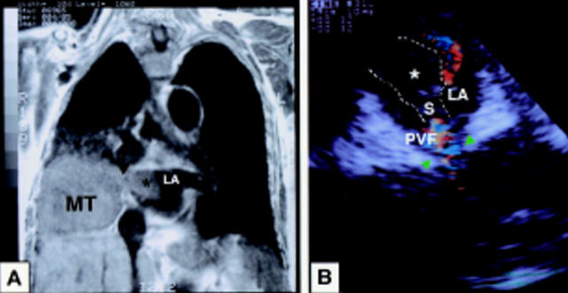
Figure 1 .
(A) MRI shows that the main tumour (MT) in the right middle lobe seems to extend directly into the left atrium (LA) through the right pulmonary vein via a stalk-like projection (arrow) and to make an intra-atrial abnormal invasive mass (*). (B) TOE shows the mass (*) in the LA measures about 2.5 cm in diameter. Colour Doppler imaging reveals the mass to have a stalk (S) derived from the right pulmonary vein (arrowhead) which appears to provide its blood flow (PVF). The broken line outlines the edge of the invasive mass
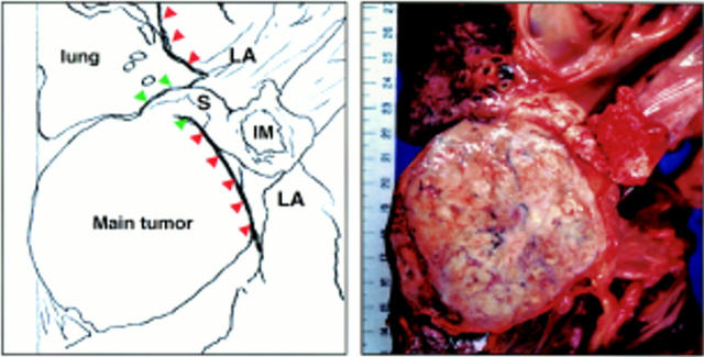
Figure 2 .
Necropsy findings. The primary tumour in the lung (main tumour) directly extends into the right middle pulmonary vein (green arrowheads) to form a tumour stalk (S) through the vein as well as the invasive mass (IM) in the left atrium (LA). The primary tumour does not directly invade through the LA wall, which forms a clear border (red arrowheads) between the main tumour and the LA lumen.


