Abstract
Polymorphous low grade adenocarcinomas (PLGAs) are thought to be indolent tumours that are localised preferentially to the palate and affect the minor salivary glands almost exclusively. Metastases to locoregional lymph nodes occur in only 6–10% of cases. Recently, two cases of PLGA with microscopically confirmed distant metastases have been reported. This study reports a third case of PLGA with histologically and immunohistochemically confirmed distant metastases. It is the first case with multiple pleural, as well as pulmonary parenchymal, metastases and metastases in cervical and para-oesophageal lymph nodes. In most cases, PLGAs are salivary gland tumours with limited potential to metastasise and a good prognosis after local treatment. However, the recently reported cases reveal that the tumour can give rise to widely spread metastases. To obtain more information about the incidence of distant metastases, periodic chest x ray examination during follow up is desirable.
Key Words: polymorphous low grade adenocarcinoma • neoplasm metastasis • comparative genomic hybridisation • human chromosome pair 6 • human chromosome pair 11
Full Text
The Full Text of this article is available as a PDF (194.0 KB).

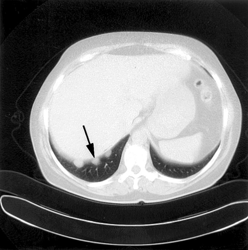
Figure 1 Thoraco-abdominal computed tomography scan showing three pleural metastases in the right sinus (arrow).

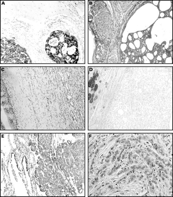
Figure 2 Section from the palatal tumour (A–D). (A) Polymorphous low grade adenocarcinoma (PLGA) infiltrating the palatal bone and exhibiting S-100 positivity. (B) PLGA with perineural spread and cribriform growth pattern (haematoxylin and eosin stained). (C) PLGA with cord-like growth and Indian files, next to pre-existing respiratory epithelium from the nasal cavity (haematoxylin and eosin stained). (D) PLGA with ductular growth, no Alcian blue positivity in tumour cells, but Alcian blue positivity in pre-existing salivary tissue. Section from the lung metastasis (E and F). (E) PLGA metastasis with solid fields and some ductular growth (haematoxylin and eosin stained). (F) PLGA metastasis with Indian file configuration (haematoxylin and eosin stained).
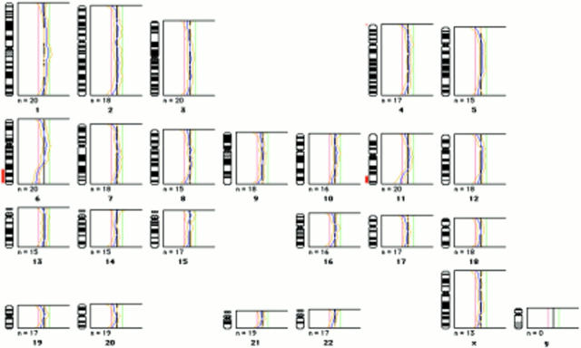
Figure 3 Comparative genomic hybridisation (CGH) analysis of the pleural metastases. Red to green ratio profiles obtained from CGH analysis of the pleural metastases. Losses on 6q and 11q are indicated by vertical lines to the left of the chromosome ideograms.


