Abstract
Aim—To develop a simple method of recording digital images of surgical specimens on to a personal computer (PC) for use in presentations for teaching and reporting of their pathology.
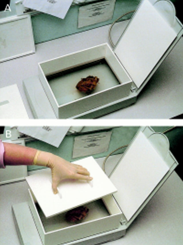
Methods—A perspex box was constructed to international A4 size 100 mm deep. This box had a base of 3 mm clear perspex with sides and top of 5 mm white perspex. This box was partially filled with distilled water and a specimen immersed in it. It was then placed on top of a standard A4 scanner. The specimen was then scanned into a PC using image capture software.
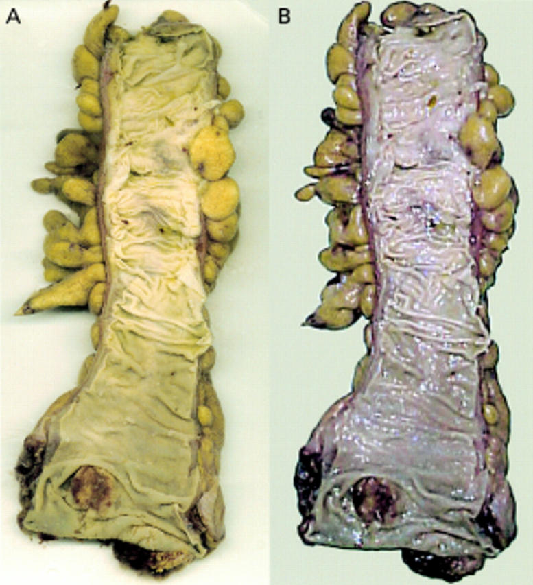
Results—The images produced showed noticeable improvement over normal photographs, especially with specimens prone to wet highlights.
Conclusions—The method has proved to be a rapid and efficient means of producing macroscopic images of surgical specimens.
Key Words: scanning • personal computer • macroscopic images • surgical specimens
Full Text
The Full Text of this article is available as a PDF (122.1 KB).

Figure 1 (A) The perspex box mounted on the scanner. (B) Application of the heavy sliding insert.

Figure 2 (A) Scanned opened bowel with small carcinoma near distal margin. (B) Same specimen photographed using digital camera showing severe highlights and poor colour reproduction. Both digital images were subjected to the same image manipulation.

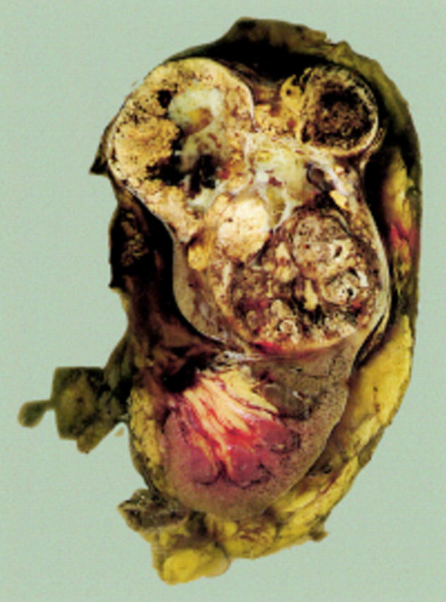
Figure 3 Kidney slice with large carcinoma in upper pole.


