Abstract
Objective: To determine if the distribution of synovitis is the same in osteoarthritis (OA) using sensitive measures of inflammation derived from dynamic, contrast enhanced magnetic resonance imaging (DEMRI).
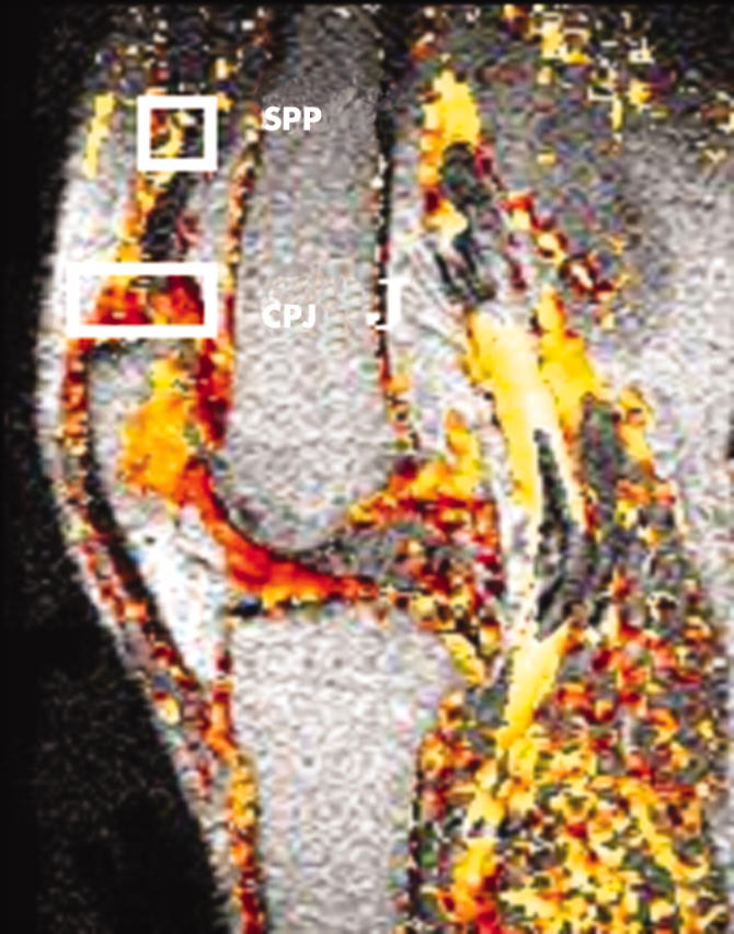
Methods: 20 subjects with established OA of the knee were recruited. Conventional MR images together with the DEMRI measurements were obtained. Areas of synovitis at the CPJ region and at a distant site in the SPP were calculated; differences in CPJ and SPP synovitis were determined using DEMRI parameters: the initial rate of contrast enhancement (IRE) and maximal enhancement (ME).
Results: The area of synovitis was significantly greater adjacent to the CPJ than in the SPP. IRE and ME measures were greater at the CPJ than the SPP.
Conclusions: The magnitude of synovitis at the CPJ is not disease-specific and applies across the spectrum of degenerative disease as well as inflammatory diseases.
Full Text
The Full Text of this article is available as a PDF (161.7 KB).
Figure 1.

Conventional MR image acquired from the knee with superimposed colour data showing values of the IRE—pixels shown in yellow represent high IRE values while those in red show relatively lower values. Data were analysed in two regions of interest represented by white rectangles and located adjacent to the CPJ and the proximal SPP.
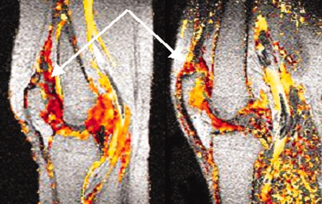
Figure 2.
Comparison of CPJ and SPO-CPJ displacement of synovial tissue by superior patella osteophyte.


