Full text
PDF
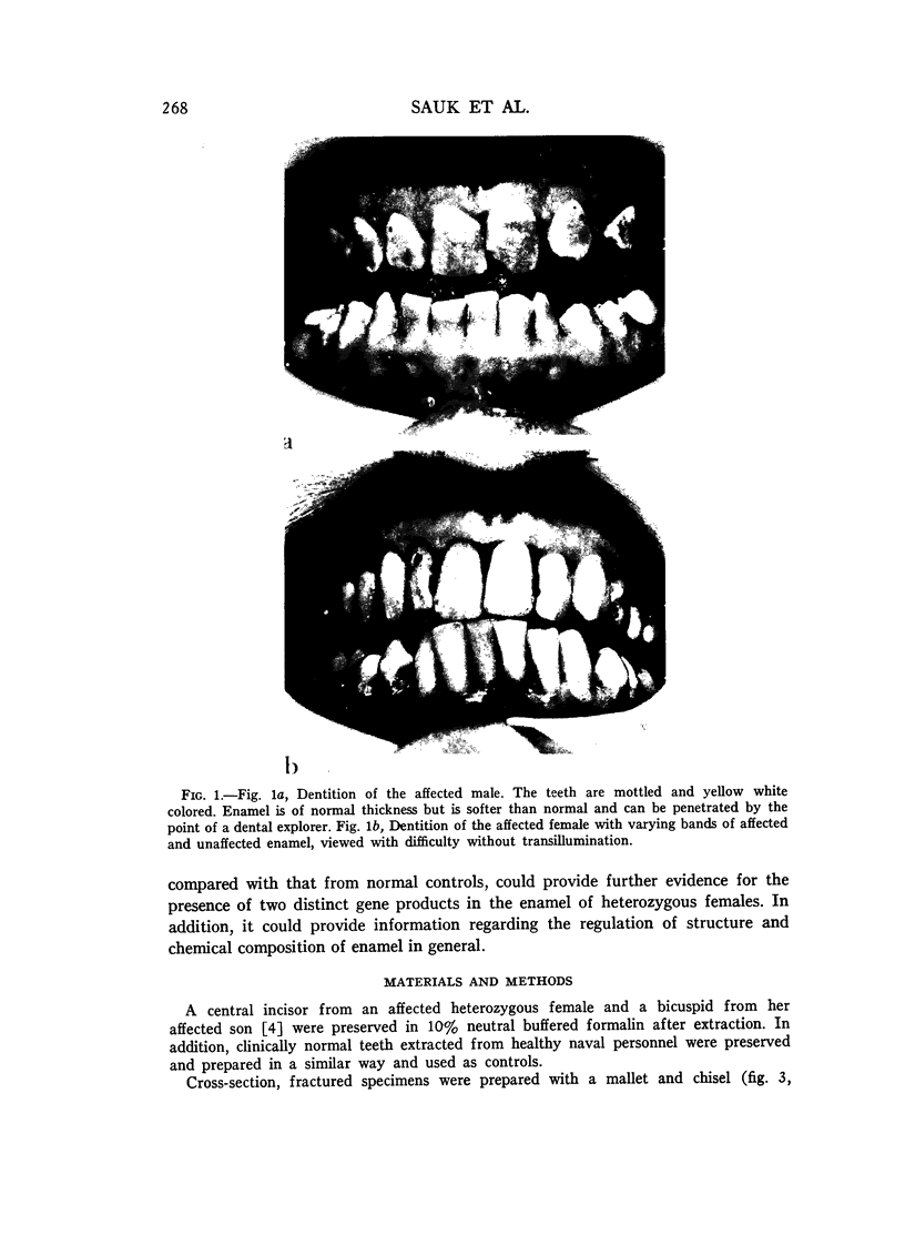
Images in this article
Selected References
These references are in PubMed. This may not be the complete list of references from this article.
- Besic F. C., Knowles C. R., Wiemann M. R., Jr, Keller O. Electron probe microanalysis of noncarious enamel and dentin and calcified tissues in mottled teeth. J Dent Res. 1969 Jan-Feb;48(1):131–139. doi: 10.1177/00220345690480010501. [DOI] [PubMed] [Google Scholar]
- Boyde A. The development of enamel structure. Proc R Soc Med. 1967 Sep;60(9):923–928. doi: 10.1177/003591576706000965. [DOI] [PMC free article] [PubMed] [Google Scholar]
- Hoffman S., Mc Ewan W. S., Drew C. M. Scanning electron microscope studies of EDTA-treated enamel. J Dent Res. 1969 Nov-Dec;48(6):1234–1242. doi: 10.1177/00220345690480062501. [DOI] [PubMed] [Google Scholar]
- Hoffman S., McEwan W. S., Drew C. M. Scanning electron microscope studies of dental enamel. J Dent Res. 1969 Mar-Apr;48(2):242–250. doi: 10.1177/00220345690480021301. [DOI] [PubMed] [Google Scholar]
- LYON M. F. Gene action in the X-chromosome of the mouse (Mus musculus L.). Nature. 1961 Apr 22;190:372–373. doi: 10.1038/190372a0. [DOI] [PubMed] [Google Scholar]
- Poole D. F., Johnson N. W. The effects of different demineralizing agents on human enamel surfaces studied by scanning electron microscopy. Arch Oral Biol. 1967 Dec;12(12):1621–1634. doi: 10.1016/0003-9969(67)90196-3. [DOI] [PubMed] [Google Scholar]
- RUSHTON M. A. HEREDITARY ENAMEL DEFECTS. Proc R Soc Med. 1964 Jan;57:53–58. [PMC free article] [PubMed] [Google Scholar]
- SCHULZE C. Erbbedingte Strukturanomalien menschlicher Zahne. Acta Genet Stat Med. 1957;7(1):231–235. [PubMed] [Google Scholar]
- WITKOP C. J. Hereditary defects in enamel and dentin. Acta Genet Stat Med. 1957;7(1):236–239. doi: 10.1159/000150974. [DOI] [PubMed] [Google Scholar]
- Witkop C. J., Jr Partial expression of sex-linked recessive amelogenesis imperfecta in females compatible with the Lyon hypothesis. Oral Surg Oral Med Oral Pathol. 1967 Feb;23(2):174–182. doi: 10.1016/0030-4220(67)90092-8. [DOI] [PubMed] [Google Scholar]


