Abstract
An implantable loop recorder (ILR) was implanted in a 45 year old man with recurrent syncope. A subsequent episode of injurious syncope led to performance of a cranial and shoulder magnetic resonance imaging (MRI). An artefact mimicking both wide and narrow complex tachycardias was recorded by the ILR during the shoulder MRI but not the cranial MRI. Caution should be used when interpreting the ECGs of ILRs in patients who have undergone MRI.
Keywords: loop recorder, magnetic resonance imaging, electromagnetic interference
The Reveal Plus (Medtronic Inc, Minneapolis, Minnesota, USA) implantable loop recorder (ILR) is commercially available and has been implanted in approximately 20 000 patients.1 Smaller than a pack of gum, this leadless, battery powered, subcutaneously placed electronic loop recorder stores multiple instances of up to 42 minutes of high fidelity time and date stamped ECG for review. With a battery life of up to 14 months, it is ideally suited for long term outpatient evaluation of syncope of suspected arrhythmic origin.2,3 While direct patient activation of the ILR through a small hand held pager-like device is the primary means to capture a symptomatic arrhythmic event, the Reveal Plus can automatically detect significant bradycardic and tachycardic events to supplement patient activation.
Magnetic resonance imaging (MRI) has now become the procedure of choice for a variety of central nervous system disorders and is becoming increasingly important in cardiovascular imaging.4 It is often used to further define a suspected musculoskeletal abnormality.5
While adverse interactions between pacemakers and implantable cardioverter defibrillators in the MRI environment have been recognised for years,6,7 substantially less has been reported regarding the interaction between the ILR and MRI scanners.8 We report the ECG recordings of an implantable loop after a 45 year old man underwent MRI of the brain and then later an MRI of the right shoulder.
CASE REPORT
A 45 year old man presented with recurrent episodes of syncope, several of which were injurious. After the history and physical examination, ECG, multiple external loop recordings, tilt table testing, electrophysiological testing, cranial MRI, and electroencephalography failed to disclose the cause of the patient's recurrent syncope, an ILR (Reveal Plus) was implanted in his left parasternal region.
Four months later, the patient fell off the roof of his single story home while attempting to “rescue his cat”. The patient lost consciousness and injured his right shoulder. The ILR was interrogated in the emergency room after the fall and nothing had been recorded in the memory of the ILR. During a cranial MRI, one of the authors (JRG) attended to the patient, clearing the ILR before MRI and interrogating and downloading its contents after the MRI. Nothing had been recorded by the ILR during the cranial scan.
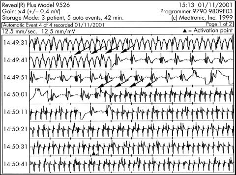
The following day, an orthopaedic consultant obtained an MRI of the patient's right shoulder. A 1.5 T MRI was used (T2 fast-spin echo, echo time 34–85 ms, repetition time 2500–4000 ms, 5 mm slice thickness, 15 slices). The ILR was verified to be functioning properly immediately before the patient's MRI and its memory was cleared. The contents of the ILR were downloaded immediately after the MRI. While not monitored with ECG or pulse oximetry, the patient did not experience any cardiopulmonary or neurological symptoms during the scan. Figure 1 shows what appears to be a wide complex tachycardia followed by sinus rhythm and then a narrow complex tachycardia. The time–date stamp feature of the ILR confirmed that both episodes of “tachycardia” were recorded by the ILR while the patient was undergoing MRI of the right shoulder.
Figure 1.
Output of a Reveal Plus implantable loop recorder after the patient underwent right shoulder magnetic resonance imaging showing wide complex tachycardia followed by sinus rhythm and then narrow complex tachycardia. The arrows above the rhythm strip (lines 2 and 4) indicate the patient's native QRS within the artefact. The cycle length of the artefact is 260 ms.
DISCUSSION
The one to one correlation of the presence of an arrhythmia and associated symptoms provides the intellectual underpinnings justifying the choice of treatment. It is well known that “electrocardiographic artifact can simulate ventricular tachycardia”9 leading to inappropriate diagnostic testing and treatment. “Artefactual atrioventricular block” has also been reported to lead to inappropriate drug treatment in the intensive care unit setting.10 As reported by others, during artefact mimicking tachycardia the patient's native QRS complex is visible within the “tachycardia” sequence.9 Thus, in our patient no specific treatment was directed towards treating the patient's “arrhythmia”.
It is well known that electromagnetic interference can degrade the performance of electronic medical devices.11 Time varying magnetic forces and radiofrequency waves are two types of electromagnetic energy. The powerful gradient magnetic fields and the radiofrequency energies required for MRI applied across the body of the ILR undoubtedly produced the artefact that was observed during shoulder MRI. Because the cranial MRI was done with a “head coil”, thereby focusing the radiofrequency and gradient magnetic energy on the region of interest, it is likely that the strength of the applied radiofrequency and magnetic gradients were insufficient to create the artefact seen only during the shoulder scan.
This case adds to the body of previously recognised ECG artefacts. Because patients with injurious syncope who have ILRs may undergo MRI, care should be taken to exclude artefact when interpreting the ECG recordings of the ILR after MRI.
Abbreviations
ILR
implantable loop recorder
MRI
magnetic resonance imaging
REFERENCES
- 1.Medtronic Technical Support: March 20, 2002.
- 2.Kenny RA, Krahn AD. Implantable loop recorder: evaluation of unexplained syncope. Heart 1999:81:431–3. [DOI] [PMC free article] [PubMed] [Google Scholar]
- 3.Kennedy HL, Podrid PJ. Role of Holter monitoring and exercise testing for arrhythmia assessment and management. In: Podrid PJ, Kowey PR, eds. Cardiac arrhythmia: mechanisms, diagnosis, and management. Philadelphia: Lippincott Williams and Wilkins, 2001:165–93.
- 4.Weiss RG. Evolving cardiovascular applications for magnetic resonance imaging. Cleve Clin J Med 2001;68:238–42. [DOI] [PubMed] [Google Scholar]
- 5.Shellock FG, Bert JM, Fritts HM, et al. Evaluation of the rotator cuff and glenoid labrum using a 0.2-Tesla extremity magnetic resonance (MR) system: MR results compared to surgical findings. J Magn Reson Imaging 2001;14:763–70. [DOI] [PubMed] [Google Scholar]
- 6.Gimbel JR. Implantable pacemaker and defibrillator safety in the MR environment: new thoughts for the new millenium. In: 2001 syllabus, special cross-specialty categorical course in diagnostic radiology: practical MR safety considerations for physicians, physicists, and technologists. Oak Brook, Illinois: Radiological Society of North America, 2001:69–76.
- 7.Goldschlager N, Epstein A, Friedman P, et al. Environmental and drug effects on patients with pacemakers and implantable cardioverter/defibrillators: a practical guide to patient treatment. Arch Intern Med 2001;161:649–55. [DOI] [PubMed] [Google Scholar]
- 8.De Cock CC, Spruijt HJ, Van Campen LMC, et al. Electromagnetic interference of an implantable loop recorder by commonly encountered electronic devices. Pacing Clin Electrophysiol 2000;23:1516–8. [DOI] [PubMed] [Google Scholar]
- 9.Knight BP, Pelosi F, Michaud GF, et al. Clinical consequences of electrocardiographic artifact mimicking ventricular tachycardia. N Engl J Med 1999;341:1270–4. [DOI] [PubMed] [Google Scholar]
- 10.Littman L, Humphrey SS, Monroe MH: Consult for “heart block”: what is the rhythm? J Cardiovasc Electrophysiol 2001;12:1429–30. [DOI] [PubMed] [Google Scholar]
- 11.Silberberg, JL. Performance degradation of electronic medical devices due to electromagnetic interference. Compliance Eng 1993;fall:1–8.


