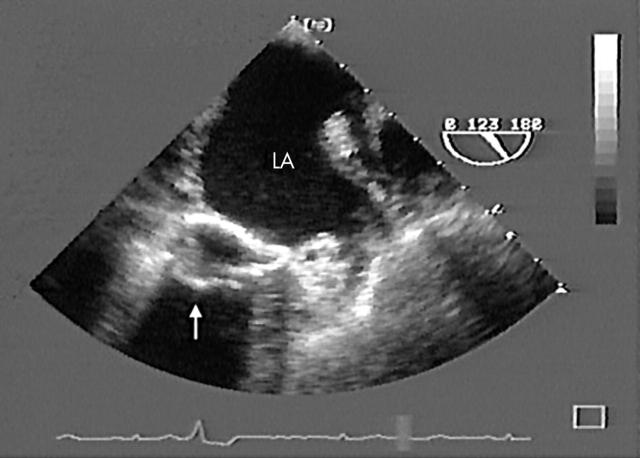
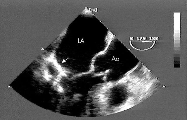
A 74 year old woman was found to have a calcified mass on a plain chest radiograph. Follow up studies identified a large, densely calcified mitral periannular mass, consistent with mitral annular calcification (MAC). Three months later, she presented with the acute onset of lethargy. Initial laboratory studies were remarkable only for an elevated serum calcium concentration of 14.8 mg/dl (normal range, 8.5–10.5 mg/dl). She was treated with intravenous fluids and pamidronate and, over the following 48 hours, her neurologic status and serum calcium concentration returned to baseline with no residual sequelae.
Metabolic evaluation revealed parathyroid hormone values to be mildly reduced, with normal 1,25 dihydroxyvitamin D and serum phosphorous concentrations. No evidence of neoplasm could be identified upon computed tomography, bone scanning, and bronchoscopy. Repeat echocardiography revealed a more heterogeneous appearance of the mitral periannular mass with a clear, central echolucent zone (panels below, Ao, aorta; LA, left atrium). Whether the hypercalcaemic state and mitral annular changes were directly or causally related to the caseous transformation of MAC is unclear.
Caseous calcification of the mitral annulus is a relatively rare variant with an echocardiographic prevalence of 0.6% in patients with MAC and 0.06–0.07% in large series of patients of all ages. Caseous calcification is defined echocardiographically as a large, round or semilunar, echodense mass in the periannular mitral region, which contains central areas of echolucencies resembling liquefaction. As the prevalence of caseous calcification in echocardiographic studies is less than that of necropsy series, it is likely that the widespread use and improvements in echocardiography may increasingly uncover this condition, which has been greatly underappreciated and, at times, unrecognised.


