Abstract
A 73 year old woman presented with profound central cyanosis and a history of a minor stroke. She had normal heart morphology, normal pulmonary artery pressure, and a normal coronary angiography. A patent foramen ovale (PFO) with a massive right to left shunt was demonstrated at the atrial level, with normal pulmonary venous saturations and Po2 values. This rare, age related case of right ventricular diastolic dysfunction in a normotensive patient revealed a generous PFO allowing a pronounced right to left shunt.
Keywords: patent foramen ovale, right ventricular diastolic dysfunction, cyanosis, right to left shunt
A 73 year old woman presented with recent onset cyanosis and a past history of a minor stroke. Room air arterial Po2 was 40 mm Hg. Transoesophageal echocardiography revealed a patent foramen ovale (PFO) with a large right to left shunt. No other anomaly was seen.
Cardiac catheterisation disclosed normal coronary arteries and normal pulmonary pressure. Right ventricular (RV) diastolic pressure was slightly higher than left ventricular (LV) diastolic pressure. On 100% Fio2, pulmonary venous Po2 was 540 mm Hg and the left atrial Po2 was 97 mm Hg. Balloon occlusion of the PFO served to test the tolerance of PFO occlusion and to measure the effective stretched defect size. Immediate post-transcatheter PFO closure using a 24 mm atrial septal defect (ASD) Amplatzer occluder resulted in a rise of arterial Po2 to 320 mm Hg. Other causes of the atrial level right to left shunt such as pulmonary disease, pulmonary vascular disease, RV hypertrophy, RV systolic dysfunction, right atrial myxoma, tricuspid valve disease, and pericardial effusion were excluded.
This rare, age related case of RV diastolic dysfunction in a normotensive patient revealed a generous PFO allowing a pronounced right to left shunt.
DISCUSSION
A persistent right to left shunt at the atrial level across a PFO has previously been described in relation to RV dysfunction or elevation of the pulmonary pressure in conditions such as chronic obstructive pulmonary disease, or pulmonale, recurrent pulmonary embolism, tricuspid valve dysfunction, RV infarction, and right sided myxoma.1 A significant right to left shunt via PFO has rarely been described without underlying cardiac or pulmonary disease.2–4 Several theoretical pathophysiological explanations were offered to explain such a phenomenon. Preferential streaming of the blood from the inferior vena cava across the PFO and transatrial septal pressure gradients were suggested as the likely cause for the haemodynamic state.
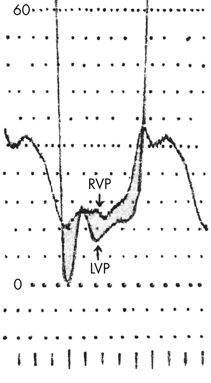
This is a case report of a 73 year old woman who presented with profound central cyanosis and a history of a minor stroke. She had normal heart morphology, normal pulmonary artery pressure and a normal coronary angiography. A massive right to left shunt was demonstrated at atrial level with normal pulmonary venous saturations and Po2 values. The reason for this huge right to left shunt at the PFO level is illustrated by the diastolic pressure curves representing compliance differences between right and left ventricles (fig 1). This is probably an age related phenomenon in the presence of normal LV compliance and function of this normotensive woman. Age related reduction of ventricular compliance is a frequently described finding. This is a rare case in which this age related phenomenon affected mostly the right ventricle, and in the presence of a large PFO allowed this pronounced right to left flow at the atrial level with profound cyanosis. The patient’s oxygenation improved dramatically following the transient balloon occlusion of the defect. Testing the tolerance of this procedure and measuring the defect size (22 mm) was followed by transcatheter PFO closure by a 24 mm Amplatzer ASD occluder. Physical rehabilitation to normal function followed.
Figure 1.

A simultaneous recording of diastolic left ventricular pressure (LVP) and diastolic right ventricular pressure (RVP). This illustrates that the diastolic RVP is slightly greater than the LVP throughout the diastole.
Abbreviations
ASD, atrial septal defect
LV, left ventricular
PFO, patent foramen ovale
RV, right ventricular
REFERENCES
- 1.Kerut EK, Norfleet WT, Plotnick GD, et al. Patent foramen ovale: a review of associated conditions and the impact of physiological size. J Am Coll Cardiol 2000;38:613–23. [DOI] [PubMed] [Google Scholar]
- 2.Ciafone RA, Anaesty JM, Weintraub RM, et al. Cyanosis in uncomplicated atrial septal defect with normal cardiac and pulmonary arterial pressures. Chest 1998;74:596–9. [DOI] [PubMed] [Google Scholar]
- 3.Thomas JD, Tabakin BS, Ittleman FP. Atrial septal defect with right to left shunt despite normal pulmonary artery pressure. J Am Coll Cardiol 1987;9:221–4. [DOI] [PubMed] [Google Scholar]
- 4.Godart F, Rey C, Prat A, et al. Atrial right-to-left shunting causing severe hypotemia despite normal right-sided pressure; report of 11 consecutive cases corrected by percutaneous closure. Eur Heart J 2000;21:483–9. [DOI] [PubMed] [Google Scholar]


