Abstract
Objective: To evaluate the comparative diagnostic value of harmonic imaging (HI) in the assessment of patients with suspected infective endocarditis (IE).
Setting: Tertiary referral centre.
Design: 139 consecutive patients were evaluated with three imaging modalities: transthoracic echocardiography with fundamental imaging (FI); HI; and transoesophageal echocardiography (TOE). Image quality was assessed for each modality by semiquantitative scoring (0, poor, to 3, excellent). Presence, dimension, and characteristics of vegetations were assessed separately for each imaging modality, as well as presence of abscesses.
Results: 35 patients had definite IE. TOE was positive in 33 patients, HI in 28, and FI in 12 (p < 0.001 for FI v HI and v TOE). Mean image quality was 1.4 (0.7) for FI, 2.1 (0.6) for HI (p < 0.01 v FI), and 2.6 (0.4) for TOE (p < 0.001 v HI). The association between FI and TOE findings was Φ = 0.35 (χ2 = 17.57, p = 0.0014) and between HI and TOE it was Φ = 0.95 (χ2 = 125.72, p < 0.0001; p < 0.0001 v FI). The global echo score of vegetations was 7.1 (3.3) with FI, 8.5 (3.4) with HI, and 11.3 (3.9) with TOE (p < 0.001 v HI). Compared with TOE, FI identified only one of seven abscesses (sensitivity 14%) and HI identified two of seven abscesses (sensitivity 28%).
Conclusions: HI provides an accurate assessment of suspected IE. TOE achieves superior definition of IE related abnormalities.
Keywords: diagnosis, echocardiography, harmonic imaging, infective endocarditis
Despite advances in diagnosis and treatment, infective endocarditis (IE) still has high morbidity and mortality often due to delayed diagnosis.1 To improve the outcome of patients with IE, a high level of clinical suspicion is needed together with good access to echocardiography, which is pivotal in the diagnostic investigation.1,2 Transoesophageal echocardiography (TOE) has proved to be superior to transthoracic echocardiography (TTE) for the detection of vegetations and cardiac complications that result from IE.3–6 However, owing to the semi-invasive nature and cost of TOE, its indiscriminate use to rule out IE seems neither justified nor cost effective.2
Harmonic imaging (HI) is an imaging modality that is based on the principle of receiving double the emitted ultrasound frequency (second harmonic).7 It was initially used to improve the transthoracic visualisation of left heart echo contrast agents, but it was subsequently used to improve the imaging quality of heart structures without the use of contrast.8 Compared with fundamental imaging (FI), HI has a better signal to noise ratio between the cavity and wall with a reduction in near field clutter leading to improved endocardial border delineation and higher image resolution of valve structures.7 We therefore hypothesised that HI can be used to obtain an accurate identification of vegetations and a precise assessment of IE related complications.
METHODS
One hundred and thirty nine consecutive patients with a mean (SD) age of 46.1 (28.4) years referred to our echocardiographic laboratory for suspected IE made up the study population. There were 83 men and 56 women. All patients gave informed consent to participate in the study, the protocol for which was approved by the ethics committee of the hospital. Echocardiography was indicated after excluding on the basis of thorough clinical examination and rapidly available tests easily detectable infections, in the presence of one or more of the following situations: (1) fever of unknown origin; (2) cardiac murmur; (3) history of heart valve disease; (4) embolic phenomena; (5) immunological phenomena; and (6) blood cultures positives for pathogens typically involved in IE. Patients with a mechanical valve prosthesis for whom TOE was considered a first choice study were excluded. IE was diagnosed according to the Duke criteria.9 Diagnosis at discharge was obtained from the clinical records.
Echocardiography
Transthoracic echocardiography
All studies were performed with a Sequoia 256 system (Acuson, Mountain View, California, USA) with a 3V transducer. Each cardiac valve was examined in detail by M mode, two dimensional, and Doppler colour flow mapping at minimum depth setting. TTE images were acquired in the FI mode at the highest possible transducer frequency that still allowed clear delineation of valve structures. Gain settings were adjusted individually for each patient to visualise valve structures optimally. After all cardiac valves had been examined the transducer was switched into the harmonic mode with a transmitting frequency of 1.75 MHz. The receiver gain was again adjusted individually for each patient to obtain the best visualisation of the cardiac valves. All echocardiographic studies were recorded on S-VHS videotape. Four 3 second clips with the best achieved image resolution were acquired for each valve and digitally stored for offline analysis.
Transoesophageal echocardiography
TOE studies were performed after precordial examination by the same operator. Patients were positioned in the left lateral decubitus position after topical anaesthesia of the pharynx with lidocaine. An omniplane transducer with 5–7 MHz transmitting frequency was used. Valves were imaged in all available imaging planes at the highest possible frequency. Each valve was examined by M mode echocardiography at 100 cm/s sweep, two dimensional cross sectional echocardiography, and colour Doppler. As with TTE, four 3 second clips with the best achieved image resolution were acquired for each valve and were stored digitally.
Data analysis
All studies were analysed off line from the video clips. To account for possible bias the TTE clips from each patient in either the fundamental or harmonic mode were copied in random order from the original studies to a new compact disc. Each study was assigned a number that was used for patient and modality identification after data analysis. Therefore, the observers were completely blinded to the imaging mode of the video sequences and to the patient data by obscuring the display of the technical instrument settings and patient data. The video clips were analysed by two experienced observers. In case of uncertainty or divergent results, the studies were reviewed by a third observer and consensus interpretation was achieved. The video data were analysed as follows:
Identification of vegetations—A vegetation was diagnosed when an endocardial structure with echogenicity distinct from that of the adjacent endocardium and displaying motion distinct from the cardiac structures was seen attached to an endocardial surface.10 In the presence of inadequate image quality or when abnormalities only partially typical for vegetations (echogenicity similar to that of the endocardial surface, moving after the cardiac cycle, etc) were visible, the study was considered indeterminate.
Assessment of the characteristics of the vegetations—The echocardiographic appearance of vegetations was characterised by a modification of the method described by Sanfilippo and colleagues11 considering in a semiquantitative way (grades 1–4) the extent, mobility, and consistency of vegetations. These characteristics were combined with the maximum length of vegetations (grade 1, < 0.5 mm; grade 2, 0.5–1 mm; grade 3, 1–2 mm; grade 4, > 2 mm) to generate a global score ranging from 4–16.
Identification of paravalvar complications—An abscess was considered to be present when echolucent cavities within the valve anulus or adjacent myocardium were found in the setting of valve infection.6 Fistulas were diagnosed with colour Doppler by the presence of abnormal flow communicating cardiac chambers.
Assessment of image quality—Considering the visibility of aortic, mitral, and tricuspid valve structures, four levels were defined: 0, poor image quality (no clear visualisation of valve structures); 1, suboptimal image quality (hazy delineation of valve structures); 2, good image quality (adequate visualisation of at least two valve structures with minimum clutter); 3, optimal image quality (complete visualisation of all valve structures without clutter).
Statistical analysis
Continuous measures were expressed as mean (SD). The Wilcoxon paired test was used to compare differences in mean image quality and vegetation characteristics. All assessments of significance between diagnostic techniques used two tailed tests. Analysis of the degree of association between pairs of variables was accomplished with the Φ coefficient for paired dichotomies.12 The Fisher r to z transformation was used to test the null hypothesis that the association between HI and TOE was not significantly different from the association between FI findings and TOE results. The criterion of significance was the probability of a type 1 error of 0.05 or less. The χ2 sampling distribution was used as the approximate sampling distribution to assess the significance of Φ coefficients.
RESULTS
Table 1 lists reasons for referral to echocardiography and diagnosis at discharge.
Table 1.
Frequency of reasons for referral to echocardiography and diagnosis at discharge
| Reason for referral | |
| FUO without evident extracardiac infection | 50 |
| FUO with blood cultures positive for germs typical for IE | 18 |
| FUO in recent cardiac surgery | 14 |
| FUO with predisposing heart disease | 26 |
| FUO in injecting drug users | 10 |
| Recurrent embolic phenomena | 12 |
| FUO with immunological phenomena | 5 |
| Other | 4 |
| Diagnosis at discharge | |
| Pneumonia | 4 |
| Connective tissue disease | 8 |
| Meningitis | 7 |
| Splenic abscess | 8 |
| Viral illness | 11 |
| Stroke | 9 |
| Pericarditis | 10 |
| Cerebral abscess | 8 |
| Malignancy | 11 |
| Line sepsis | 4 |
| Bacteraemia of unknown origin | 14 |
| Other | 10 |
| Total IE | 35 |
FUO, fever of unknown origin; IE, infective endocarditis.
Thirty five patients had definite IE. Diagnosis was made at surgery (n = 13), necropsy (n = 6), or during clinical and echocardiographic follow up (n = 16).
Detection of vegetations
Table 2 summarises the vegetations identified by echocardiography according to location and imaging modality.
Table 2.
Detection of vegetations according to location and imaging modality
| FI | HI | TOE | |
| Aortic valve | 5 | 10 | 11 |
| Mitral valve | 4 | 9 | 10 |
| Tricuspid valve | 2 | 3 | 3 |
| Aortic prosthesis | 0 | 2 | 3 |
| Mitral prosthesis | 1 | 4 | 6 |
| Total | 12 | 28 | 33 |
FI, fundamental imaging; HI, harmonic imaging; TOE, transoesophageal echocardiography.
The difference between TOE and HI for the detection of vegetations was not significant (33 v 28). In contrast, results were significantly different between TOE and FI (33 v 12, p < 0.0001) and between HI and FI (28 v 12, p = 0.0003). TOE study was negative in 104 patients. In these patients FI was negative in 73 (70.1%) and HI excluded the presence of vegetations in 96 (92.3%, p < 0.01 for HI v FI). There were two false negatives by TOE: one resulted from previous embolisation of vegetations and the other from early infection on an aortic prosthesis. In both cases repeat TOE proved diagnostic.
Concordance of TTE findings with TOE
Findings of FI agreed with TOE in 86 patients (61.8%). The association between FI interpretation and TOE findings was Φ = 0.35 (χ2 = 17.57, df = 4, r = 0.29 with p = 0.0014). Findings of HI agreed with TOE in 125 patients (89.9%). The association between HI interpretation and TOE results was Φ = 0.95 (χ2 = 125.72, df = 4, r = 0.85 with p < 0.0001). The p value of the difference between the Φ coefficients for FI and HI (0.35 and 0.95, respectively) was estimated to be < 0.0001.
Table 3 shows the calculated sensitivity and specificity for FI and HI, assuming TOE as the reference standard.
Table 3.
Diagnostic performance of transthoracic echocardiography for the detection of vegetations, according to imaging modality, assuming TOE as reference standard
| FI | HI | |
| Sensitivity | 36.4% | 81.8% |
| Specificity | 80.2% | 98.1% |
| Positive predictive value | 36.4% | 93.1% |
| Negative predictive value | 80.2% | 94.5% |
| Accuracy | 69.7% | 94.2% |
FI, fundamental imaging; HI, harmonic imaging.
Impact of image quality on diagnostic accuracy
Mean image quality was 1.4 (0.7) for FI, 2.1 (0.6) for HI, and 2.6 (0.4) for TOE and was significantly different between the three imaging modalities (p < 0.001 for FI v HI, HI v TOE, and FI v TOE).
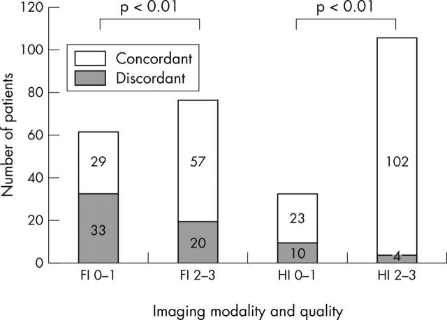
The number of studies with poor or moderate image quality (score 0 and 1) was significantly lower for TOE (n = 2) (1.4%) than for HI (n = 33) (23.7%, p < 0.001 v TOE) and for FI (n = 62) (44.6%, p < 0.001 v FI). The concordance of FI with TOE findings was lower in the presence of poor or moderate image quality (46.7%) than with good or excellent imaging quality FI studies (74%, p < 0.001) (fig 1). The same applied to HI findings (69.6% for 0–1 imaging quality v 96.2% for 2–3 imaging quality; p < 0.001).
Figure 1.
Bar graph showing the number of studies concordant with transoesophageal echocardiographic (TOE) findings, according to imaging modality (FI, fundamental imaging; HI, harmonic imaging) and image quality (0–1, poor to moderate image quality; 2–3, good to excellent image quality).
For patients with a negative FI study but a positive HI study (fig 2), the number of studies with low image quality (0–1) with the FI modality (n = 12) (8.6%) was significantly higher than the number of studies with low image quality with HI (n = 2) (1.4%, p < 0.001). The difference in image quality between TOE and HI was not significant in the studies in which vegetations were detected by both techniques. Among the five patients with positive TOE in whom HI did not identify the presence of vegetations, three had 0–1 image quality and two had a prosthesis in the mitral position.
Figure 2.
Example of a patient with a vegetation attached to the posterior leaflet of the mitral valve. No valve abnormalities are evident at FI. HI shows an irregularly shaped structure with uneven margins attached to the atrial aspect of the posterior leaflet of the mitral valve (arrow), indicative of vegetation. TOE depicts a large mitral vegetation with an echogenic core and a hypodense surface, attached on the atrial aspect of the posterior mitral leaflet (arrow). AO, aorta; LA, left atrium; LV, left ventricle.
Qualitative assessment of vegetations and paravalvar involvement
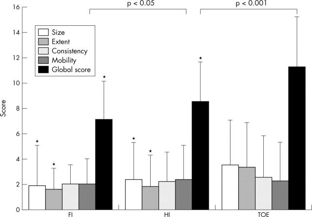
The global echo score of vegetations was 7.1 (3.3) with FI, 8.5 (3.4) with HI (p = 0.03 v FI), and 11.3 (3.9) with TOE (p < 0.001 v HI) (fig 3). The size and extent of vegetations were significantly smaller with both FI and HI than with TOE, whereas vegetation mobility and consistency did not differ significantly. TOE disclosed seven abscesses and two fistulas. FI identified only one abscess (sensitivity 14%) and one fistula, and HI failed to identify five abscesses (sensitivity 28%) and one fistula.
Figure 3.
Bar graph showing the echocardiographic characteristics of vegetations assessed by the three imaging modalities. The lines above the bars indicate the standard deviation. *p<0.05 v TOE.
DISCUSSION
Echocardiography is the primary technique for the detection of vegetations and cardiac complications resulting from IE. The diagnostic role of echocardiography in the setting of IE is so important that diagnostic criteria for IE have been revised to include echocardiographic findings as a major criterion.9 The key advance has been made by TOE. In fact, TOE has been reported to have an 87–100% sensitivity for the detection of vegetations when compared with 30–63% sensitivity for TTE.3,4 TTE does not detect vegetations that are < 3 mm in diameter and underestimates the size of larger vegetations because of its lower image resolution. Moreover, TTE may provide technically inadequate imaging in patients with poor acoustic window secondary to pulmonary disease, obesity, or recent thoracotomy.
HI has recently emerged as a promising additional modality in echocardiography.7 It exploits the gradual generation of harmonic energy as ultrasound waves propagate through tissue. Two aspects of harmonic generation are critical to the improved imaging obtained by this technique. Firstly, harmonic energy increases with the distance the ultrasound wave propagates, so there is little HI in the near field, thus limiting near field (chest wall) artefacts. Secondly, harmonic amplitude varies with the square of the fundamental amplitude, and thus most harmonics result from the central ultrasound beam rather than the weaker side lobe artefacts.7
HI has been primarily used to enhance left ventricular endocardial border delineation and it has been proved useful to increase accuracy and interobserver agreement of stress echocardiography.13 HI has proved accurate for the assessment of abnormalities such as left atrial appendage anatomy,14 rheumatic mitral stenosis,15 right to left atrial shunting,16 and aortic arch atheromas,17 which have traditionally been considered indications for TOE.
The key factor in determining the accuracy of echocardiography in the assessment of IE is the image quality. In our experience both FI and HI have a significantly greater concordance with TOE (as the reference method) in the presence of good or optimal image quality. However, the advantage of HI over FI resides in the significantly lower percentage of patients with suboptimal image quality. Although HI is used regularly by most operators, in many laboratories systems provided with HI still coexist with equipment functioning on an FI basis. It is therefore recommended that all patients being examined for suspected IE should be imaged with HI to increase the accuracy of TTE.
The pre-test likelihood in the study population and the negative predictive value are critical when evaluating the screening value of a diagnostic technique. According to the definition of Lindner and colleagues18 our patients had an intermediate to high pre-test probability of IE. In our patients the negative predictive value of TOE was 98%, which was higher than that found by Sochowski and colleagues(93%).19 This discrepancy may result from the different TOE technologies (single plane versus multiplane) used. Image quality influenced the negative predictive value of both transthoracic imaging modalities in a critical way. In fact both FI and HI were accurate in ruling out IE in the presence of adequate image quality. This was consistent with the study of Irani and colleagues,20 who found that in the presence of good image quality a negative FI study could obviate the need for TOE in patients with suspected endocarditis. In their retrospective study, however, 51% of TTE studies were non-diagnostic because of either inadequate image quality (25%) or indeterminate findings (26%). Moreover, TOE studies were performed in 48% of patients with monoplane technology, patients with prosthesis were excluded, and characteristics of vegetations and subvalvar involvement were not considered. In our study HI provided adequate image quality in 76.2% of patients; for these patients the negative predictive value of the technique was 94%, which seems adequate for clinical use.
When considering the results of our study it must be remembered that patients with mechanical prosthesis were excluded, no pacemaker leads were infected, and the relatively young study population might have had better average transthoracic image quality and less frequent degenerative changes on valve structures.
Moreover, although HI had increased sensitivity for detection of IE on bioprosthesis, when compared with FI, the number of patients with valve prosthesis was too small to lead to a definite conclusion. In our opinion TOE remains the first choice for such patients to save time and money and especially to avoid the potentially devastating consequences of missing the diagnosis of prosthetic IE.
The American Heart Association/American College of Cardiology guidelines recommend that TTE should be carried out in all patients with a high or even low probability of IE.2 This approach has been considered cost ineffective either because it can result in an overuse of echocardiography, which seems inappropriate in a cost conscious era,21,22 or because a significant number of cases may go undetected, given the reported low sensitivity of TTE in unselected populations.23 In this respect it may be valid to implement HI because it may provide an accurate screening tool for patients with suspected IE at reasonable costs.
In patients with a positive HI, TOE has not only proved to be a confirmatory test but it has also provided much adjunctive information that may be critical in guiding the treatment strategy. Several studies have shown that larger vegetations are associated with increased embolic risk.24,25 Moreover, if the size of vegetations is noted to increase appreciably during antibiotic treatment the patient is more likely to develop clinically significant emboli and may benefit from early surgery.26
Perianular extension of endocarditis is associated with more complications, including heart failure, atrioventricular block, need for surgery, and adverse prognosis, with mortality ranging between 40–90%.1 In our study HI tended to underestimate the width of vegetations and did not provide any significant improvement over FI in the detection of abscesses. It should therefore be emphasised that HI did not adequately assess certain complications of IE, such as perianular abscesses, which may have a tremendous impact on prognosis and management and are themselves considered to be a major diagnostic criterion.9
Study limitations
Complete blinding of transthoracic studies was not possible because HI images can be distinguished by an experienced observer. However, investigators were not aware of a patient’s identity during the analysis. Moreover, comparison between FI, HI, and TOE studies on the same patient was impossible because of the random order of the clips stored on the disc. Evaluation of the agreement between the three imaging modalities should have obviated such a limitation.
Although we studied consecutive patients, selection bias is probable in our study for several reasons. Firstly, ours is a tertiary care centre, and patients seen at our institution do not necessarily reflect those seen in community hospitals. Secondly, most patients were referred from the division of infectious disease of our hospital, which has now been involved in an endocarditis project for some 10 years. The awareness of the referring physicians of suspected IE is thus likely to be higher than is generally seen. Thirdly, the type of IE seen at our institution is different from that seen in large metropolitan medical centres (the rate of tricuspid endocarditis is lower than reported from high volume city hospitals). Most of our patients had an intermediate to high pre-test probability of IE on the basis of clinical criteria. The results of the present study should therefore not be extrapolated to populations with different clinical characteristics.
Conclusions
HI has greatly improved the diagnostic value of TTE in patients with suspected native IE, by ameliorating imaging quality and reducing the number of indeterminate studies. In particular a negative good quality HI has a high negative predictive value and may reduce the need for TOE in patients with suspected IE. In the presence of indeterminate HI, TOE seems indicated. In the presence of positive HI, TOE may be useful to improve identification of the characteristics of the vegetation, the subvalvar involvement, and the presence of abscesses, all of which may be important in treatment decisions.
Abbreviations
FI, fundamental imaging
HI, harmonic imaging
IE, infective endocarditis
TOE, transoesophageal echocardiography
TTE, transthoracic echocardiography
REFERENCES
- 1.Erbel R, Liu F, Ge J, et al. Identification of high-risk subgroups in infective endocarditis and the role of echocardiography. Eur Heart J 1995;16:588–602. [DOI] [PubMed] [Google Scholar]
- 2.Bayer AS, Bolger AF, Taubert KA, et al. AHA scientific statement. Diagnosis and management of infective endocarditis and its complications. Circulation 1998;98:2936–48. [DOI] [PubMed] [Google Scholar]
- 3.Shively BK, Gurule FT, Roldan CA, et al. Diagnostic value of transesophageal compared with transthoracic echocardiography in infective endocarditis. J Am Coll Cardiol 1991;18:391–7. [DOI] [PubMed] [Google Scholar]
- 4.Erbel R, Rohmann S, Drexler M, et al. Improved diagnostic value of echocardiography in patients with infective endocarditis by transesophageal echocardiography: a prospective study. Eur Heart J 1988;9:43–53. [PubMed] [Google Scholar]
- 5.Karalis DG, Bansal RC, Hauck AJ, et al. Transesophageal echocardiographic recognition of subaortic complications in aortic valve endocarditis: clinical and surgical implications. Circulation 1992;86:353–62. [DOI] [PubMed] [Google Scholar]
- 6.Daniel WG, Mügge A, Martin RP, et al. Improvement in the diagnosis of abscesses associated with endocarditis by transesophageal echocardiography. N Engl J Med 1991;324:795–800. [DOI] [PubMed] [Google Scholar]
- 7.Thomas JD, Rubin DN. Tissue harmonic imaging: why does it work? J Am Soc Echocardiogr 1998;11:803–8. [DOI] [PubMed] [Google Scholar]
- 8.Spencer KT, Bednarz J, Rafter PG, et al. Use of harmonic imaging without echocardiographic contrast to improve two-dimensional image quality. Am J Cardiol 1998;82:794–9. [DOI] [PubMed] [Google Scholar]
- 9.Durack DT, Lukes AS, Bright DK. New criteria for diagnosis of infective endocarditis: utilization of specific echocardiographic findings. Am J Med 1994;96:200–9. [DOI] [PubMed] [Google Scholar]
- 10.Chirillo F, Bruni A, Giujusa T, et al. Echocardiography in infective endocarditis: reassessment of the diagnostic criteria of vegetation as evaluated from the precordial and transoesophageal approach. Am J Card Imaging 1995;9:174–9. [PubMed] [Google Scholar]
- 11.Sanfilippo AJ, Picard MH, Newell JB, et al. Echocardiographic assessment of patients with infectious endocarditis: prediction of risk for complications. J Am Coll Cardiol 1991;18:1191–9. [DOI] [PubMed] [Google Scholar]
- 12.Dawson B, Trapp RG. Basic & clinical biostatistics. New York: Lange Medical Books/McGraw-Hill, 2001:187–9.
- 13.Hoffmann R, Marwick TH, Poldermans D, et al. Refinements in stress echocardiographic techniques improve inter-institutional agreement in interpretation of dobutamine stress echocardiograms. Eur Heart J 2002;23:821–9. [DOI] [PubMed] [Google Scholar]
- 14.Ono M, Asanuma T, Tanabe K, et al. Improved visualization of the left atrial appendage by transthoracic two-dimensional tissue harmonic compared with fundamental echocardiographic imaging. J Am Soc Echocardiogr 1998;11:1044–9. [DOI] [PubMed] [Google Scholar]
- 15.Prior DL, Jaber WA, Homa DA, et al. Impact of the harmonic imaging on the assessment of rheumatic mitral stenosis. Am J Cardiol 2000;86:573–6. [DOI] [PubMed] [Google Scholar]
- 16.Kühl HP, Hoffmann R, Merx MV, et al. Transthoracic echocardiography using second harmonic imaging: diagnostic alternative to transesophageal echocardiography for the detection of atrial right to left shunts in patients with cerebral embolic events. J Am Coll Cardiol 1999;34:1823–30. [DOI] [PubMed] [Google Scholar]
- 17.Schwammenthal E, Schwammenthal Y, Tanne D, et al. Transcutaneous detection of aortic arch atheromas by suprasternal harmonic imaging. J Am Coll Cardiol 2002;39:1127–32. [DOI] [PubMed] [Google Scholar]
- 18.Lindner JR, Case A, Dent JM, et al. Diagnostic value of echocardiography in suspected endocarditis: an evaluation based on the pretest probability of disease. Circulation 1996;93:730–6. [DOI] [PubMed] [Google Scholar]
- 19.Sochowski RA, Chan K. Implication of negative results on a monoplane transesophageal echocardiographic study in patients with suspected infective endocarditis. J Am Coll Cardiol 1993;21:216–21. [DOI] [PubMed] [Google Scholar]
- 20.Irani WN, Grayburn PA, Afridi I. A negative transthoracic echocardiogram obviates the need for transesophageal echocardiography in patients with suspected native valve active infective endocarditis. Am J Cardiol 1996;78:101–3. [DOI] [PubMed] [Google Scholar]
- 21.Kuruppu JC, Corretti M, Mackowiak P, Overuse of transthoracic echocardiography in the diagnosis of native valve endocarditis, et al. Arch Intern Med 2002;162:1715–20. [DOI] [PubMed] [Google Scholar]
- 22.Greaves K, Mou D, Patel A, et al. Clinical criteria and appropriate use of transthoracic echocardiography for the exclusion of infective endocarditis. Heart 2003;89:273–5. [DOI] [PMC free article] [PubMed] [Google Scholar]
- 23.Heidenreich PA, Masoudi FA, Maini B, et al. Echocardiography in patients with suspected endocarditis: a cost-effectiveness analysis. Am J Med 1999;107:198–208. [DOI] [PubMed] [Google Scholar]
- 24.Mügge A, Daniel WG, Frank G, et al. Echocardiography in infective endocarditis: reassessment of prognostic implications of vegetation size determined by the transthoracic and the transesophageal approach. J Am Coll Cardiol 1989;14:631–8. [DOI] [PubMed] [Google Scholar]
- 25.Di Salvo G, Habib G, Pergola V, et al. Echocardiography predicts embolic events in infective endocarditis. J Am Coll Cardiol 2001;37:1669–76. [DOI] [PubMed] [Google Scholar]
- 26.Rohmann S, Erbel R, Darius H, et al. Prediction of rapid versus prolonged healing of infective endocarditis by monitoring vegetation size. J Am Soc Echocardiogr 1991;4:465–74. [DOI] [PubMed] [Google Scholar]


