Abstract
To determine whether established CD8+ T cell memory to an epitope prominent during the replicative phase of a γ-herpesvirus infection protects against subsequent challenge, mice were primed with a recombinant vaccinia virus expressing the p56 peptide and then boosted by intranasal exposure to an influenza A virus incorporating p56 in the neuraminidase protein. Clonally expanded populations of functional, p56-specific CD8+ T cells were present at high frequency in both the lung and the lymphoid tissue 1 month later, immediately before respiratory challenge with γHV-68. This prime-and-boost regime led to a massive reduction of productive γHV-68 infection in the respiratory tract and, initially, to much lower levels of latency in both the regional lymph nodes and the spleen. The CD8+ T cell response to another epitope (p79) was diminished, there was less evidence of B cell activation, and the onset of the CD4+ T cell-dependent splenomegaly was delayed. Within 3–4 weeks of the γHV-68 challenge, however, the extent of latent infection in the lymph nodes and spleen was equivalent, and both groups developed the prominent infectious mononucleosis-like syndrome that is characteristic of this infection. The reverse protocol (influenza then vaccinia) seemed to be slightly less effective. Even though immune CD8+ T cells may be present at the time and site of virus challenge, establishing a high level of CD8+ T cell memory to lytic-phase epitopes alone does not protect against the longer-term consequences of this γHV infection.
The availability of a γ-herpesvirus (γHV)-68 that infects mice (1–3) is facilitating the rapid dissection of immunity to these persistent pathogens, which cause productive, lytic infection of epithelia and are then maintained in a latent form in B lymphocytes and other cell types (3–7). The analysis to date with γHV-68 indicates that every component of the specific host response plays a part in the control of these large DNA viruses. Both CD8+ effectors and CD4+ T cells producing IFN-γ are involved in limiting the acute phase of infection in the lung after respiratory exposure (8, 9), whereas antibody is thought to modulate the consequences of reactivation from latency (10, 11).
The two γHVs known to infect humans, Epstein–Barr virus (EBV) and human herpesvirus 8, are generally well controlled through life (4, 5). These viruses can, however, cause major problems under the conditions of massive immunosuppression that are classically associated with AIDS and organ transplantation. The extent of EBV shedding from the oropharynx may be greatly enhanced, whereas both EBV-associated lymphomas and the human herpesvirus 8-associated Kaposi’s sarcoma are well recognized components of the AIDS complex (4, 5, 12, 13). The problem of EBV reactivation in pediatric bone marrow transplant patients can be controlled by transferring a mixture of in vitro-expanded virus-specific CD4+ and CD8+ T cell lines (14), but this expensive and technically demanding procedure will always be of limited application. Efforts are also being made to develop a peptide-based vaccine for EBV, the idea being that a primed CD8+ T cell response will help to limit the initial phase of the infectious process and allow the rapid emergence of immune CD4+ T cells and antibody (15, 16). Even if such a vaccine does not provide sterilizing immunity, it might serve to boost CD8+ T cell memory before transplant (5, 15, 16) and to prevent the debilitating infectious mononucleosis (IM) syndrome (17, 18) seen characteristically when EBV is first encountered during adolescence. This immunization strategy is tested here with the γHV-68 model (19, 20).
MATERIALS AND METHODS
Mice, γHV-68 Challenge, and Sampling.
Female C57BL/6J (B6) mice were purchased from The Jackson Laboratory and kept under specific-pathogen-free conditions at St. Jude Children’s Research Hospital. They were anesthetized with Avertin and challenged intranasally (i.n.) with 600 plaque-forming units of γHV-68 grown on Owl Monkey Kidney cells (21). Bronchoalveolar lavage (BAL), spleen, and mediastinal lymph node (MLN) populations were obtained from anesthetized, exsanguinated mice. Peripheral blood lymphocytes (PBL) were purified (20) from heparinized blood by centrifugation on Ficoll (Fisher).
Vaccinia and Influenza Viruses Expressing γHV-68 Epitopes.
Oligonucleotides encoding the AGPHNDMEI (p56) or NTSINFVKI (p79 plus the NH2-terminal flanking asparagine) peptides of γHV-68 (19) were phosphorylated with polynucleotide kinase (Promega) and ligated (Rapid ligation kit, Boehringer Mannheim) into pSC11-ES. This derivative of pSC11 has a cloning site 3′ of an adenovirus E19 leader sequence to deliver cytotoxic T lymphocyte epitope minigenes into the endoplasmic reticulum (22) and was kindly provided by J. Bennink (National Institute of Allergy and Infectious Diseases, National Institutes of Health, Bethesda, MD). Proper insertion of the minigene oligonucleotides was confirmed by DNA sequence. Plasmids pSC11-ES-p56 or pSC11-ES-p79 were transfected (Lipofectin, Life Technologies, Gaithersburg, MD) into thymidine kinase-deficient (TK−) 143 cells infected with vaccinia virus WR (23). The recombinant vaccinia viruses (vacc-p56 and vacc-p79) were then plaque-purified by using TK− selection with bromodeoxyuridine and β-galactosidase detection with 5-bromo-4-chloro-3-indolyl β-d-galactoside (Sigma). The B6 mice were infected intraperitoneally (i.p.) with 3 × 10⁷ plaque-forming units of vacc-p56 or vacc-p79 or with a control vaccinia recombinant that expresses an H-2Db-restricted lymphocytic choriomeningitis virus (vacc-LCMV) nucleoprotein peptide (24).
An oligonucleotide encoding the p56 epitope was inserted into the stalk of a cloned form of the influenza A/WSN neuraminidase gene and used to generate a recombinant virus (WSN-p56) by reverse genetics (25, 26). The WSN-p56 was plaque-purified on MDCK cells, with the presence of the AGPHNDMEI epitope being confirmed by sequencing PCR-amplified cDNA. An A/WSN virus incorporating an H-2Ld-restricted lymphocytic choriomeningitis virus nucleoprotein peptide (WSN-NP) was used as an irrelevant control (26). Mice were primed by i.n. challenge with 104 plaque-forming units of WSN-p56 or WSN-NP.
Titrating γHV-68.
The limiting-dilution assay was based on that of Weck et al. (27). The lungs were homogenized and freeze-thawed. Mouse embryonic fibroblasts (2214-CRL, American Type Culture Collection) were seeded (104 per well) overnight into 96-well plates to give monolayers. Dilutions of homogenate (2- to 3-fold) in DMEM + 0.1% BSA were added to 16 replicate microcultures, maintained for 12–13 days in DMEM supplemented with penicillin (100 units/ml), streptomycin (100 μg/ml), gentamicin (10 μg/ml), glutamine (2 mM), and 2.5% FCS (HyClone), and then fixed in methanol, stained with Giemsa, and observed for cytopathic effect. Titers were calculated by regression plot of the fraction of wells with no cytopathic effect versus the logarithm of dilution. Virus titers in lymphoid tissue were determined similarly, except that dilutions of single-cell suspensions were added to the monolayers without disruption, and cultures were maintained for 17–18 days. The majority (if not all) of the virus detected by this approach will have reactivated from latency, whereas the assay of lung homogenates measures only lytic virus.
Flow Cytometry.
Lymphocytes were suspended in ice-cold PBS/BSA (0.1%)/azide (0.01%), and then stained (19, 20) with conjugated mAbs that (unless stated otherwise) were supplied by PharMingen. The B cells were characterized with CD19-phycoerythrin and CD69-fluorescein isothiocyanate, and the T cells with CD62L-fluorescein isothiocyanate, CD8-tricolor (Caltag, South San Francisco, CA) and T cell receptor Vβ4-phycoerythrin (19). The cells were washed once after a 30-minute incubation on ice and analyzed on a FACScan by using cellquest software (Becton-Dickinson). Virus-specific CD8+ T cells were identified by staining for 30 minutes at room temperature with tetrameric complexes (20, 28) of phycoerythrin-labeled H-2Db+p56 (p56Db) or H-2Kb+p79 (p79Kb), followed by CD62L-fluorescein isothiocyanate and CD8-tricolor on ice. Lymphocytes from the MLN and spleen were enriched for CD8+ T cells before assay by in vitro depletion with mAbs to I-Ab (M5/114.15.2) and CD4 (GK1.5) followed by sheep-anti-rat Ig and sheep-anti-mouse Ig-coated magnetic beads (Dynal, Oslo, Norway).
Peptide Stimulation and Staining Cytoplasmic IFN-γ.
Cell populations were enriched for the CD8+ set (as above) and cultured for 5 hours in RPMI medium 1640 supplemented with penicillin (100 units/ml), streptomycin (100 μg/ml), glutamine (2 mM), FCS (10%), and 5 μg/ml brefeldin A (Epicentre Technologies, Madison, WI), in the presence or absence of 1 μM specific peptide (20, 28, 29). The cells were then stained for CD8-fluorescein isothiocyanate, fixed in 1% formaldehyde, permeabilized with 0.5% saponin, and stained for IFN-γ–phycoerythrin. The percent CD8+IFN-γ+ without peptide (<0.5%) was subtracted from the percent CD8+IFN-γ+ with peptide to give the percent specific CD8+ T cells.
RESULTS
The strategy used in these experiments was to prime B6 mice with recombinant viruses incorporating CD8+ T cell epitopes derived from prominent, lytic-phase γHV-68 proteins. Clonal expansion of CD8+ T cells specific for the p56 peptide is most evident after the initial virus challenge, whereas p79 is thought to be expressed largely when γHV-68 reactivates in latently infected B lymphocytes (20, 28). Virus-specific CD8+ T cell responses were monitored by staining with the p56Db and p79Kb tetramers, by the detection of cytoplasmic IFN-γ after in vitro stimulation with p56 or p79, and by determination of virus titers in the lung and spleen (20, 27, 28).
Single Immunization with Vacc-p56, Vacc-p79, or WSN-p56.
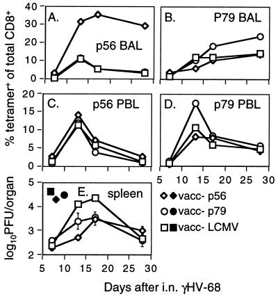
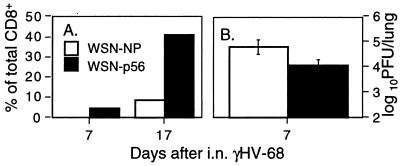
Minimal evidence was found for any recall response elicited by the vacc-p56 (Fig. 1 A and C), vacc-p79 (Fig. 1 B and D), or WSN-p56 (Fig. 2A) viruses at day 7 after i.n. challenge with γHV-68, and no significant effect was seen on virus titers in the lung (Figs. 1E and 2B). The homologous priming regime was, however, associated with substantially increased prevalence of both the p56Db+CD8+ set in the BAL (Figs. 1A and 2A) and the p79Kb+CD8+ PBL population (Fig. 1D) later in the course of the disease process. Furthermore, immunization with both vacc-p56 and vacc-p79 caused a significant (P < 0.005) fall in peak virus titers (day 17) in the spleen, although there was no permanent effect on latency, because the difference between the immune groups and the mice given the recombinant control vaccinia was no longer apparent after another10 days (Fig. 1E). Thus, although a measure of protection is afforded by single priming with the p56 and p79 epitopes, the resultant secondary CD8+ T cell responses do not limit either the early growth of 231 çHV-68 in the lung or the long-term establishment of persistence in the lymphoid tissue.
Figure 1.
The virus-specific CD8+ T cell response in mice primed with recombinant vaccinia viruses expressing the p56 and p79 epitopes of γHV-68. The B6 mice were infected i.p. with vacc-p56, vacc-p79, or the control vacc-LCMV 1 month before i.n. challenge with γHV-68. Pooled BAL or PBL populations from three to six mice were then analyzed for the prevalence of p56Db+CD8+ (A and C) or p79Kb+CD8+ (B and D) T cells. Virus titers (mean of three to six samples) were determined by limiting-dilution assay for lungs (closed symbols) taken on day 7 and for spleens (open symbols) sampled through the course of the experiment (E). Both priming regimes significantly decreased (P < 0.005) the level of infection in the spleen at the day-17 time point.
Figure 2.
The secondary response in mice primed with an influenza A virus incorporating the p56 epitope. The B6 mice were infected i.n. with the WSN-p56 or control WSN-NP viruses and then challenged i.n. 1 month later with γHV-68. The prevalence of p56Db+CD8+ the cells in the BAL is shown in A, whereas the levels of γHV-68 in the lung on day 7 are shown in B. The difference in titers was not significant (P < 0.06).
Maximizing p56-Specific CD8+ T Cell Numbers.
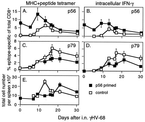
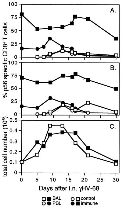
These experiments were designed to establish whether priming virus-specific CD8+ T cells can usefully modify either the acute or long-term consequences of a γHV infection. Because the single-immunization regimes proved of relatively little value (Figs. 1 and 2), the next step was to determine whether greater numbers of CD8+ memory T cells might be more effective. Mice were thus infected i.p. with vacc-p56 and then boosted i.n. with WSN-p56 4 weeks later. This massively increased the prevalence of the p56-specific CD8+ set in the spleen, PBL, and BAL populations (day 0, Fig. 3 A and B; Fig. 4 A and B). More than 70% of the few CD8+ T cells that could be recovered by BAL at 1 month after infection with the recombinant influenza A virus stained with the p56Db tetramer (Fig. 4 A–C). Respiratory challenge at this stage with γHV-68 greatly increased the numbers of p56-specific CD8+ T cells in all sites, giving a much more substantial recall response (Fig. 3 A and B; Fig. 4 A–C) than that achieved by single priming with either recombinant virus (Figs. 1 and 2). The primary response to p79 in these p56-boosted mice was, however, reduced in magnitude (Fig. 3 C and D). Similarly, the characteristic CD4+ T cell-dependent splenomegaly (21, 30) developed late (Fig. 3E), although we did not analyze the virus-specific CD4+ T cell response (31) directly in these experiments.
Figure 3.
The response in the spleen after respiratory γHV-68 challenge of mice primed with vacc-p56 and boosted with WSN-p56. The B6 mice were infected i.p. with vacc-p56 and i.n. with WSN-p56 1 month later. After another month, a few were sampled for analysis (day 0 time point), whereas a majority were challenged i.n. with γHV-68. The numbers of CD8+ T cells specific for the p56 (A and B) or p79 (C and D) epitopes were determined by staining with the p56Db (A) or p79Kb tetramers (C) or by in vitro stimulation with the p56 (B) or p79 (D) peptides in the presence of brefeldin A followed by staining for cytoplasmic IFN-γ. The kinetics of the splenomegaly that develops in these mice is illustrated in E.
Figure 4.
Characteristics of the host response in the blood and lung of doubly primed mice following γHV-68 challenge. See Fig. 3. for experimental details. Shown are the percent p56-specific CD8+ T cells (A and B) determined by staining with the p56Db tetramer (A) or subsequent to peptide stimulation in vitro and staining for cytoplasmic IFN-γ (B) and the total cell counts in the inflammatory population recovered by BAL (C).
The Effect on Virus Titers and the IM-Like Syndrome.
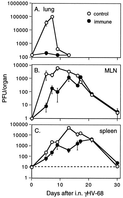
The establishment of γHV-68 in the respiratory tract was almost totally prevented by the vacc-p56/WSN-p56 prime and boost regime. (Fig. 5A). Even so, evidence of latent infection was detected in the MLN and spleen of the immune mice from day 5 and day 7 after infection, although at a lower level than in the controls (Fig. 5 B and C). This decrease in the virus load was presumably responsible for the diminished CD8+ T cell response to the p79 epitope (Fig. 3 C and D).
Figure 5.
Virus titers in the lung and lymphoid tissue of mice that were doubly immunized with the p56 epitope before γHV-68 challenge. The priming and challenge schedules are described in Fig. 3. The results for the lung (A) and spleen (C) are mean ± SEM for six individuals, whereas the values for the MLN (B) are mean ± SE from three pairs. Titers were significantly lower in the spleens of p56-immune mice at day 13 after infection (P < 0.02, Student’s t test) but not by day 21 (P > 0.75). Significantly lower titers in the immune mice were also apparent in lungs at days 5 (P < 0.05) and 7 (P < 0.01), the spleens at days 7 (P < 0.01) and 17 (P < 0.03), and the MLN at day 9 (P < 0.05).
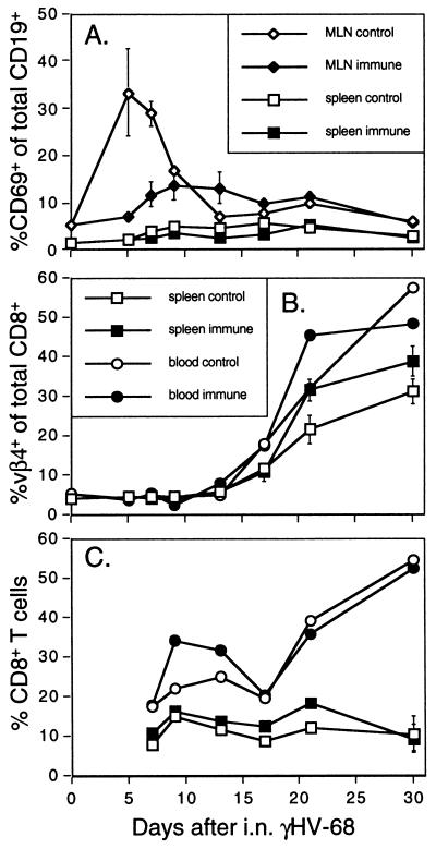
Any protection against the establishment of latency proved, however, to be transient, with the levels of virus detected in lymphoid tissue being equivalent for the control and the immune mice within 20 days of challenge (Fig. 5 B and C). Furthermore, although immunization diminished the number of activated B cells (CD69hiCD19+) in the MLN (Fig. 6A), the later B cell-dependent expansion of Vβ4+CD8+ T cells characteristic of the IM-like disease caused by γHV-68 (19) was not significantly decreased in magnitude (Fig. 6B). Also, although the profiles for total CD8+ T cell numbers in the PBL (Fig. 6C) showed evidence of enhanced, early kinetics as a consequence of prior antigenic exposure, the later stages were comparable for the two groups of mice (Fig. 6C). It thus seems that priming with a lytic-phase γHV-68 epitope cannot prevent either the establishment of latency or the emergence of the characteristic IM-like syndrome, even though immune T cells are present at high frequency in both the lymphoid tissue and the lung before virus challenge (Figs. 3 and 4).
Figure 6.
Consequences for B cell activation and the development of the IM-like disease. The proportion of CD19+ B cells expressing the early activation marker CD69 was determined for the MLN and spleen (A), whereas the frequency of Vβ4+CD8+ T cells was measured for the spleen and PBL compartments (B). Total CD8+ T cell numbers in the PBL is shown in C. The mice had been doubly primed with vacc-p56 and WSN-p56 before the γHV-68 challenge (see Fig. 3 legend). Earlier studies have established that these Vβ4+CD8+ T cells are not specific for the known peptide epitopes of γHV-68 (19, 20).
Consequences of Reversing the Prime and Boost Regime.
Earlier studies directed at promoting the CD8+ T cell response to the invading, sporozoite stage of the Plasmodium yoelii malaria parasite (32) indicated that it was more effective to give a modified influenza virus followed by a vaccinia recombinant. We thus repeated the analysis by using a WSN-p56/vacc-p56 regime but found that, although the titers of lytic virus in the lung at day 7 after the γHV-68 challenge were greatly reduced (Table 1), the diminution was less than that seen with the vacc-p56/WSN-p56 protocol (Fig. 5). Comparing the two sets of data indicates that the later i.n. exposure to WSN-p56 may promote a greater secondary response in the respiratory tract (Fig. 4, Table 1), a factor that would be irrelevant with P. yoelii malaria (32). Again, there was no protection against the longer-term consequences of γHV-68 infection (Table 1).
Table 1.
Consequences of priming with WSN-p56 and boosting with vacc-p56
| Organ | Days after γHV-68 | Group† | % in the CD8+ set*
|
Log10PFU/Organ* | ||
|---|---|---|---|---|---|---|
| p56Db+ | p79Kb+ | Vβ4+ | ||||
| Blood and MLN‡ | 7 | C | 0.9 | 1.2 | 4.3 | 3.0 ± −0.1 |
| P | 31.4 | 6.8 | 3.7 | 0.6 ± 0.3 | ||
| 17 | C | 10.7 | 10.4 | 19.3 | 2.5 ± 0.6 | |
| P | 17.6 | 6.8 | 20.4 | 2.6 ± 0.5 | ||
| 27 | C | 5.0 | 5.1 | 43.3 | 1.4 ± 0.1 | |
| P | 5.0 | 2.8 | 47.2 | 1.6 ± 0.2 | ||
| Spleen | 7 | C | 0.9 | 0.9 | 4.2 ± 0.3 | 2.7 ± 0.6 |
| P | 9.8 | 0.7 | 4.1 ± 0.1 | 0.4 ± 0.4 | ||
| 17 | C | 6.5 | 4.8 | 14.9 ± 0.1 | 4.1 ± 0.3 | |
| P | 13.2 | 3.8 | 13.9 ± 1.9 | 1.3 ± 0.6 | ||
| 27 | C | 2.8 | 4.5 | 25.5 ± 3.3 | 3.1 ± 0.2 | |
| P | 5.3 | 2.8 | 34.0 ± 3.4 | 2.7 ± 0.2 | ||
| BAL and Lung§ | 7 | C | 0.5 | 1.1 | ND | 5.3 ± 0.1 |
| P | 33.5 | 2.3 | ND | 2.5 ± 0.1 | ||
| 17 | C | 8.1 | 15 | ND | ND | |
| P | 73.9 | 8 | ND | ND | ||
| 28 | C | 7.3 | 14.4 | ND | ND | |
| P | 63.8 | 9.9 | ND | ND | ||
The data is either a single value for pooled samples, or the mean ± SE for groups of six mice.
The immune (P) B6 mice were first primed i.n. with WSN-p56 and then were given vacc-p56 i.p. one month later, followed by i.n. challenge with γHV-68 after a further month. The controls (C) were given WSN-NP and vacc-LCMV.
The CD8+ T cell phenotypes are for the blood, while the virus titers are for the MLN.
The CD8+ T cell phenotypes are for the BAL, while the virus titers are for the lung. The lung titers for the control and immune groups were significantly different (p < 0.0003).
ND, not done.
DISCUSSION
The level of protection against the respiratory phase of γHV-68 infection in the vacc-p56/WSN-p56-immune mice was at least as impressive as anything achieved previously for other experimental systems directed at promoting CD8+ T cell memory to readily eliminated (33) or persistent (32) pathogens. The comparison between the single- and double-priming regimes with recombinant viruses expressing either p56 or p79 would suggest that the efficacy of the recall response is very much a function of memory CD8+ T cell numbers. It is difficult to see how any vaccination protocol could improve on the expansion of the virus-specific CD8+ set achieved by this recent, double exposure to live virus vectors, particularly when these immune CD8+ T cells were present at high frequency in the lung before virus challenge. The results raise real concern about whether we can hope to defeat any pathogen that normally establishes persistence in the face of a naive host response by simply priming the CD8+ T cell compartment.
The presence of CD8+ memory T cells specific for the most prominent epitope (p56) expressed in the lytic phase of the infectious process (20) delayed, or diminished, the early establishment of γHV-68 persistence in the lymphoid tissue. This effect was apparent for more than 10 days, reflecting, perhaps, that virus distributed from the infected lung is a primary determinant of the initial extent of latency. Furthermore, although single immunization with the p79 epitope that is thought to be expressed optimally on B cells reverting to lytic cycle was associated at first with less evidence of viral persistence, the effect was not sustained.
Although γHV-68 latency can be demonstrated for other cell types, the establishment of persistent infection in the lymphoid compartment (MLN and spleen) is clearly a function of B cell involvement (8). The γ-HVs are considered to exploit normal germinal center B lymphocyte function (34, 35). Much of the splenomegaly associated with γHV-68 infection is a consequence of expanded B cell numbers (21, 30) and, although it is by no means established that this reflects virus-specific CD4+ T help (31), the effect is not seen in the absence of the CD4+ subset (21, 30). The absence of splenic enlargement in the primed mice could reflect that the CD4+ T cell response is delayed (or diminished) because of a decrease in the antigen load during the lytic phase of the infectious process. An alternative possibility is that splenomegaly is a direct consequence of the extent of persistent infection in the B cells, which then stimulate the CD4+ T cells.
Progeny virus produced by respiratory epithelial cells could infect B cells directly in the bronchus-associated lymphoid tissue or travel via afferent lymph to the regional MLN. The findings in the immune animals may reflect that the low level of virus replication in the lung is sufficient to achieve this level of seeding. However, it is also possible that small numbers of B lymphocytes in the mucosa-associated lymphoid tissue of the upper respiratory tract are infected directly at the time of virus challenge. It is difficult to see how any strategy based solely on priming CD8+ T cells could control such early events in a mucosal site. A vaccination approach designed to promote the neutralizing antibody response (36) also failed to prevent the establishment of γHV-68 latency. The problem here may be the maintenance of sufficient levels of virus-specific Ig at a mucosal surface.
Despite some effort, we have yet to identify a significant major histocompatibility complex class I-restricted peptide derived from any of the viral proteins that are thought to be expressed during latency (37). Priming against such an epitope might well improve vaccine efficacy. The present results do establish, however, that any immunization strategy for the γHVs directed solely at CD8+ T cell epitopes expressed during the lytic phase of infection is not likely to be particularly useful and may even depress other elements of the specific host response. Once viral latency is established in even a small population of B lymphocytes, the numbers can increase to the levels found in naive mice after the resolution of the acute phase of the immune response. It is very clear that γHV-68, at least, has a very robust strategy for colonizing its host that readily compensates for lack of early, lytic-cycle replication.
Acknowledgments
We thank Dr. Satvir S. Tevethia and Professor A. C. Minson for reading this paper and Vicki Henderson for help with the manuscript. The experiments were supported by Public Health Service Grants CA 21765, AI38359, and AI42373 and by the American Lebanese-Syrian Associated Charities. G.T.B. is a C. J. Martin Fellow of the Australian National Health and Medical Research Council (Reg. Key. 997 309).
ABBREVIATIONS
- BAL
bronchoalveolar lavage
- EBV
Epstein–Barr virus
- γHV
γ-herpesvirus
- IM
infectious mononucleosis
- i.n.
intranasal
- i.p.
intraperitoneal
- MLN
mediastinal lymph node
- p56Db
p79Kb, tetrameric complexes of major histocompatibility complex class I glycoprotein+γHV peptide
- PBL
peripheral blood lymphocyte
- vacc
vaccinia virus
- WSN
A/WSN/33 influenza virus
References
- 1.Efstathiou S, Ho Y M, Hall S, Styles C J, Scott S D, Gompels U A. J Gen Virol. 1990;71:1365–1372. doi: 10.1099/0022-1317-71-6-1365. [DOI] [PubMed] [Google Scholar]
- 2.Virgin H W, IV, Latrielle P, Wamsley P, Hallsworth K, Weck K E, Dal Canto A J, Speck S H. J Virol. 1997;71:5894–5904. doi: 10.1128/jvi.71.8.5894-5904.1997. [DOI] [PMC free article] [PubMed] [Google Scholar]
- 3.Nash A A, Sunil-Chandra N P. Curr Opin Immunol. 1994;6:560–563. doi: 10.1016/0952-7915(94)90141-4. [DOI] [PubMed] [Google Scholar]
- 4.Rickinson A B, Kieff E. In: Virology. Third Ed. Fields B N, Knipe D M, Howley P M, editors. Philadelphia: Lippincott; 1996. pp. 2343–2396. [Google Scholar]
- 5.Rickinson A B, Moss D J. Annu Rev Immunol. 1997;14:405–431. doi: 10.1146/annurev.immunol.15.1.405. [DOI] [PubMed] [Google Scholar]
- 6.Sunil-Chandra N P, Efstathiou S, Nash A A. J Gen Virol. 1992;73:3275–3279. doi: 10.1099/0022-1317-73-12-3275. [DOI] [PubMed] [Google Scholar]
- 7.Weck K E, Kim S S, Virgin H W, Speck S H. J Virol. 1999;73:3273–3283. doi: 10.1128/jvi.73.4.3273-3283.1999. [DOI] [PMC free article] [PubMed] [Google Scholar]
- 8.Ehtisham S, Sunil-Chandra N P, Nash A A. J Virol. 1993;67:5247–5252. doi: 10.1128/jvi.67.9.5247-5252.1993. [DOI] [PMC free article] [PubMed] [Google Scholar]
- 9.Christensen J P, Cardin R D, Branum K C, Doherty P C. Proc Natl Acad Sci USA. 1999;96:5135–5140. doi: 10.1073/pnas.96.9.5135. [DOI] [PMC free article] [PubMed] [Google Scholar]
- 10.Stevenson P G, Cardin R D, Christensen J P, Doherty P C. J Gen Virol. 1999;80:477–483. doi: 10.1099/0022-1317-80-2-477. [DOI] [PubMed] [Google Scholar]
- 11.Weck K E, Kim S S, Virgin H W, Speck S H. J Virol. 1999;73:4651–4661. doi: 10.1128/jvi.73.6.4651-4661.1999. [DOI] [PMC free article] [PubMed] [Google Scholar]
- 12.Yao Q Y, Rickinson A B, Gaston J S, Epstein M A. Int J Cancer. 1985;35:43–49. doi: 10.1002/ijc.2910350108. [DOI] [PubMed] [Google Scholar]
- 13.Ganem D. Curr Clin Top Infect Dis. 1998;18:237–251. [PubMed] [Google Scholar]
- 14.Heslop H E, Brenner M K, Rooney C. Hum Gene Ther. 1994;5:381–397. doi: 10.1089/hum.1994.5.3-381. [DOI] [PubMed] [Google Scholar]
- 15.Thomson S A, Khanna R, Gardner J, Burrows S R, Coupar B, Moss D J, Suhrbier A. Proc Natl Acad Sci USA. 1998;92:5845–5849. doi: 10.1073/pnas.92.13.5845. [DOI] [PMC free article] [PubMed] [Google Scholar]
- 16.Thomson S A, Elliott S L, Sherritt M A, Sproat K W, Coupar B E H, Scalzo A A, Forbes C A, Ladhams A M, Mo X Y, Tripp R A, et al. J Immunol. 1996;157:822–826. [PubMed] [Google Scholar]
- 17.Callan M F C, Steven N, Krausa P, Wilson J D, Moss P A H, Gillespie G M, Bell J I, McMichael A J. Nat Med. 1996;2:906–911. doi: 10.1038/nm0896-906. [DOI] [PubMed] [Google Scholar]
- 18.Henle G, Henle W, Diehl V. Proc Natl Acad Sci USA. 1968;59:94–101. doi: 10.1073/pnas.59.1.94. [DOI] [PMC free article] [PubMed] [Google Scholar]
- 19.Tripp R A, Hamilton-Easton A M, Cardin R D, Nguyen P, Behm F G, Woodland D L, Doherty P C, Blackman M A. J Exp Med. 1997;185:1641–1650. doi: 10.1084/jem.185.9.1641. [DOI] [PMC free article] [PubMed] [Google Scholar]
- 20.Stevenson P G, Belz G T, Altman J D, Doherty P C. Eur J Immunol. 1999;29:1059–1067. doi: 10.1002/(SICI)1521-4141(199904)29:04<1059::AID-IMMU1059>3.0.CO;2-L. [DOI] [PubMed] [Google Scholar]
- 21.Cardin R C, Brooks J W, Sarawar S R, Doherty P C. J Exp Med. 1996;184:863–871. doi: 10.1084/jem.184.3.863. [DOI] [PMC free article] [PubMed] [Google Scholar]
- 22.Restifo N P, Bacik I, Irvine K R, Yewdell J W, McCabe B J, Anderson R W, Eisenlohr L C, Rosenberg S A, Bennink J R. J Immunol. 1995;154:4414–4422. [PMC free article] [PubMed] [Google Scholar]
- 23.Mackett M, Smith G L, Moss B. J Virol. 1984;49:857–864. doi: 10.1128/jvi.49.3.857-864.1984. [DOI] [PMC free article] [PubMed] [Google Scholar]
- 24.Yanagi Y, Tishon A, Lewicki H, Cubbitt B A, Oldstone M B. J Virol. 1992;66:2527–2531. doi: 10.1128/jvi.66.4.2527-2531.1992. [DOI] [PMC free article] [PubMed] [Google Scholar]
- 25.Enami M, Luytjes W, Krystal M, Palese P. Proc Natl Acad Sci USA. 1990;87:3802–3805. doi: 10.1073/pnas.87.10.3802. [DOI] [PMC free article] [PubMed] [Google Scholar]
- 26.Castrucci M R, Hou S, Doherty P C, Kawaoka Y. J Virol. 1994;68:3486–3490. doi: 10.1128/jvi.68.6.3486-3490.1994. [DOI] [PMC free article] [PubMed] [Google Scholar]
- 27.Weck K E, Barkon M L, Yoo L I, Speck S H, Virgin H W. J Virol. 1996;70:6775–6780. doi: 10.1128/jvi.70.10.6775-6780.1996. [DOI] [PMC free article] [PubMed] [Google Scholar]
- 28.Stevenson P G, Belz G T, Altman J D, Doherty P C. Proc Natl Acad Sci USA. 1998;95:15565–15570. doi: 10.1073/pnas.95.26.15565. [DOI] [PMC free article] [PubMed] [Google Scholar]
- 29.Murali-Krishna K, Altman J D, Suresh M, Sourdive D, Zajac A, Miller J, Slansky J, Ahmed R. Immunity. 1998;8:177–187. doi: 10.1016/s1074-7613(00)80470-7. [DOI] [PubMed] [Google Scholar]
- 30.Usherwood E J, Ross A J, Allen D J, Nash A A. J Gen Virol. 1996;77:627–630. doi: 10.1099/0022-1317-77-4-627. [DOI] [PubMed] [Google Scholar]
- 31.Christensen J P, Doherty P C. J Virol. 1999;73:4279–4283. doi: 10.1128/jvi.73.5.4279-4283.1999. [DOI] [PMC free article] [PubMed] [Google Scholar]
- 32.Li S, Rodrigues M, Rodriguez D, Rodriguez J R, Esteban M, Palese P. Proc Natl Acad Sci USA. 1993;90:5214–5218. doi: 10.1073/pnas.90.11.5214. [DOI] [PMC free article] [PubMed] [Google Scholar]
- 33.Flynn K J, Belz G T, Altman J D, Ahmed R, Woodland D L, Doherty P C. Immunity. 1998;8:683–691. doi: 10.1016/s1074-7613(00)80573-7. [DOI] [PubMed] [Google Scholar]
- 34.Bowden R J, Simas J P, Davis A J, Efstathiou S. J Gen Virol. 1997;78:1675–1687. doi: 10.1099/0022-1317-78-7-1675. [DOI] [PubMed] [Google Scholar]
- 35.Stevenson P G, Doherty P C. J Virol. 1999;73:1075–1079. doi: 10.1128/jvi.73.2.1075-1079.1999. [DOI] [PMC free article] [PubMed] [Google Scholar]
- 36.Stewart J P, Micali N, Usherwood E J, Bonina L, Nash A A. Vaccine. 1999;17:152–157. doi: 10.1016/s0264-410x(98)00190-x. [DOI] [PubMed] [Google Scholar]
- 37.Virgin H W, Presti R M, Li X Y, Liu C, Speck S H. J Virol. 1999;73:2321–2332. doi: 10.1128/jvi.73.3.2321-2332.1999. [DOI] [PMC free article] [PubMed] [Google Scholar]


