Abstract
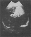
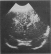
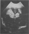
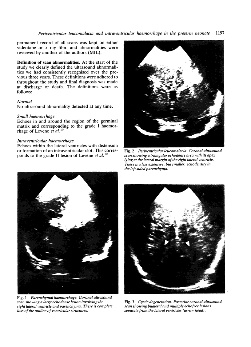
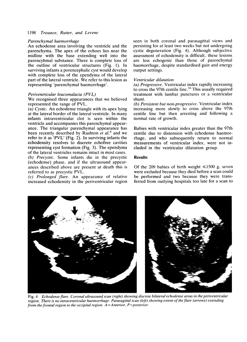
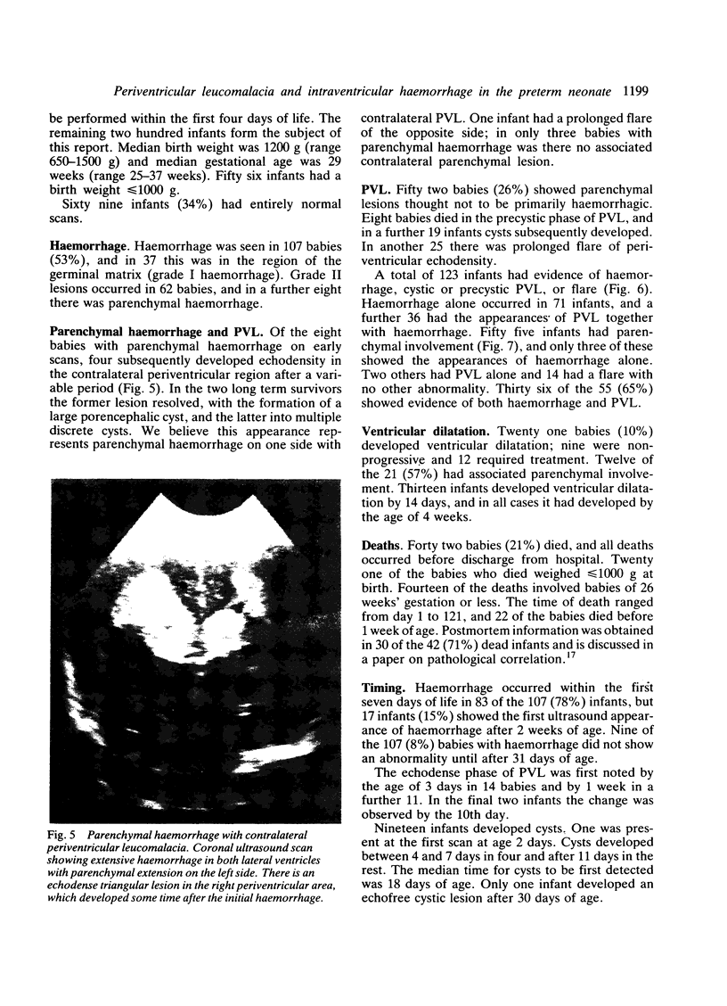
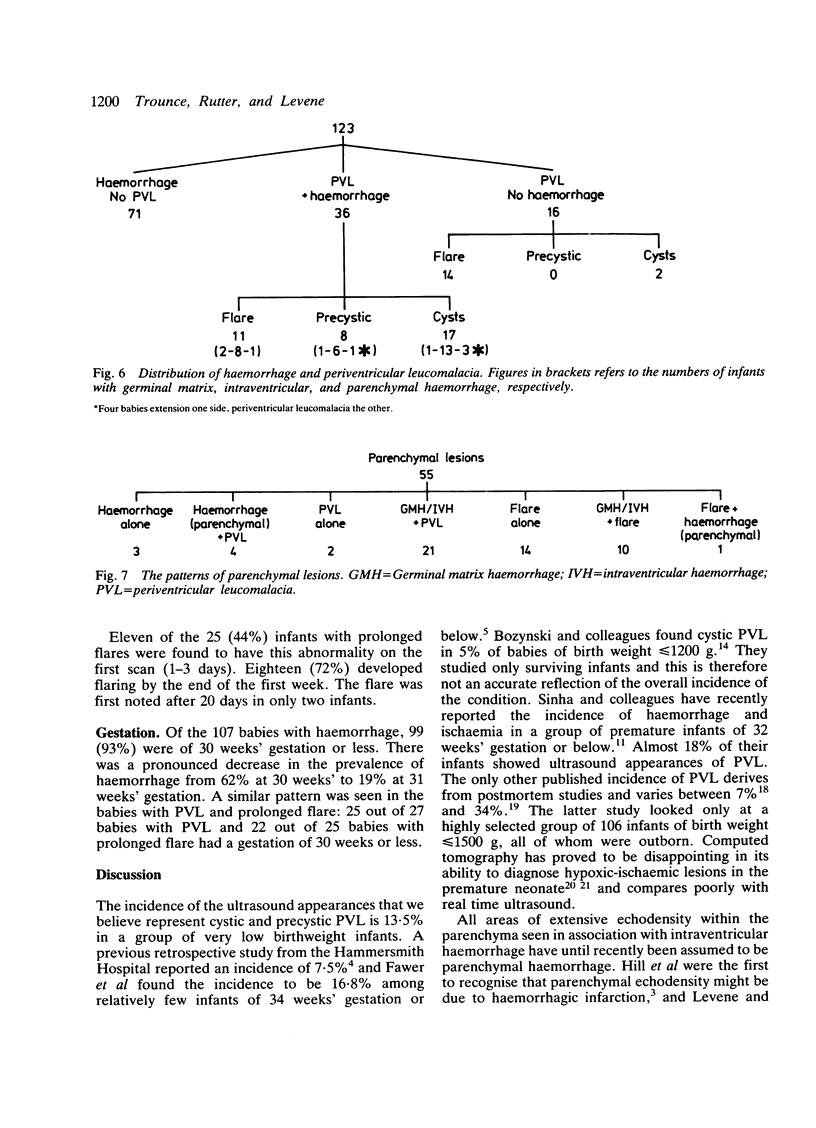
Two hundred very low birthweight infants were prospectively scanned to ascertain the incidence of periventricular leucomalacia (PVL) and haemorrhage. Before collection of data, clear definitions of ultrasound abnormalities believed to represent PVL and intraventricular haemorrhage were described. These referred to small and moderate intraventricular haemorrhage, paenchymal haemorrhage, and PVL, including prolonged flare (echoes in the periventricular region lasting for two weeks or more and not becoming cystic). Sixty nine infants (34%) had no abnormality on ultrasound scans. Intraventricular haemorrhage occurred in 107 babies (37 grade I and 62 grade II), and only eight infants were thought to have true parenchymal haemorrhage. Ultrasound appearances of PVL were seen in 27 infants, 19 of whom developed cysts and eight died in the precystic stage. Prolonged flare occurred in another 25 babies. Unilateral parenchymal haemorrhage occurred in four infants who subsequently developed cystic PVL in the contralateral hemisphere. Twenty one infants developed ventricular dilatation, 12 of whom had associated parenchymal lesions. Haemorrhage, PVL, and flare occurred commonly in infants of 30 weeks' gestation and below and became markedly less common in more mature infants. We believe prolonged flare represents a form of PVL, and in this study a total of 52 (26%) infants had an ultrasound appearance of periventricular leucomalacia, an incidence considerably higher than previously reported.
Full text
PDF
Images in this article
Selected References
These references are in PubMed. This may not be the complete list of references from this article.
- Armstrong D., Norman M. G. Periventricular leucomalacia in neonates. Complications and sequelae. Arch Dis Child. 1974 May;49(5):367–375. doi: 10.1136/adc.49.5.367. [DOI] [PMC free article] [PubMed] [Google Scholar]
- BANKER B. Q., LARROCHE J. C. Periventricular leukomalacia of infancy. A form of neonatal anoxic encephalopathy. Arch Neurol. 1962 Nov;7:386–410. doi: 10.1001/archneur.1962.04210050022004. [DOI] [PubMed] [Google Scholar]
- Bowerman R. A., Donn S. M., DiPietro M. A., D'Amato C. J., Hicks S. P. Periventricular leukomalacia in the pre-term newborn infant: sonographic and clinical features. Radiology. 1984 May;151(2):383–388. doi: 10.1148/radiology.151.2.6709907. [DOI] [PubMed] [Google Scholar]
- Bozynski M. E., Nelson M. N., Matalon T. A., Genaze D. R., Rosati-Skertich C., Naughton P. M., Meier W. A. Cavitary periventricular leukomalacia: incidence and short-term outcome in infants weighing less than or equal to 1200 grams at birth. Dev Med Child Neurol. 1985 Oct;27(5):572–577. [PubMed] [Google Scholar]
- Clark C. E., Clyman R. I., Roth R. S., Sniderman S. H., Lane B., Ballard R. A. Risk factor analysis of intraventricular hemorrhage in low-birth-weight infants. J Pediatr. 1981 Oct;99(4):625–628. doi: 10.1016/s0022-3476(81)80276-4. [DOI] [PubMed] [Google Scholar]
- Dubowitz L. M., Bydder G. M., Mushin J. Developmental sequence of periventricular leukomalacia. Correlation of ultrasound, clinical, and nuclear magnetic resonance functions. Arch Dis Child. 1985 Apr;60(4):349–355. doi: 10.1136/adc.60.4.349. [DOI] [PMC free article] [PubMed] [Google Scholar]
- Estrada M., El Gammal T., Dyken P. R. Periventricular low attenuations: a normal finding in computerized tomographic scans of neonates? Arch Neurol. 1980 Dec;37(12):754–756. doi: 10.1001/archneur.1980.00500610034004. [DOI] [PubMed] [Google Scholar]
- Fawer C. L., Calame A., Perentes E., Anderegg A. Periventricular leukomalacia: a correlation study between real-time ultrasound and autopsy findings. Periventricular leukomalacia in the neonate. Neuroradiology. 1985;27(4):292–300. doi: 10.1007/BF00339560. [DOI] [PubMed] [Google Scholar]
- Flodmark O., Becker L. E., Harwood-Nash D. C., Fitzhardinge P. M., Fitz C. R., Chuang S. H. Correlation between computed tomography and autopsy in premature and full-term neonates that have suffered perinatal asphyxia. Radiology. 1980 Oct;137(1 Pt 1):93–103. doi: 10.1148/radiology.137.1.7422867. [DOI] [PubMed] [Google Scholar]
- Grant E. G., Schellinger D., Richardson J. D., Coffey M. L., Smirniotopoulous J. G. Echogenic periventricular halo: normal sonographic finding or neonatal cerebral hemorrhage. AJR Am J Roentgenol. 1983 Apr;140(4):793–796. doi: 10.2214/ajr.140.4.793. [DOI] [PubMed] [Google Scholar]
- Hecht S. T., Filly R. A., Callen P. W., Wilson-Davis S. L. Intracranial hemorrhage: late onset in the preterm neonate. Radiology. 1983 Dec;149(3):697–699. doi: 10.1148/radiology.149.3.6647846. [DOI] [PubMed] [Google Scholar]
- Hill A., Melson G. L., Clark H. B., Volpe J. J. Hemorrhagic periventricular leukomalacia: diagnosis by real time ultrasound and correlation with autopsy findings. Pediatrics. 1982 Mar;69(3):282–284. [PubMed] [Google Scholar]
- Levene M. I., Fawer C. L., Lamont R. F. Risk factors in the development of intraventricular haemorrhage in the preterm neonate. Arch Dis Child. 1982 Jun;57(6):410–417. doi: 10.1136/adc.57.6.410. [DOI] [PMC free article] [PubMed] [Google Scholar]
- Levene M. I. Measurement of the growth of the lateral ventricles in preterm infants with real-time ultrasound. Arch Dis Child. 1981 Dec;56(12):900–904. doi: 10.1136/adc.56.12.900. [DOI] [PMC free article] [PubMed] [Google Scholar]
- Levene M. I., Wigglesworth J. S., Dubowitz V. Hemorrhagic periventricular leukomalacia in the neonate: a real-time ultrasound study. Pediatrics. 1983 May;71(5):794–797. [PubMed] [Google Scholar]
- Nwaesei C. G., Pape K. E., Martin D. J., Becker L. E., Fitz C. R. Periventricular infarction diagnosed by ultrasound: a postmortem correlation. J Pediatr. 1984 Jul;105(1):106–110. doi: 10.1016/s0022-3476(84)80372-8. [DOI] [PubMed] [Google Scholar]
- Pape K. E., Armstrong D. L., Fitzhardinge P. M. Central nervous system patholgoy associated with mask ventilation in the very low birthweight infant: a new etiology for intracerebellar hemorrhages. Pediatrics. 1976 Oct;58(4):473–483. [PubMed] [Google Scholar]
- Rushton D. I., Preston P. R., Durbin G. M. Structure and evolution of echo dense lesions in the neonatal brain. A combined ultrasound and necropsy study. Arch Dis Child. 1985 Sep;60(9):798–808. doi: 10.1136/adc.60.9.798. [DOI] [PMC free article] [PubMed] [Google Scholar]
- Sinha S. K., Davies J. M., Sims D. G., Chiswick M. L. Relation between periventricular haemorrhage and ischaemic brain lesions diagnosed by ultrasound in very pre-term infants. Lancet. 1985 Nov 23;2(8465):1154–1156. doi: 10.1016/s0140-6736(85)92680-7. [DOI] [PubMed] [Google Scholar]
- Trounce J. Q., Fagan D., Levene M. I. Intraventricular haemorrhage and periventricular leucomalacia: ultrasound and autopsy correlation. Arch Dis Child. 1986 Dec;61(12):1203–1207. doi: 10.1136/adc.61.12.1203. [DOI] [PMC free article] [PubMed] [Google Scholar]
- Volpe J. J., Herscovitch P., Perlman J. M., Raichle M. E. Positron emission tomography in the newborn: extensive impairment of regional cerebral blood flow with intraventricular hemorrhage and hemorrhagic intracerebral involvement. Pediatrics. 1983 Nov;72(5):589–601. [PubMed] [Google Scholar]
- de Crespigny L. C., Mackay R., Murton L. J., Roy R. N., Robinson P. H. Timing of neonatal cerebroventricular haemorrhage with ultrasound. Arch Dis Child. 1982 Mar;57(3):231–233. doi: 10.1136/adc.57.3.231. [DOI] [PMC free article] [PubMed] [Google Scholar]
- de Vries L. S., Dubowitz L. M., Dubowitz V., Kaiser A., Lary S., Silverman M., Whitelaw A., Wigglesworth J. S. Predictive value of cranial ultrasound in the newborn baby: a reappraisal. Lancet. 1985 Jul 20;2(8447):137–140. doi: 10.1016/s0140-6736(85)90237-5. [DOI] [PubMed] [Google Scholar]


