Abstract
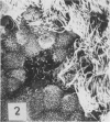
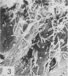
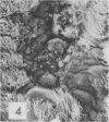
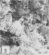
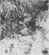
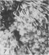
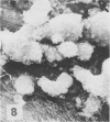
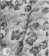
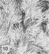
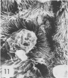
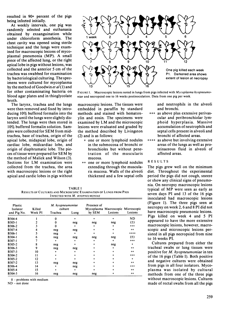
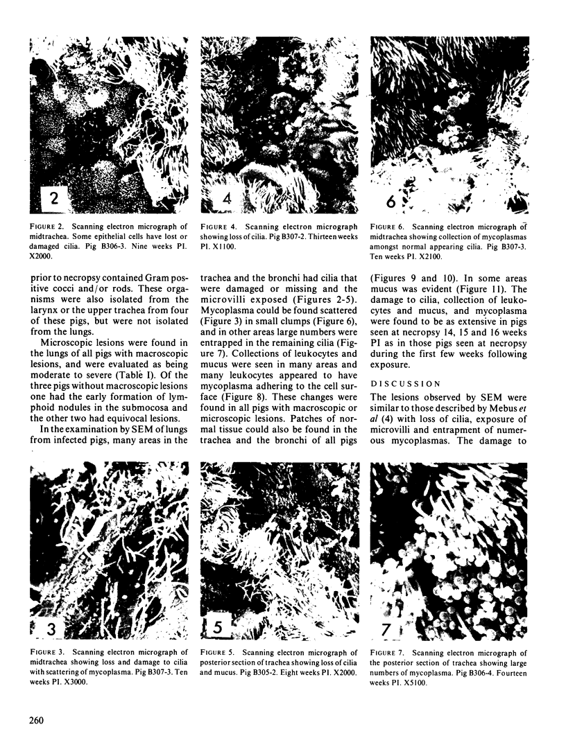
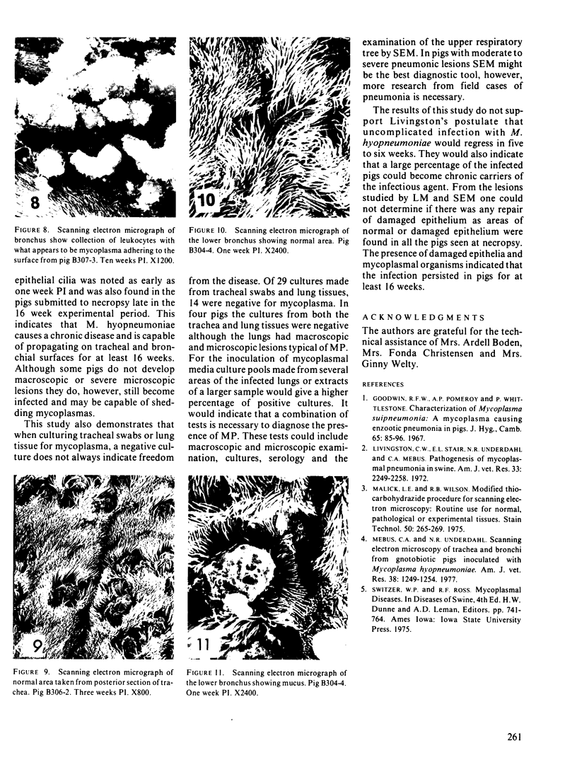
Sixteen gnotobiotic pigs raised in flexible plastic isolators (four pigs per isolator) were inoculated with a culture of Mycoplasma hyopneumoniae. One pig was killed and underwent necropsy at weekly intervals for the following 16 weeks. Macroscopic lesions were observed in the lungs of 13 of 16 pigs and microscopic lesions were found in 14 of 16 pigs. Mycoplasma hyopneumoniae was cultured from the trachea or lungs from 10 of the 16 pigs. Scanning electron microscope studies showed areas of damage to the cilia, collections, of leucocytes and mucus, and mycoplasma in the trachea as well as the bronchi. These conditions were found in all the pigs seen at necropsy from nine to 16 weeks postinoculation and there was no evidence of noticeable regression or recovery during this 16 week period.
Full text
PDF
Images in this article
Selected References
These references are in PubMed. This may not be the complete list of references from this article.
- Livingston C. W., Jr, Stair E. L., Underdahl N. R., Mebus C. A. Pathogenesis of mycoplasmal pneumonia in swine. Am J Vet Res. 1972 Nov;33(11):2249–2258. [PubMed] [Google Scholar]
- Malick L. E., Wilson R. B. Modified thiocarbohydrazide procedure for scanning electron microscopy: routine use for normal, pathological, or experimental tissues. Stain Technol. 1975 Jul;50(4):265–269. doi: 10.3109/10520297509117069. [DOI] [PubMed] [Google Scholar]
- Mebus C. A., Underdahl N. R. Scanning electron microscopy of trachea and bronchi from gnotobiotic pigs inoculated with Mycoplasma hyopneumoniae. Am J Vet Res. 1977 Aug;38(8):1249–1254. [PubMed] [Google Scholar]


