

Abstract
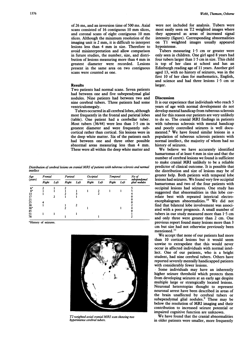
The pattern of cerebral hamartomas among a population of patients with tuberous sclerosis and normal intellect was determined. All patients with tuberous sclerosis over 5 years old with normal intellect who were resident in the Bath health district were offered cranial scanning by magnetic resonance imaging. Cerebral axial and coronal images were obtained in 10 mm contiguous sections with a Picker 0.5 tesla magnetic resonance imaging unit. The number, size, and distribution of lesions found was recorded. Eleven of 13 eligible patients underwent scanning. Two patients had normal scans. Seven patients had between one and five subependymal nodules. Nine patients had between two and nine cerebral tubers best seen on T2 weighted images. Our findings suggest that the wrong conclusions may be drawn if the number of lesions alone is used to predict neurological outcome in tuberous sclerosis.
Full text
PDF

Images in this article
Selected References
These references are in PubMed. This may not be the complete list of references from this article.
- Altman N. R., Purser R. K., Post M. J. Tuberous sclerosis: characteristics at CT and MR imaging. Radiology. 1988 May;167(2):527–532. doi: 10.1148/radiology.167.2.3357966. [DOI] [PubMed] [Google Scholar]
- Inoue Y., Nakajima S., Fukuda T., Nemoto Y., Shakudo M., Murata R., Matsuoka O., Takemoto K., Matsumura Y., Onoyama Y. Magnetic resonance images of tuberous sclerosis. Further observations and clinical correlations. Neuroradiology. 1988;30(5):379–384. doi: 10.1007/BF00404101. [DOI] [PubMed] [Google Scholar]
- Kingsley D. P., Kendall B. E., Fitz C. R. Tuberous sclerosis: a clinicoradiological evaluation of 110 cases with particular reference to atypical presentation. Neuroradiology. 1986;28(1):38–46. doi: 10.1007/BF00341764. [DOI] [PubMed] [Google Scholar]
- Martin N., de Broucker T., Cambier J., Marsault C., Nahum H. MRI evaluation of tuberous sclerosis. Neuroradiology. 1987;29(5):437–443. doi: 10.1007/BF00341739. [DOI] [PubMed] [Google Scholar]
- Nixon J. R., Miller G. M., Okazaki H., Gomez M. R. Cerebral tuberous sclerosis: postmortem magnetic resonance imaging and pathologic anatomy. Mayo Clin Proc. 1989 Mar;64(3):305–311. doi: 10.1016/s0025-6196(12)65250-1. [DOI] [PubMed] [Google Scholar]
- Osborne J. P., Fryer A., Webb D. Epidemiology of tuberous sclerosis. Ann N Y Acad Sci. 1991;615:125–127. doi: 10.1111/j.1749-6632.1991.tb37754.x. [DOI] [PubMed] [Google Scholar]
- Roach E. S., Williams D. P., Laster D. W. Magnetic resonance imaging in tuberous sclerosis. Arch Neurol. 1987 Mar;44(3):301–303. doi: 10.1001/archneur.1987.00520150047020. [DOI] [PubMed] [Google Scholar]
- Tamaki K., Okuno T., Ito M., Asato R., Konishi J., Mikawa H. Magnetic resonance imaging in relation to EEG epileptic foci in tuberous sclerosis. Brain Dev. 1990;12(3):316–320. doi: 10.1016/s0387-7604(12)80313-5. [DOI] [PubMed] [Google Scholar]
- Webb D. W., Osborne J. P. Non-penetrance in tuberous sclerosis. J Med Genet. 1991 Jun;28(6):417–419. doi: 10.1136/jmg.28.6.417. [DOI] [PMC free article] [PubMed] [Google Scholar]