Abstract
Monoclonal antibodies (MAbs) against Francisella tularensis were obtained. Three MAbs specifically reacted with F. tularensis, while four MAbs reacted with other members of the genus Francisella as well. Fluorescent isothiocyanate-conjugated MAbs unequivocally stained bacterial cells in specimens from experimentally infected mice. Two MAbs agglutinated F. tularensis antigen in the agglutination tests. These MAbs should improve methods for detection and identification of F. tularensis.
Francisella tularensis is a gram-negative coccobacillus that causes tularemia in humans and animals. Tularemia is traditionally diagnosed by the isolation of F. tularensis or the detection of specific antibodies. Isolated bacteria were subsequently identified by slide agglutination or immunofluorescence tests using anti-F. tularensis immune serum. Specific antibodies are frequently detected by the microagglutination test (18) in most clinical laboratories. However, because such antibodies cross-react with other bacteria (3), there is a need for an improved method for the serodiagnosis of tularemia. Antigenic analysis of F. tularensis as well as other members of the genus is important because Francisella novicida and Francisella philomiragia have biochemical and genetic properties similar to those of F. tularensis (9), although they rarely cause tularemia-like diseases (13, 22). Monoclonal antibodies (MAbs) are a useful tool for analyzing the antigenic properties of bacteria (15) because they recognize a single epitope with high specificity. Although some MAbs against F. tularensis lipopolysaccharide (LPS) have been produced (5, 10), MAbs against other antigenic components are not available commercially. In this study, we obtained seven MAbs that recognize at least five different epitopes carried by F. tularensis. Four MAbs reacted with F. novicida and F. philomiragia as well. These MAbs can be used for antigenic analyses of Francisella organisms as well as for the diagnosis of tularemia and tularemia-like diseases.
Twenty-six F. tularensis strains (15 Japanese strains and 11 non-Japanese strains), the F. novicida U112 strain, and the F. philomiragia 029 strain were kindly provided by H. Fujita, Ohara Research Laboratory, Fukushima, Japan. Two F. philomiragia strains (ATCC 25017 and ATCC 25018), and Brucella abortus, Brucella melitensis, Brucella suis, Escherichia coli, Haemophilus influenzae, Klebsiella pneumoniae subsp. pneumoniae, Pasteurella aerogenes, Yersinia enterocolitica, and Yersinia pseudotuberculosis were propagated in our laboratory. All F. tularensis strains were propagated on Difco Eugon agar (Becton, Dickinson and Company, Sparks, MD) with chocolatized 8% sheep blood in a biosafety level-3 laboratory. The MAb against F. tularensis LPS (FB11) (Biodesign International, Saco, ME) was used as a reference, and fluorescent isothiocyanate (FITC)-labeled antirabies virus monoclonal antibody (Fujirebio Diagnostics, Inc. Malvern, PA) was used as an isotype control. All animal experiments were approved by the animal research committee of the National Institute of Infectious Diseases.
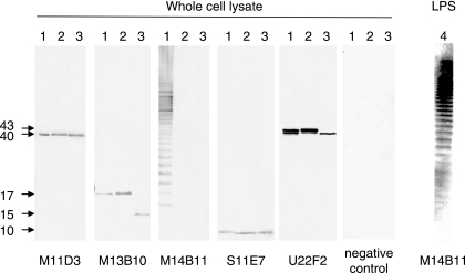
Hybridoma clones secreting MAbs (M11D3, M11H7, M13B10, M14B11, M15C6, S11E7, and U22F2) were obtained by the fusion of mouse myeloma cells (P3-X63-Ag8.653) and spleen cells from BALB/c mice, which had been immunized with the formalin-inactivated F. tularensis GIEM Miura (Japanese) strain, the Schu (non-Japanese) strain, or the F. novicida U112 strain, as described elsewhere (14). Characteristics of the MAbs (Table 1) were based on MAbs obtained from hybridoma supernatant or mice ascitic fluids. Western blotting following sodium dodecyl sulfate-polyacrylamide gel electrophoresis (SDS-PAGE) revealed that the MAbs recognized at least five different epitopes carried by F. tularensis LVS (Fig. 1). The banding patterns obtained with the Schu and GIEM Miura strains were not different from those obtained with the LVS strain (data not shown). MAb M14B11 stained ladder-like bands having molecular masses greater than 15 kDa. Identical ladder-like bands were obtained with MAbs M11H7 and M15C6 (data not shown). These three MAbs also reacted with purified LPS (Fig. 1), a major protective antigen of F. tularensis (17). On the other hand, MAb M11D3, M13B10, and S11E7 reactions produced single bands with molecular masses of 40, 17, and 10 kDa, respectively, while MAb U22F2 reactions produced 41- and 43-kDa bands (Fig. 1). These four MAbs did not react with proteinase K-digested antigen (data not shown), suggesting that the MAbs recognized protein components. F. tularensis proteins of 10, 17, 40, 41, and 43 kDa were found to be recognized by the sera from tularemia patients (4, 12). In addition, immunoreactive membrane components of F. tularensis might play important roles in both the invasion of host cells and escape from phagolysososmes (6, 11). Although it is unclear whether our MAbs recognize these essential components, they may help to analyze the pathogenicity of F. tularensis. We are presently attempting to determine the epitopes recognized by these MAbs.
TABLE 1.
Summary of the characteristics of monoclonal antibodies
| MAb | Immunized antigena | Antigen reacted (kDa)b | Reaction against species (no. of strains tested)c
|
Agglutination activityd | Ig isotypee | ||
|---|---|---|---|---|---|---|---|
| F. tularensis (26) | F. novicida (1) | F. philomiragia (3) | |||||
| M11D3 | GIEM Miura | 40 | + | + | + | − | M |
| M11H7 | GIEM Miura | >15g | + | − | − | − | G3 |
| M13B10 | GIEM Miura | 17 | + | + | + | − | G1 |
| M14B11 | GIEM Miura | >15g | + | − | − | + | G2a |
| M15C6 | GIEM Miura | >15g | + | − | − | + | M |
| S11E7 | Schu | 10 | + | + | + | − | G1 |
| U22F2 | U112 | 41, 43 | + | + | − | − | G1 |
| FB11f | 15 | >15g | + | − | − | − | G2a |
Francisella strains used for immunization of mice.
Molecular mass of F. tularensis antigen appeared in Western blotting following SDS-PAGE.
Determined by indirect fluorescence assay: +, positive; −, negative.
Determined by microagglutination test: +, positive; −, negative.
Determined with a mouse monoclonal antibody isotyping test kit (Serotec, Oxford, United Kingdom).
Reference MAb purchased commercially.
Ladder-like bands of molecular mass greater than 15 kDa.
FIG. 1.
Reactions of MAbs shown by Western blots following SDS-PAGE. Bacterial lysates from F. tularensis LVS, F. novicida U112, and F. philomiragia 029 (lanes 1 to 3, respectively) were reacted with MAbs M11D3, M13B10, M14B11, S11E7, and U22F2 and normal mouse serum (negative control). The reaction of MAb M14B11 against purified LPS from F. tularensis Schu (lane 4) is also shown. The positions of the molecular size markers are indicated (in kilodaltons).
All MAbs reacted with all Japanese and non-Japanese F. tularensis strains but did not react with B. abortus, B. melitensis, B. suis, Y. enterocolitica, Y. pseudotuberculosis, E. coli, H. influenzae, K. pneumoniae subsp. pneumoniae or P. aerogenes by indirect fluorescence assay. Since cross-reactions among F. tularensis, Brucella spp., and Yersinia spp. have been discussed by many researchers (3, 19), reactions of the MAbs against B. abortus, Y. enterocolitica, and Y. pseudotuberculosis were further analyzed by Western blotting. The results indicated that our MAbs did not react with the antigens of these three bacteria (data not shown). MAbs M11H7, M14B11, and M15C6 did not react with F. novicida or F. philomiragia (Fig. 1), indicating that these three MAbs were specific for F. tularensis. On the other hand, MAbs M11D3, M13B10, S11E7, and U22F2 appeared to recognize the conserved epitopes among F. tularensis, F. novicida, and F. philomiragia (Fig. 1). Since the antigens of F. philomiragia recognized by MAbs M13B10 and U22F2 migrated differently than those from F. tularensis and F. novicida, F. philomiragia seemed to be more distantly related to F. tularensis and F. novicida. This finding seems to be in good agreement with the view that F. novicida should be classified as a subspecies of F. tularensis (9, 13, 20). Although the numbers of strains tested were limited, it should be possible to use the MAbs to differentiate among Francisella species. Unusual Francisella organisms, including symbionts of ticks, have been found worldwide (2, 21). Although the antigenic properties of these unusual Francisella organisms are mostly unknown, our MAbs might help to characterize the relationships among the different Francisella organisms.
F. tularensis antigen was agglutinated by MAbs M14B11 and M15C6 in both the microagglutination and the slide agglutination tests (Table 1). In the slide agglutination test, a solution containing MAb M14B11 (0.2 mg/ml of purified immunoglobulin G [IgG]) agglutinated an equal volume of F. tularensis whole-cell suspension (an optical density at 600 nm of 1.8), while solutions containing MAb M11H7 or FB11 (in excess of 0.8 mg/ml of purified IgG) did not show any agglutination at all (data not shown). Thus, F. tularensis could be rapidly identified by a simple slide agglutination test using MAb M14B11.
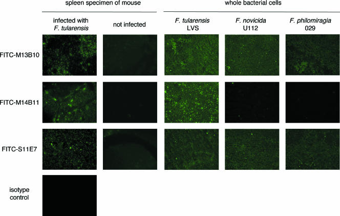
We next determined whether the MAbs could be used to identify F. tularensis in the tissue of infected animals by using a direct immunofluorescent assay (DFA). IgG MAbs purified with a protein G Sepharose column (Amersham Biosciences AB, Uppsala, Sweden) were conjugated with FITC with a Fluoro Taq FITC conjugation kit column (Sigma-Aldrich Co., St. Louis, MO) according to the manufacturer's protocol. When impression smears of the spleens from mice infected with the Yama strain were reacted with FITC-labeled MAbs M14B11, M13B10, and S11E7, bacterial cells were readily identified by fluorescence microscopy. FITC-labeled MAbs M13B10 and S11E7 also stained bacterial cells of F. novicida and F. philomiragia (Fig. 2). These results suggest that FITC-labeled MAbs can be used to detect and identify Francisella organisms from clinical samples.
FIG. 2.
Reactions of MAbs shown by DFA. FITC-labeled MAbs M13B10, M14B11, and S11E7 were reacted with the impression smears of the spleens from a mouse infected with F. tularensis Yama and an uninfected mouse and whole-bacteria cells of F. tularensis LVS, F. novicida U112, and F. philomiragia 029. FITC-labeled antirabies virus monoclonal antibody was used as an isotype control.
Tularemia has been considered to be a disease confined to the northern hemisphere and most frequently in Scandinavia, North America, Japan, and Russia (7). However, it has emerged in other geographic locations recently (16). The prevalence and distribution of F. tularensis have received much attention because of fears that the organisms could be used as a bioterrorism agent. Furthermore, F. tularensis is associated with protozoa (1) and might reside in the environment in a viable but nonculturable form (8). Therefore, it would be very useful to have a method for detecting F. tularensis in environmental samples such as soil and water. The MAbs obtained here appear to be ideal tools for identifying not only F. tularensis but also F. novicida and F. philomiragia for ecological and epidemiological studies as well as for antigenic analyses of Francisella organisms, the pathogens of tularemia or tularemia-like diseases.
Acknowledgments
We thank H. Fujita (Ohara Research Laboratory) for providing the bacterial strains. We also thank K. Imaoka (Department of Veterinary Science, National Institute of Infectious Diseases) for helping with the cultivation of bacteria.
This work was supported by Health and Labor Science Research Grants for Research on Emerging and Re-emerging Infectious Diseases from the Ministry of Health, Labor and Welfare in Japan.
Footnotes
Published ahead of print on 22 November 2006.
REFERENCES
- 1.Abd, H., T. Johansson, I. Golovliov, G. Sandström, and M. Forsman. 2003. Survival and growth of Francisella tularensis in Acanthamoeba castellanii. Appl. Environ. Microbiol. 69:600-606. [DOI] [PMC free article] [PubMed] [Google Scholar]
- 2.Barns, S. M., C. C. Grow, R. T. Okinaka, P. Keim, and C. R. Kuske. 2005. Detection of diverse new Francisella-like bacteria in environmental samples. Appl. Environ. Microbiol. 71:5494-5500. [DOI] [PMC free article] [PubMed] [Google Scholar]
- 3.Behan, K. A., and G. C. Klein. 1982. Reduction of Brucella species and Francisella tularensis cross-reacting agglutinins by dithiothreitol. J. Clin. Microbiol. 16:756-757. [DOI] [PMC free article] [PubMed] [Google Scholar]
- 4.Bevanger, L., J. A. Maeland, and A. I. Naess. 1989. Competitive enzyme immunoassay for antibodies to a 43,000-molecular-weight Francisella tularensis outer membrane protein for the diagnosis of tularemia. J. Clin. Microbiol. 27:922-926. [DOI] [PMC free article] [PubMed] [Google Scholar]
- 5.Bhatti, A. R., J. P. Wong, and D. E. Woods. 1993. Production and partial characterization of hybridoma clones secreting monoclonal antibodies against Francisella tularensis. Hybridoma 12:197-202. [DOI] [PubMed] [Google Scholar]
- 6.Clemens, D. L., B.-Y. Lee, and M. A. Horwitz. 2005. Francisella tularensis enters macrophages via a novel process involving pseudopod loops. Infect. Immun. 73:5892-5902. [DOI] [PMC free article] [PubMed] [Google Scholar]
- 7.Ellis, J., P. C. Oyston, M. Green, and R. W. Titball. 2002. Tularemia. Clin. Microbiol. Rev. 15:631-646. [DOI] [PMC free article] [PubMed] [Google Scholar]
- 8.Forsman, M., E. W. Henningson, E. Larsson, T. Johansson, and G. Sandström. 2000. Francisella tularensis does not manifest virulence in viable but non-culturable state. FEMS Microbiol. Ecol. 31:217-224. [DOI] [PubMed] [Google Scholar]
- 9.Forsman, M., G. Sandström, and A. Sjöstedt. 1994. Analysis of 16S ribosomal DNA sequences of Francisella strains and utilization for determination of the phylogeny of the genus and for identification of strains by PCR. Int. J. Syst. Bacteriol. 44:38-46. [DOI] [PubMed] [Google Scholar]
- 10.Fulop, M. J., T. Webber, R. J. Manchee, and D. C. Kelly. 1991. Production and characterization of monoclonal antibodies directed against the lipopolysaccharide of Francisella tularensis. J. Clin. Microbiol. 29:1407-1412. [DOI] [PMC free article] [PubMed] [Google Scholar]
- 11.Golovliov, I., V. Baranov, Z. Krocova, H. Kovarova, and A. Sjöstedt. 2003. An attenuated strain of the facultative intracellular bacterium Francisella tularensis can escape the phagosome of monocytic cells. Infect. Immun. 71:5940-5950. [DOI] [PMC free article] [PubMed] [Google Scholar]
- 12.Havlasova, J., L. Hernychova, P. Halada, V. Pellantova, J. Krejsek, J. Stulik, A. Macela, P. R. Jungblut, P. Larsson, and M. Forsman. 2002. Mapping of immunoreactive antigens of Francisella tularensis live vaccine strain. Proteomics 2:857-867. [DOI] [PubMed] [Google Scholar]
- 13.Hollis, D. G., R. E. Weaver, A. G. Steigerwalt, J. D. Wenger, C. W. Moss, and D. J. Brenner. 1989. Francisella philomiragia comb. nov. (formerly Yersinia philomiragia) and Francisella tularensis biogroup novicida (formerly Francisella novicida) associated with human disease. J. Clin. Microbiol. 27:1601-1608. [DOI] [PMC free article] [PubMed] [Google Scholar]
- 14.Hotta, A., M. Kawamura, H. To, M. Andoh, T. Yamaguchi, H. Fukushi, and K. Hirai. 2002. Phase variation analysis of Coxiella burnetii during serial passage in cell culture by use of monoclonal antibodies. Infect. Immun. 70:4747-4749. [DOI] [PMC free article] [PubMed] [Google Scholar]
- 15.Hotta, A., G. Q. Zhang, M. Andoh, T. Yamaguchi, H. Fukushi, and K. Hirai. 2004. Use of monoclonal antibodies for analyses of Coxiella burnetii major antigens. J. Vet. Med. Sci. 66:1289-1291. [DOI] [PubMed] [Google Scholar]
- 16.Petersen, J. M., and M. E. Schriefer. 2005. Tularemia: emergence/re-emergence. Vet. Res. 36:455-467. [DOI] [PubMed] [Google Scholar]
- 17.Sandström, G., A. Sjöstedt, T. Johansson, K. Kuoppa, and J. C. Williams. 1992. Immunogenicity and toxicity of lipopolysaccharide from Francisella tularensis LVS. FEMS Microbiol. Immunol. 5:201-210. [DOI] [PubMed] [Google Scholar]
- 18.Sato, T., H. Fujita, Y. Ohara, and M. Homma. 1990. Microagglutination test for early and specific serodiagnosis of tularemia. J. Clin. Microbiol. 28:2372-2374. [DOI] [PMC free article] [PubMed] [Google Scholar]
- 19.Schmitt, P., W. Splettstosser, M. Porsch-Ozcurumez, E. J. Finke, and R. Grunow. 2005. A novel screening ELISA and a confirmatory Western blot useful for diagnosis and epidemiological studies of tularemia. Epidemiol. Infect. 133:759-766. [DOI] [PMC free article] [PubMed] [Google Scholar]
- 20.Sjöstedt, A. B. 2005. Family XVII. FRANCISELLAE, genus I. Francisella Dorofe'ev 1947, 176AL, p. 200-210. In D. J. Brenner, N. R. Krieg, J. T. Staley, and G. M. Garrity (ed.), The proteobacteria, part B. Bergey's manual of systematic bacteriology, 2nd ed., vol. 2. Springer-Verlag, New York, NY. [Google Scholar]
- 21.Sun, L. V., G. A. Scoles, D. Fish, and S. L. O'Neill. 2000. Francisella-like endosymbionts of ticks. J. Invertebr. Pathol. 76:301-303. [DOI] [PubMed] [Google Scholar]
- 22.Whipp, M. J., J. M. Davis, G. Lum, J. de Boer, Y. Zhou, S. W. Bearden, J. M. Petersen, M. C. Chu, and G. Hogg. 2003. Characterization of a novicida-like subspecies of Francisella tularensis isolated in Australia. J. Med. Microbiol. 52:839-842. [DOI] [PubMed] [Google Scholar]


