Abstract
GAIP (G α interacting protein) is a member of the RGS (regulators of G protein signaling) family and accelerates the turnover of GTP bound to Gαi, Gαq, and Gα13. There are two pools of GAIP—a soluble and a membrane-anchored pool. The membrane-anchored pool is found on clathrin-coated vesicles (CCVs) and pits in rat liver and AtT-20 pituitary cells. By treatment of a GAIP-enriched rat liver fraction with alkaline phosphatase, we found that membrane-bound GAIP is phosphorylated. By immunoprecipitation carried out on [32P]orthophosphate-labeled AtT-20 pituitary cells stably expressing GAIP, 32P-labeling was associated exclusively with the membrane pool of GAIP. Phosphoamino acid analysis revealed that phosphorylation of GAIP occurred largely on serine residues. Recombinant GAIP could be phosphorylated at its N terminus with purified casein kinase 2 (CK2). It could also be phosphorylated by isolated CCVs in vitro. Phosphorylation was Mn2+-dependent, using both purified CK2 and CCVs. Ser-24 was identified as one of the phosphorylation sites. Our results establish that GAIP is phosphorylated and that only the membrane pool is phosphorylated, suggesting that GAIP can be regulated by phosphorylation events taking place at the level of clathrin-coated pits and vesicles.
Keywords: G proteins, regulator of G protein signaling, casein kinase 2
Signal transduction components and G-protein coupled receptor (GPCR) signaling pathways in particular undergo phosphorylation and dephosphorylation events that control their effects on multiple physiological processes (1). For example, GPCR are phosphorylated by G protein-coupled receptor kinases (GRKs) (2, 3), and several G proteins undergo phosphorylation in vivo on their α (4–6) and γ (7) subunits, which may regulate their membrane targeting (8) and/or interaction with partners (9–12)
Recently, a family of more than 20 members, the RGS proteins (for regulators of G protein signaling), have been identified that act as GTPase activating proteins (GAPs) for Gα subunits (for reviews see refs. 13 and 14). We localized one member of the RGS family, RGS-GAIP (G α interacting protein), by immunocytochemistry and found it to be associated with clathrin-coated pits and vesicles (15, 16) and further demonstrated that CCVs isolated from rat liver possess GAP activity (16).
Clathrin-coated vesicles (CCVs) are known to play a role in G-protein coupled receptor- (2) and receptor-mediated endocytosis at the plasma membrane and in sorting of lysosomal enzymes in the trans-Golgi network (TGN) (17–19). Formation of CCVs is known to be tightly regulated by phosphorylation and dephosphorylation of adapters and coat proteins (20–24).
GAIP contains nine putative phosphorylation sites—two for protein kinase C (PKC) and seven for casein kinase 2 (CK2) (25). These consensus sites are distributed over all three domains of GAIP: (i) its highly conserved RGS domain (amino acids 86–205) required for GAP activity (26), (ii) its N terminus (amino acids 1–85) most likely responsible for membrane anchoring of GAIP by palmitoylation (27), and (iii) the 12-aa C terminus (amino acids 206–217), which is unique and interacts with GIPC, a PDZ domain-containing protein (28). Up to now, no information has been available on whether GAIP or any other mammalian RGS protein undergoes phosphorylation.
We report here the identification of a phosphorylated pool of GAIP that is membrane-anchored in rat hepatocytes and AtT-20 pituitary cells. We also demonstrate that GAIP can be phosphorylated by purified CK2 and isolated CCVs in vitro, and we identified Ser-24 as one of the residues that is phosphorylated. These findings suggest that GAIP is phosphorylated at its N terminus by CK2 on CCVs.
Experimental Procedures
Materials.
Easytag (a mixture of [35S]methionine and [35S]cysteine) was obtained from DuPont/NEN. Phorbol myristate acetate (PMA) was purchased from Calbiochem, cellulose plates from Analtech, and [14C]methylated protein molecular weight markers from Amersham Pharmacia. Mutant GAIP (S24A) was generated by a PCR according to the manufacturer's instructions by using a QuikChange mutagenesis kit (Stratagene), pET28a GAIP1–217 (16), and the following primers: 5′-GCCCCCTTCAATGGCCAGTCATGATACAGCC and 3′-GGCTGTATCATACTGGCCATTGAAGGGGGC. Mutation was verified by automatic sequencing. The EcoRI/SmaI fragment containing the mutation was subcloned into the wild-type template. His6 GAIP proteins were expressed in BL-21 (DE 3), purified, and dialysed as described (16). Protein was determined by using the BCA assay (Pierce).
Antibodies.
Anti-hemagglutinin (HA) mAb 16B12 was purchased from Babco (Richmond, CA). Anti-GAIP (N), GAIP (23–217), and anti-GAIP (C) were characterized previously (15).
Preparation and Alkaline Phosphatase Treatment of Liver Fractions.
Residual microsomes (RM) and CCV-enriched fractions were obtained from rat liver as described (15, 16). A total of 3.5 or 7 μl RM fraction (14 mg/ml) was resuspended in 100 μl of CIP buffer [50 mM Tris⋅HCl (pH 9)/1 mM EDTA/100 mM ZnCl2/1 mM spermidine) in the presence of protease inhibitors and 300 μM PMSF. Each concentration was then split into two microfuge tubes, one containing 30 units alkaline phosphatase (Roche Molecular Biochemicals) and incubated at 30°C for 3 h. Reactions were diluted with 10 μl 1 M Tris⋅HCl (pH 6.8) and stopped by boiling in SDS-sample buffer. Proteins were separated by 12.5% SDS/PAGE, transferred to poly(vinylidene difluoride) membranes, and immunoblotted with anti-GAIP (N) detected by enhanced chemiluminescence.
Biosynthetic Labeling and Immunoprecipitation.
AtT-20#1 cells stably expressing GAIP were prepared as described previously (25, 27) and labeled for 4 h with either 100 μCi/ml Easytag or 1 mCi/ml [32P]orthophosphate. 35S labeling was performed in Cys- and Met-free DMEM supplemented with 4% dialysed FCS. 32P labeling was carried out in phosphate-free DMEM supplemented with 4% FCS. Cells were rinsed three times with ice-cold TBS [50 mM Tris⋅HCl (pH 8)/150 mM NaCl] and homogenized in the presence of 50 mM β-glycerophosphate, 10 mM NaF, and 1 mM NaVO4 (27). Cytosolic (100,000 × g supernatants) and crude membrane (100,000 × g pellet) fractions were prepared by centrifugation of postnuclear supernatants (27), solubilized in RIPA buffer (TBS/1% Triton X-100/0.5% Na deoxycholate/0.1% SDS) at 4°C for 1 h. Lysates were precleared with 20 μl protein A/G-Sepharose (anti-HA mAb) or protein A-Sepharose (rabbit antisera). GAIP was precipitated with anti-HA (5 μg/ml), or anti-GAIP (N), anti-GAIP (23-217), or anti-GAIP (C) antisera diluted 1:333, and protein A/G Sepharose (anti-HA mAb) or protein A-Sepharose beads were added for 90 min. Beads were boiled, and immune complexes were separated by 10% SDS/PAGE, exposed for autoradiography, and quantified by densitometry using alpha imager software.
HEK293T cells were transiently transfected (calcium phosphate method) with 20 μg of either pCDNA3 GAIP (1–217) or pCDNA3 (mock vector). Twenty hours after transfection, cells were serum-starved overnight in DMEM (containing 0.1% FCS and 10 mM Hepes), incubated in phosphate-free DMEM containing 0.1% FCS for 30 min, and labeled for 2 h in the same medium containing 1.67 mCi/ml of [32P]orthophosphate. Immunoprecipitation was carried out as described above for AtT-20#1 cells.
Phosphoamino Acid Analysis.
Immunoprecipitates from control and PMA-treated cells were transferred to Immobilon-P membranes (Millipore), the radioactive GAIP bands from the cytosolic and membrane fractions were cut out, pooled and subjected to hydrolysis (6 M HCl) at 106°C for 1 h. The samples were pelleted in a microfuge, and supernatants were lyophilized and resuspended in 10 μl formic acid/acetic acid buffer (pH 1.9) and phosphoamino acid carriers were added (29). Amino acids were separated by horizontal, one-dimensional thin layer electrophoresis on cellulose-coated plates for 40 min. Phosphoamino acid standards were stained with ninhydrin, and radiolabeled phosphoamino acids were detected by autoradiography.
Phosphorylation of GAIP by Purified CK2.
A total of 1 μg GST-furin cd and 5 μg His6 GAIP or GAIP (S24A) were incubated with CK2 purified from bovine testis (30) in reaction buffer [50 mM Tris (pH 7.5)/150 mM KCl/2 mM MgCl2] and 0.1 mM ATP (containing 7.28 μCi [γ-32P]ATP) at 30°C for 1 h in a final volume of 20 μl as described (31). Reactions were stopped by boiling in SDS-sample buffer, proteins were resolved by 12% SDS/PAGE, and the gel was dried and submitted to autoradiography. The bands corresponding to the phosphorylated GAIP proteins were excised, and the extent of phosphorylation was determined by scintillation counting.
Phosphorylation of Recombinant GAIP Domains by Isolated CCVs.
A total of 1 μg recombinant GAIP proteins was phosphorylated in the presence of 50 μM ATP containing 8 μCi [γ-32P]ATP (7,000 Ci/mmol) in 50 μl kinase buffer [50 mM Hepes (pH 7.2)/150 mM KCl] and 0, 0.3, 3, or 30 mM MgCl2; 0.3, 3, or 20 mM ZnCl2; or 0.1, 0.3, 1, 3, or 30 mM MnCl2. Reactions were started by addition of 5 μl CCV-enriched fraction (1.2 mg/ml), incubated at 25°C for 30 min, and stopped by addition of 10 μl −2% SDS containing 100 μg/ml BSA. Proteins were precipitated with ice-cold acetone, separated by SDS/PAGE, and detected by autoradiography.
Results
RGS-GAIP Is Present in Cytosolic and Membrane Fractions of Stably Transfected AtT-20 Cells.
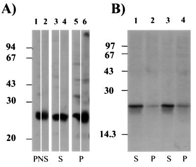
As reported earlier (27), in mouse AtT-20#1 cells stably expressing GAIP, two pools of GAIP were found—one cytosolic and one membrane associated. By immunoblotting, the amount of GAIP found in the two pools was similar (Fig. 1A). However, after biosynthetic labeling with [35S]met/cys and immunoprecipitation with GAIP antibodies, labeling of GAIP was detected mainly (≈90%) in the cytosolic fraction by autoradiography (Fig. 1B). These results show that in AtT-20 #1 cells the majority of the newly synthesized GAIP is cytosolic. We conclude that the membrane pool of GAIP turns over much more slowly than the cytosolic pool. Alternatively, there could be incomplete solubilization of the membrane pool.
Figure 1.
Distribution of GAIP in AtT-20#1 cells. (A) AtT-20 cells stably expressing GAIP (clone #1) were homogenized, and the postnuclear supernatant (PNS) was centrifuged at 100,000 × g to yield crude membrane (P) and cytosolic fractions (S). Then, 25 μg of protein/lane were separated by SDS/PAGE, cut in two strips, and immunoblotted with anti-GAIP (N) (lanes 1, 3, and 5) or anti-HA (lanes 2, 4, and 6) and detected by enhanced chemiluminescence. GAIP is found in the PNS (lanes 1 and 2) and in both cytosolic (lanes 3 and 4) and membrane (lanes 5 and 6) fractions. (B) Cells were biosynthetically labeled for 4 h with Easytag, homogenized and fractionated as in A, and solubilized in RIPA buffer. GAIP was immunoprecipitated from the cytosolic (lanes 1 and 3) or crude membrane (lanes 2 and 4) fraction with anti-GAIP (23-217) (lanes 1 and 2) or anti-GAIP (C) (lanes 2 and 3). Immune complexes were separated by SDS/PAGE and detected by autoradiography. Newly synthesized, [35S]GAIP was found mainly in the cytosolic fraction.
The Membrane Pool of GAIP Is Phosphorylated.
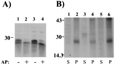
Previously, we reported that after immunoblotting rat liver fractions for GAIP, two bands, 26.5 and 28 kDa, were seen in the starting homogenate, in carrier vesicles, and in RM fractions (15, 16). This suggested that GAIP might be posttranslationally modified, perhaps by phosphorylation. To investigate this possibility, RM (the most abundant of these fractions) were digested with alkaline phosphatase and immunoblotted for GAIP. After alkaline phosphatase digestion, a single 26.5-kDa GAIP band was seen by immunoblotting (Fig. 2A), indicating that the upper band in the RM fraction is phosphorylated. Interestingly, in the liver, the phosphorylated pool represents ≈60–70% of the total GAIP in the RM fraction. The fact that a doublet was previously found in membrane but not in cytosolic fractions (16) suggested that phosphorylation might correlate with membrane association.
Figure 2.
The membrane pool of GAIP is phosphorylated. (A) A total of 25 μg (lanes 1 and 2) or 50 μg (lanes 3 and 4) protein from the RM fraction of rat liver were incubated for 3 h with 30 units alkaline phosphatase (AP), separated by SDS/PAGE, followed by immunoblotting with anti-GAIP (N) and enhanced chemiluminescence. After alkaline phosphatase digestion, the slower migrating band (26.5 kDa) of GAIP disappears and only the faster migrating band (28.5 kDa) is seen. (B) AtT-20#1 cells were labeled in vivo with [32P]orthophosphate for 4 h, and cytosolic (lanes 1, 3, and 5) and membrane fractions (lanes 2, 4, and 6) were prepared and solubilized as in Fig. 1. GAIP was immunoprecipitated using anti-HA (lanes 1 and 2), anti-GAIP (23–217) (lanes 3 and 4), or anti-GAIP (N) (lanes 5 and 6). Immune complexes were resolved by SDS/PAGE and exposed overnight for autoradiography. Phosphorylated GAIP is detected exclusively in the membrane fraction (P) with all three antibodies.
Next, we carried out immunoprecipitation for GAIP on AtT-20#1 cells after 32P labeling. 32P-labeled GAIP was detected only in the membrane fraction (Fig. 2B). Because no radioactive signal was detected in the cytosolic pool even after long exposures (up to 32 h), we conclude that, in AtT-20#1 cells, only the membrane-associated pool is phosphorylated. Thus, the results obtained on both rat liver and AtT-20#1 cells clearly indicate that phosphorylation is associated with the membrane pool of GAIP. Addition of PMA, a PKC activator, during the last 15 min of labeling did not significantly affect [32P] incorporation. This suggests that phosphorylation of GAIP in AtT-20#1 cells is not regulated by phorbol ester-sensitive PKC.
GAIP Is Phosphorylated on Serine Residues.
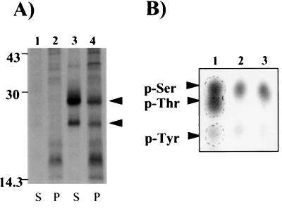
Because the quantity of labeled material available after immunoprecipitation of GAIP from AtT-20#1 cells was insufficient for phosphoamino acid determination, we shifted to HEK293T cells transiently transfected with GAIP. After 32P labeling, phosphorylated GAIP could be immunoprecipitated from both the cytosolic (60%) and membrane (40%) fractions of these cells (Fig 3A). The presence of phosphorylated GAIP in the cytosol could be because of cell type variation or, more likely, the result of saturation of the membrane targeting machinery due to overexpression. A 23.1-kDa band was also immunoprecipitated from 32P-labeled cells after transfection with GAIP (28 kDa), probably resulting from an alternative initiation start site (25). The upper labeled bands from the cytosolic and membrane fractions were combined and subjected to acid hydrolysis (29). After migration on a cellulose-coated plate in the presence of p-Ser, p-Thr, and p-Tyr as carrier, the 32P-labeled phosphoamino acids migrated mainly with the p-Ser marker. Thus, GAIP appeared to be phosphorylated mainly on Ser and sparsely on Tyr (Fig. 3B, lane 2). Again, addition of PMA during the last 15 min of 32P labeling did not affect incorporation, nor did it change the residue on which GAIP is phosphorylated (Fig. 3B, lane 3).
Figure 3.
Immunoprecipitation and phosphoamino acid analysis of 32P-labeled GAIP in transiently transfected HEK 293T cells. (A) Cells were transiently transfected with a mock pcDNA3 (lanes 1 and 2) or with pcDNA3 GAIP (lanes 3 and 4) and labeled with [32P]orthophosphate for 2 h. GAIP was immunoprecipitated from cytosolic (S) and membrane (P) fractions with anti-GAIP (C) as in Fig. 1. 32P-labeled GAIP (arrows, lanes 3 and 4) is found in both cytosolic (S) and membrane (P) fractions. A faster moving band, 23.1 kDa, is immunoprecipitated from cells labeled with [32P]orthophosphate after transfection with GAIP (28 kDa), probably resulting from an alternative initiation start site (25). (B) In the last 15 min of labeling, cells were incubated with buffer as control (lane 2) or 150 nM PMA (lane 3). The 32P-labeled GAIP bands were cut out from both the cytosolic and membrane fractions and used for one-dimensional phosphoamino acid analysis on a cellulose-coated plate as described in Experimental Procedures. Lane 1: p-Ser, p-Thr, and p-Tyr standards detected by ninhydrin staining (arrowheads, lane 1). Lanes 2 and 3: Autoradiography p-Ser was the major phosphoamino acid present in both control (lane 2) and PMA-treated cells (lane 3).
GAIP Is Phosphorylated at Its N Terminus by CK2.
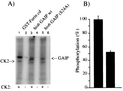
The primary sequence for GAIP contains two consensus sites for PKC and seven for CK2 (25). Among these consensus sites are four Ser—one in the N terminus, two in the RGS domain, and one in the C terminus. All four are putative CK2 phosphorylation sites, and one of those in the RGS domain is also a putative PKC phosphorylation site. Because PMA did not change the amount of radioactive phosphate incorporated, it seemed unlikely that a phorbol ester-sensitive PKC is involved in the phosphorylation of GAIP. Therefore, we turned our attention to CK2 and performed in vitro phosphorylation of GAIP in the presence or absence of purified CK2 (Fig. 4A). GAIP, as well as a positive control, GST-furin cd (a recombinant protein containing 56 amino acids of the cytoplasmic domain of furin) (31), were phosphorylated by CK2 (Fig. 4A, lanes 1 and 3). The efficiency of phosphate incorporation into GAIP was low: 1 pmol phosphate/22 pmol in the presence of Mg2+ and 1 pmol phosphate/12 pmol in the presence of Mn2+. Because we found GAIP to be phosphorylated when membrane-anchored and GAIP (27) and several other RGS proteins are thought to be anchored to membranes at their N termini (32–34), we reasoned that phosphorylation might occur at the N terminus. We therefore mutated Ser-24, the only Ser that represents a putative consensus phosphorylation site for CK2 in the N terminus of GAIP. When GAIP (S24A) was incubated in the presence of CK2, its phosphorylation was reduced (Fig. 4A, lane 5) compared with wild-type GAIP (Fig. 4A, lane 3). The reduction was estimated to be ≈50% of the wild-type GAIP (Fig. 4B). This (i) shows that GAIP is a substrate for CK2, and (ii) suggests that Ser-24 is phosphorylated by CK2.
Figure 4.
Phosphorylation of GAIP by purified CK2. (A) GST-furin cd (1 μg), His6 GAIP, or His6 GAIP (S24A) (5 μg each) were incubated for 1 h at 30°C in the presence (+) or in the absence (−) of purified CK2, [γ-32P], and 2 mM MgCl2. Both wild-type GAIP and GAIP (S24A) are phosphorylated by recombinant CK2 (arrow), but phosphorylation of the mutant GAIP is reduced. (B) Equal amounts (5 μg) of GAIP and GAIP (S24A) were incubated with CK2 and processed as in A. Bands corresponding to phosphorylated GAIP were excised, and the amount of [32P] incorporation was determined by scintillation counting. Phosphorylation of GAIP (S24A) is reduced by 50% compared with wild-type GAIP.
GAIP Is Phosphorylated by a CCV-Associated Mn2+-Dependent Kinase.
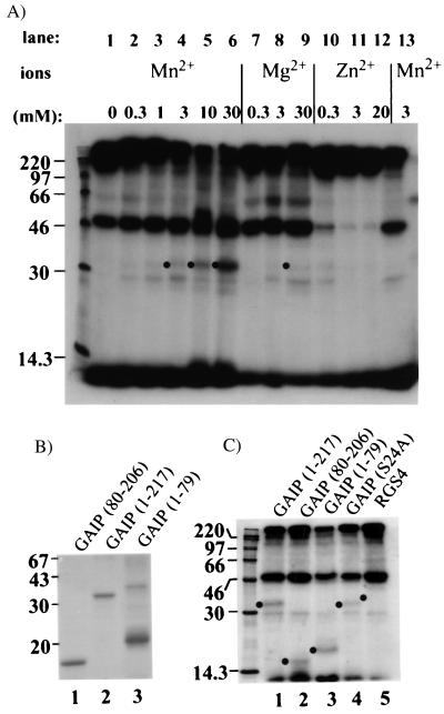
Because GAIP is localized on CCVs (15, 16) and CK2 activity has been shown to be associated with isolated CCVs (35–37), we reasoned that phosphorylation of GAIP in vivo might be mediated by a CK2 found in CCV fractions. To investigate this possibility, we performed a kinase assay in which we tested the ability of CCVs to phosphorylate recombinant GAIP and evaluated the ion dependence of the kinase activity associated with CCVs. After incubation of CCV with [γ-32P]ATP and 3 mM MnCl2, no phosphoprotein was detected in the 30- to 32-kDa region. However, several other phosphorylated proteins could be detected (Fig 5A, lane 13) at 220, 100–110, and 46–50 kDa. These bands were shown previously (38–40) to correspond to the heavy chain of clathrin, β adaptin, and μ adaptins, respectively. Dramatic variation in the phosphorylation profile of μ adaptins was seen depending on the cations used in the assay (compare Zn2+ with Mn2+ or Mg2+, Fig. 5A, lanes 10–12 vs. lanes 2–9). Mg2+-dependence was also observed for a 65-kDa phosphoprotein (Fig. 5A, lanes 7–9) that could correspond to the uncoating ATPase Hsc70 (Fig. 5A, lanes 7–9) (40). When His6 GAIP was added to the kinase assay, an additional 30- to 32-kDa band corresponding to GAIP was phosphorylated in a Mn2+-dependent manner from 1 to 30 mM (Fig. 5A, lanes 3–6), whereas little or no phosphorylation of GAIP was detected in the presence of Mg2+ or Zn2+ (compare intensities of signal in Fig. 5A, lane 6 vs. lanes 9 or 12). These results demonstrate that CCVs are able to catalyze the phosphorylation of GAIP in vitro in the presence of Mn2+.
Figure 5.
Phosphorylation of GAIP by CCVs. (A) His6 GAIP (lanes 1–12) was mixed with [γ32-P]ATP, in the absence (lane 1) or presence of increasing concentrations of Mn2+ (lanes 2–6 and 13), Mg2+ (lanes 7–9), or Zn2+ (lanes 10–12). Reactions were started by addition of CCVs and stopped with SDS/BSA. Proteins were acetone precipitated, separated by 12.5% SDS/PAGE, and exposed for autoradiography. Phosphorylation of recombinant GAIP (dots) is favored by Mn2+ (lanes 4–6). (B) Migration of recombinant GAIP. His6-tagged proteins were analyzed by SDS/PAGE on a 15% polyacrylamide gel, and stained with Coomassie blue R-250. Migration of GAIP80–206 (lane 1), wild-type GAIP (lane 2), and GAIP1–79 (lane 3) are shown. The N terminus of GAIP, GAIP1–79, migrates more slowly than expected. (C) Phosphorylations were performed as in A in the presence of 3 mM MnCl2 and with the following His6-tagged proteins: wild-type GAIP (lane 1), GAIP80–206 (lane 2), GAIP1–79 (lane 3), GAIP (S24A) (lane 4), or RGS4 (lane 5). Phosphorylated recombinant proteins are marked with a dot. Phosphorylation in the presence of CCVs occurs at the N terminus (1–79) and in the RGS domain (80–206) of GAIP. Mutation of Ser-24 reduced the phosphorylation of GAIP.
To confirm the site of phosphorylation of GAIP by CCVs, we tested the ability of CCVs to phosphorylate different domains of GAIP in the presence of Mn2+. The mobility of GAIP80–206 (Fig. 5B, lane 1) was ≈17 kDa, as expected, but GAIP1–79 had an unexpected behavior in SDS/PAGE as it migrated at 21 kDa. The autoradiogram obtained after addition of different domains to the CCV assay (Fig. 5C) shows that the core domain, GAIP80–206 (Fig. 5C, lane 2), as well as the N terminus, GAIP1–79 (Fig. 5C, lane 3), could be phosphorylated by CCVs. Interestingly, RGS4 (Fig. 5C, lane 5) was not phosphorylated in our CCV assay. GAIP (S24A) was again not as good a substrate as wild-type GAIP (Fig. 5C, compare lanes 1 and 4), as its phosphorylation was reduced by 25%. These results demonstrate that GAIP is phosphorylated on Ser-24 and suggest that additional sites on GAIP are phosphorylated by CCVs.
Discussion
Because RGS proteins can regulate the nucleotide cycle of G proteins, they are important elements regulating many signaling processes. Regulation of RGS proteins is also controlled at both the transcriptional and posttranslational levels (13, 14). As reported previously and confirmed here, two pools, GAIP-soluble and membrane-associated, are found in AtT-20 clone #1 cells stably expressing GAIP and in all cell types investigated. Previously, we demonstrated that only the membrane pool is palmitoylated, and, in this paper, we demonstrated that it is also predominantly the membrane pool that is phosphorylated. That GAIP is phosphorylated was shown in two cell types by two different methods—i.e., by alkaline phosphatase treatment of liver fractions and by 32P labeling of AtT-20#1 cells stably overexpressing GAIP. We further showed that phosphorylation occurs mainly on Ser residues, and we identified Ser-24 as one of the sites phosphorylated by CK2. We designed an assay based on our localization and biochemical data using isolated CCVs from rat liver and showed that recombinant GAIP could be phosphorylated at its N terminus by isolated CCVs in vitro. Although at present there is no consensus concerning ion requirements for CK2 activity (41–43), we showed that Mn2+ is required for phosphorylation of GAIP by CCVs. From these results, we speculate that clathrin-coated pits and vesicles could be one of the cellular compartments where GAIP is phosphorylated.
Phosphorylation events are important in regulation of signal transduction because they can affect subcellular localization and the activity of effectors, e.g., phospholipase A2, mitogen activated protein kinases, and ion channels (44–46).
RGS proteins are important inhibitory elements of signal transduction. Our findings suggest that RGS proteins may also be regulated by phosphorylation. Several RGS proteins have been shown to exist in both cytosolic and membrane forms (13, 14), but GAIP is the only RGS protein that has been localized on CCVs. A membrane form of RGS4 has been reported, and its cytosolic form has been shown to translocate to plasma membrane when activation of G proteins was mimicked (32, 33); however, no phosphorylation of recombinant RGS4 could be detected in our assay. This suggests individual variation among RGS proteins in this respect.
How phosphorylation is related to membrane association is still poorly understood. α subunits of G proteins are modified by both lipids (myristoylation, palmitoylation) and phosphate groups. Both types of modification affect their activity (11, 12), but to date no clear link has been established between these two posttranslational modifications. In the case of MARCKS proteins, membrane association is better understood, as phosphorylation contributes in a cooperative way along with electrostatic interactions and lipid modification (myristoyl-electrostatic switch) (47). A myristoylation-deficient mutant of MARCKS could not bind to the plasma membrane and failed to be phosphorylated (48). This suggests that membrane association and phosphorylation are linked, but the precise chronology of events needs to be investigated for each protein. Based on the model of the myristoyl-electrostatic switch for MARCKS protein, a cooperativity between palmitoylation and positively charged amino acids interacting with negatively charged phospholipids has been proposed to be responsible for membrane targeting of RGS 16 (34). GAIP does not fit this model because the extreme N terminus of GAIP is negatively charged, rendering membrane association by electrostatic interactions alone improbable. Palmitoylation, described for GAIP (27), by creating a high-energy thioester linkage, could facilitate membrane association and subsequent phosphorylation.
Interestingly, Ser-24, which is phosphorylated in GAIP, is not conserved among the other RGS proteins, including members of the GAIP subfamily which includes RET1-RGS and RGSZ1 (49). Therefore, the phosphorylation events we have described on Ser-24 must be specific for GAIP. This emphasizes that caution must be taken not to generalize from findings obtained on one RGS protein to others.
Phosphorylation and dephosphorylation events at the level of CCVs are a common theme for regulating protein–protein interactions that occur during endocytosis. A protein kinase able to phosphorylate in vitro the exogenous substrate casein has been shown to copurifiy with rat liver CCVs. This protein kinase has been identified biochemically as CK2 (36). We showed that GAIP could be phosphorylated by purified CK2 and rat liver CCVs on Ser-24 in vitro. Therefore, it is reasonable to assume that CK2 may be responsible for the phosphorylation of membrane-associated GAIP. Collectively, our findings suggest that the phosphorylation of GAIP may be regulated by the endocytic machinery. The functional consequences of phosphorylation remain to be understood, but appear to be tied to membrane association.
Acknowledgments
This work was supported by National Institutes of Health Grants DK17780 and CA58689 (to M.G.F.). T.F. was funded by la Fondation pour la Recherche Médicale (FRM) (1996) and the l'Association pour la Recherche sur le Cancer (ARC) (1997). E.E. is a graduate student in the Biomedical Sciences Graduate program and is supported by National Institutes of Health Training Grant CA67754.
Abbreviations
- CCV
clathrin-coated vesicle
- CK2
casein kinase 2
- GAIP
G α interacting protein
- GAP
GTPase activating protein
- PKC
protein kinase C
- PMA
phorbol myristate acetate
- RGS
regulators of G protein signaling
- HA
hemagglutinin
- RM
residual microsomes
Note Added in Proof
Phosphorylation of yeast Sst2 recently has been reported by Garrison et al (50).
References
- 1.Krebs E G, Beavo J A. Annu Rev Biochem. 1979;48:923–959. doi: 10.1146/annurev.bi.48.070179.004423. [DOI] [PubMed] [Google Scholar]
- 2.Krupnick J G, Benovic J L. Annu Rev Pharmacol Toxicol. 1998;38:289–319. doi: 10.1146/annurev.pharmtox.38.1.289. [DOI] [PubMed] [Google Scholar]
- 3.Pitcher J A, Freedman N J, Lefkowitz R J. Annu Rev Biochem. 1998;67:653–692. doi: 10.1146/annurev.biochem.67.1.653. [DOI] [PubMed] [Google Scholar]
- 4.Carlson K E, Brass L F, Manning D R. J Biol Chem. 1989;264:13298–13305. [PubMed] [Google Scholar]
- 5.Daniel-Issakani S, Spiegel A M, Strulovici B. J Biol Chem. 1989;264:20240–20247. [PubMed] [Google Scholar]
- 6.Offermanns S, Hu Y H, Simon M I. J Biol Chem. 1996;271:26044–26048. doi: 10.1074/jbc.271.42.26044. [DOI] [PubMed] [Google Scholar]
- 7.Asano T, Morishita R, Ueda H, Asano M, Kato K. Eur J Biochem. 1998;251:314–319. doi: 10.1046/j.1432-1327.1998.2510314.x. [DOI] [PubMed] [Google Scholar]
- 8.Neer E J. Cell. 1995;80:249–257. doi: 10.1016/0092-8674(95)90407-7. [DOI] [PubMed] [Google Scholar]
- 9.Fields T A, Casey P J. J Biol Chem. 1995;270:23119–23125. doi: 10.1074/jbc.270.39.23119. [DOI] [PubMed] [Google Scholar]
- 10.Umemori H, Inoue T, Kume S, Sekiyama N, Nagao M, Itoh H, Nakanishi S, Mikoshiba K, Yamamoto T. Science. 1997;276:1878–1881. doi: 10.1126/science.276.5320.1878. [DOI] [PubMed] [Google Scholar]
- 11.Glick J, Meigs T, Miron A, Casey P. J Biol Chem. 1998;273:26008–26013. doi: 10.1074/jbc.273.40.26008. [DOI] [PubMed] [Google Scholar]
- 12.Wang J, Ducret A, Tu Y, Kozasa T, Aebersold R, Ross E M. J Biol Chem. 1998;273:26014–26025. doi: 10.1074/jbc.273.40.26014. [DOI] [PubMed] [Google Scholar]
- 13.De Vries L, Farquhar M G. Trends Cell Biol. 1999;9:138–143. doi: 10.1016/s0962-8924(99)01515-9. [DOI] [PubMed] [Google Scholar]
- 14.De Vries L, Zheng B, Fischer T, Elenko E, Farquhar M G. Annu Rev Pharmacol Toxicol. 2000;40:235–271. doi: 10.1146/annurev.pharmtox.40.1.235. [DOI] [PubMed] [Google Scholar]
- 15.De Vries L, Elenko E, McCaffery J M, Fischer T, Hubler L, McQuistan T, Watson N, Farquhar M G. Mol Biol Cell. 1998;9:1123–1134. doi: 10.1091/mbc.9.5.1123. [DOI] [PMC free article] [PubMed] [Google Scholar]
- 16.Fischer T, Elenko E, McCaffery J M, DeVries L, Farquhar M G. Proc Natl Acad Sci USA. 1999;96:6722–6727. doi: 10.1073/pnas.96.12.6722. [DOI] [PMC free article] [PubMed] [Google Scholar]
- 17.Farquhar M G, Hauri H-P. In: The Golgi Apparatus. Berger E G, Roth J, editors. Basel: Birkhauser; 1997. pp. 63–129. [Google Scholar]
- 18.Schmid S L. Annu Rev Biochem. 1997;66:511–548. doi: 10.1146/annurev.biochem.66.1.511. [DOI] [PubMed] [Google Scholar]
- 19.Robinson M S. Trends Cell Biol. 1997;7:99–102. doi: 10.1016/S0962-8924(96)10048-9. [DOI] [PubMed] [Google Scholar]
- 20.Wilde A, Brodsky F M. J Cell Biol. 1996;135:635–645. doi: 10.1083/jcb.135.3.635. [DOI] [PMC free article] [PubMed] [Google Scholar]
- 21.Wilde A, Beattie E C, Lem L, Riethof D A, Liu S H, Mobley W C, Soriano P, Brodsky F M. Cell. 1999;96:677–687. doi: 10.1016/s0092-8674(00)80578-4. [DOI] [PubMed] [Google Scholar]
- 22.Hannan L A, Newmyer S L, Schmid S L. Mol Biol Cell. 1998;9:2217–2229. doi: 10.1091/mbc.9.8.2217. [DOI] [PMC free article] [PubMed] [Google Scholar]
- 23.Slepnev V I, Ochoa G C, Butler M H, Grabs D, Camilli P D. Science. 1998;281:821–824. doi: 10.1126/science.281.5378.821. [DOI] [PubMed] [Google Scholar]
- 24.Wigge P, Kohler K, Vallis Y, Doyle C A, Owen D, Hunt S P, McMahon H T. Mol Biol Cell. 1997;8:2003–2015. doi: 10.1091/mbc.8.10.2003. [DOI] [PMC free article] [PubMed] [Google Scholar]
- 25.De Vries L, Mousli M, Wurmser A, Farquhar M G. Proc Natl Acad Sci USA. 1995;92:11916–11920. doi: 10.1073/pnas.92.25.11916. [DOI] [PMC free article] [PubMed] [Google Scholar]
- 26.Popov S, Yu K, Kozasa T, Wilkie T M. Proc Natl Acad Sci USA. 1997;94:7216–7220. doi: 10.1073/pnas.94.14.7216. [DOI] [PMC free article] [PubMed] [Google Scholar]
- 27.De Vries L, Elenko E, Hubler L, Jones T L, Farquhar M G. Proc Natl Acad Sci USA. 1996;93:15203–15208. doi: 10.1073/pnas.93.26.15203. [DOI] [PMC free article] [PubMed] [Google Scholar]
- 28.De Vries L, Lou X, Zhao G, Zheng B, Farquhar M G. Proc Natl Acad Sci USA. 1998;95:12340–12345. doi: 10.1073/pnas.95.21.12340. [DOI] [PMC free article] [PubMed] [Google Scholar]
- 29.van der Geer P, Luo K, Sefton B M, Hunter T. In: Cell Biology: A Laboratory Handbook. Celis J E, editor. San Diego: Academic; 1994. pp. 422–448. [Google Scholar]
- 30.Litchfield D W, Lozeman F J, Piening C, Sommercorn J, Takio K, Walsh K A, Krebs E G. J Biol Chem. 1990;265:7638–7644. [PubMed] [Google Scholar]
- 31.Jones B G, Thomas L, Molloy S S, Thulin C D, Fry M D, Walsh K A, Thomas G. EMBO J. 1995;14:5869–5883. doi: 10.1002/j.1460-2075.1995.tb00275.x. [DOI] [PMC free article] [PubMed] [Google Scholar]
- 32.Srinivasa S P, Bernstein L S, Blumer K J, Linder M E. Proc Natl Acad Sci USA. 1998;95:5584–5589. doi: 10.1073/pnas.95.10.5584. [DOI] [PMC free article] [PubMed] [Google Scholar]
- 33.Druey K M, Sullivan B M, Brown D, Fischer E R, Watson N, Blumer K J, Gerfen C R, Scheschonka A, Kehrl J H. J Biol Chem. 1998;273:18405–18410. doi: 10.1074/jbc.273.29.18405. [DOI] [PubMed] [Google Scholar]
- 34.Chen C, Seow K T, Guo K, Yaw L P, Lin S C. J Biol Chem. 1999;274:19799–19806. doi: 10.1074/jbc.274.28.19799. [DOI] [PubMed] [Google Scholar]
- 35.Schook W J, Puszkin S. Proc Natl Acad Sci USA. 1985;82:8039–8043. doi: 10.1073/pnas.82.23.8039. [DOI] [PMC free article] [PubMed] [Google Scholar]
- 36.Bar-Zvi D, Levin A E, Branton D. J Biol Chem. 1987;262:17719–17723. [PubMed] [Google Scholar]
- 37.Morris S A, Mann A, Ungewickell E. J Biol Chem. 1990;265:3354–3357. [PubMed] [Google Scholar]
- 38.Bar-Zvi D, Mosley S T, Branton D. J Biol Chem. 1988;263:4408–4415. [PubMed] [Google Scholar]
- 39.Bar-Zvi D, Branton D. J Biol Chem. 1986;261:9614–9621. [PubMed] [Google Scholar]
- 40.Drucker M, Happel N, Robinson D G. Eur J Biochem. 1996;240:570–575. doi: 10.1111/j.1432-1033.1996.0570h.x. [DOI] [PubMed] [Google Scholar]
- 41.Gatica M, Hinrichs M V, Jedlicki A, Allende C C, Allende J E. FEBS Lett. 1993;315:173–177. doi: 10.1016/0014-5793(93)81157-u. [DOI] [PubMed] [Google Scholar]
- 42.Jimenez J S, Benitez M J, Lechuga C G, Collado M, Gonzalez-Nicolas J, Moreno F J. Mol Cell Biochem. 1995;152:1–6. doi: 10.1007/BF01076457. [DOI] [PubMed] [Google Scholar]
- 43.Karino A, Tanoue S, Fukuda M, Nakamura T, Ohtsuki K. FEBS Lett. 1996;398:317–321. doi: 10.1016/s0014-5793(96)01266-5. [DOI] [PubMed] [Google Scholar]
- 44.Lin L L, Wartmann M, Lin A Y, Knopf J L, Seth A, Davis R J. Cell. 1993;72:269–278. doi: 10.1016/0092-8674(93)90666-e. [DOI] [PubMed] [Google Scholar]
- 45.Blumer K J, Johnson G L. Trends Biochem Sci. 1994;19:236–240. doi: 10.1016/0968-0004(94)90147-3. [DOI] [PubMed] [Google Scholar]
- 46.Jan J Y, Jan Y N. Curr Opin Cell Biol. 1997;9:155–160. doi: 10.1016/s0955-0674(97)80057-9. [DOI] [PubMed] [Google Scholar]
- 47.McLaughlin S, Aderem A. Trends Biochem Sci. 1995;20:272–276. doi: 10.1016/s0968-0004(00)89042-8. [DOI] [PubMed] [Google Scholar]
- 48.Seykora J T, Myat M M, Allen L A H, Ravetch J V, Aderem A. J Biol Chem. 1996;271:18797–18802. doi: 10.1074/jbc.271.31.18797. [DOI] [PubMed] [Google Scholar]
- 49.Zheng B, De Vries L, Farquhar M G. Trends Biochem Sci. 1999;24:411–414. doi: 10.1016/s0968-0004(99)01474-7. [DOI] [PubMed] [Google Scholar]
- 50.Garrison T R, Zhang Y, Pausch M, Apanovitch D, Abersold R, Dohlman H G. J Biol Chem. 1999;247:36387–36391. doi: 10.1074/jbc.274.51.36387. [DOI] [PubMed] [Google Scholar]


