

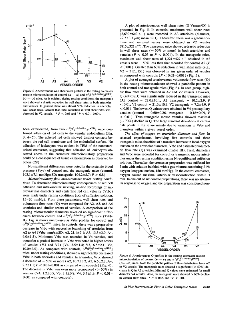
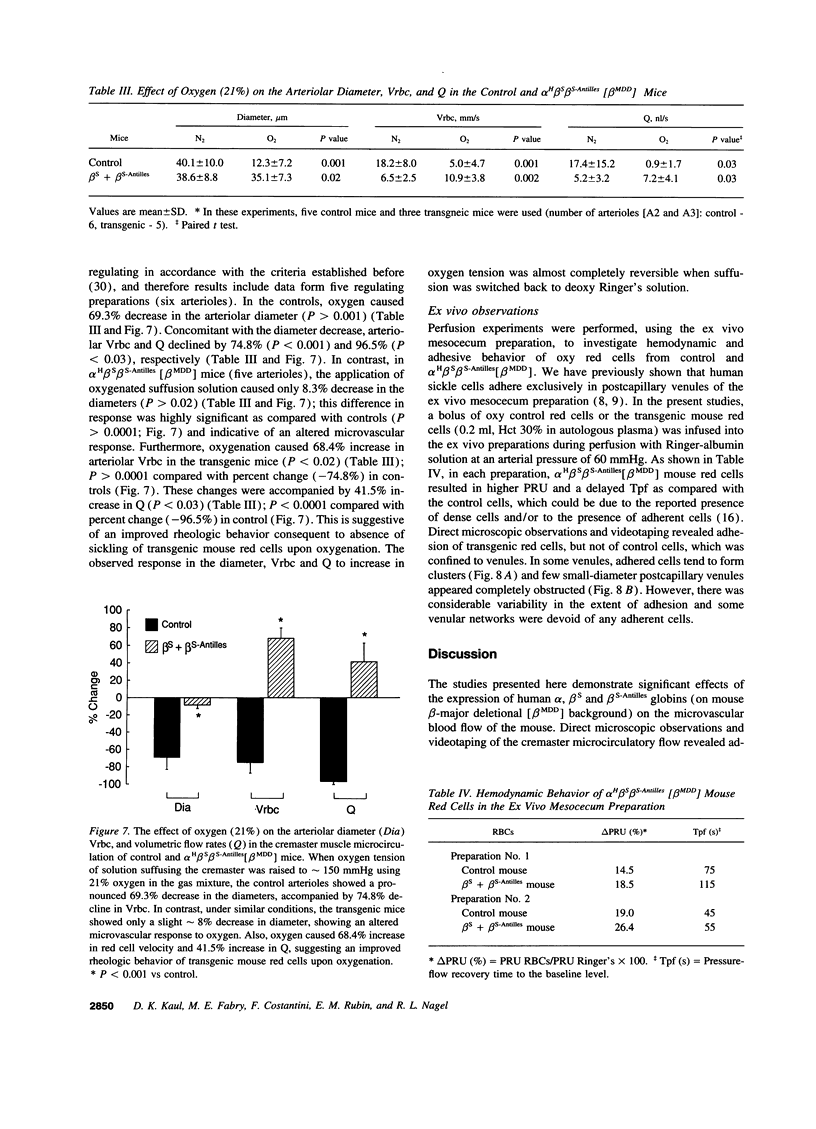
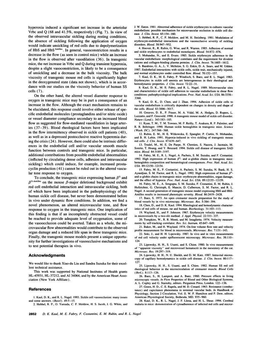
Abstract
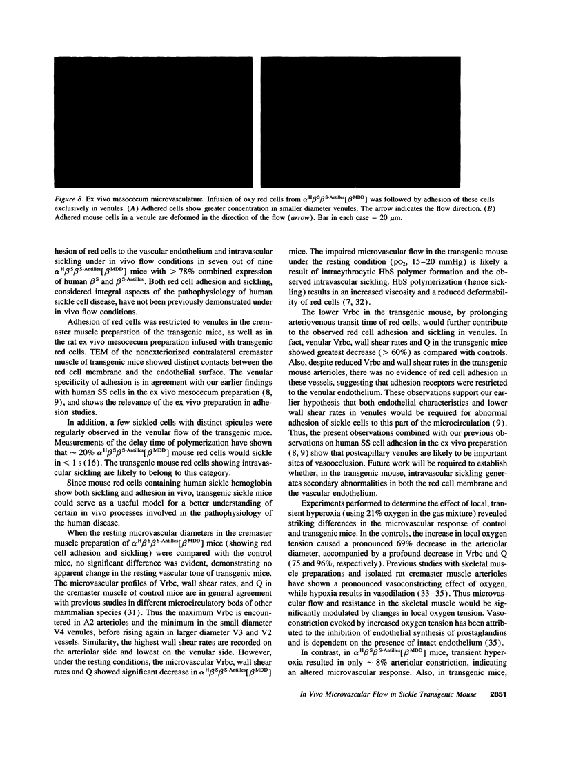
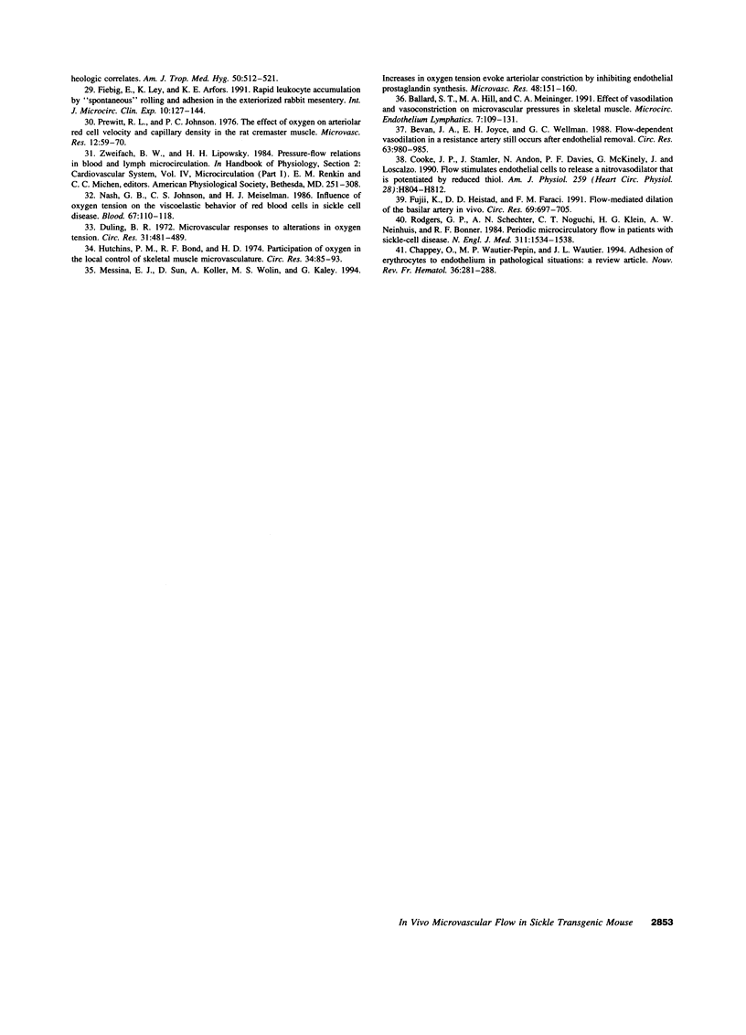
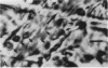
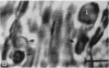
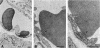
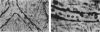
Intravascular sickling, red cell-endothelium interaction, and altered microvascular responses have been suggested to contribute to the pathophysiology of human sickle cell disease, but have never been demonstrated under in vivo flow. To address this issue, we have examined a transgenic mouse line, alphaHbetaSbetaS-Antilles [betaMDD] which has a combined high (78%) expression of beta S and beta S-Antilles globins. In vivo microcirculatory studies using the cremaster muscle preparation showed adhesion of red cells, restricted to postcapillary venules, in transgenic mice but not in control mice. Electron microscopy revealed distinct contacts between the red cell membrane and the endothelium surface. Some red cells exhibiting sickling were regularly observed in the venular flow. Infusion of transgenic mouse red cells into the ex vivo mesocecum vasculature also showed adhesion of mouse red cells exclusively in venules. Under resting conditions (pO2, 15-20 mmHg), there were no differences in the cremaster microvascular diameters of control and transgenic mice; however, transgenic mice showed a drastic reduction in microvascular red cell velocities (Vrbc) with maximal Vrbc decrease (> 60%) occurring in venules, the sites of red cell adhesion and sickling. Local, transient hyperoxia (pO2, 150 mmHg) resulted in striking differences between control and transgenic mice. In controls, oxygen caused a 69% arteriolar constriction, accompanied by 75% reduction in Vrbc. In contrast, in transgenic mice, hyperoxia resulted in only 8% decrease in the arteriolar diameter and in 68% increase in VrBC; the latter is probably due to an improved flow behavior of red cells as a consequence of unsickling. In summary, the high expression of human sickle hemoglobin in the mouse results not only in intravascular sickling but also red cell-endothelium interaction. The altered microvascular response to oxygen could be secondary to blood rheological changes, although possible intrinsic differences in the endothelial cell/vascular smooth muscle function in the transgenic mouse may also contribute. These sickle transgenic mice could serve as a useful model to investigate vasoocclusive mechanisms, as well as to test potential therapies.
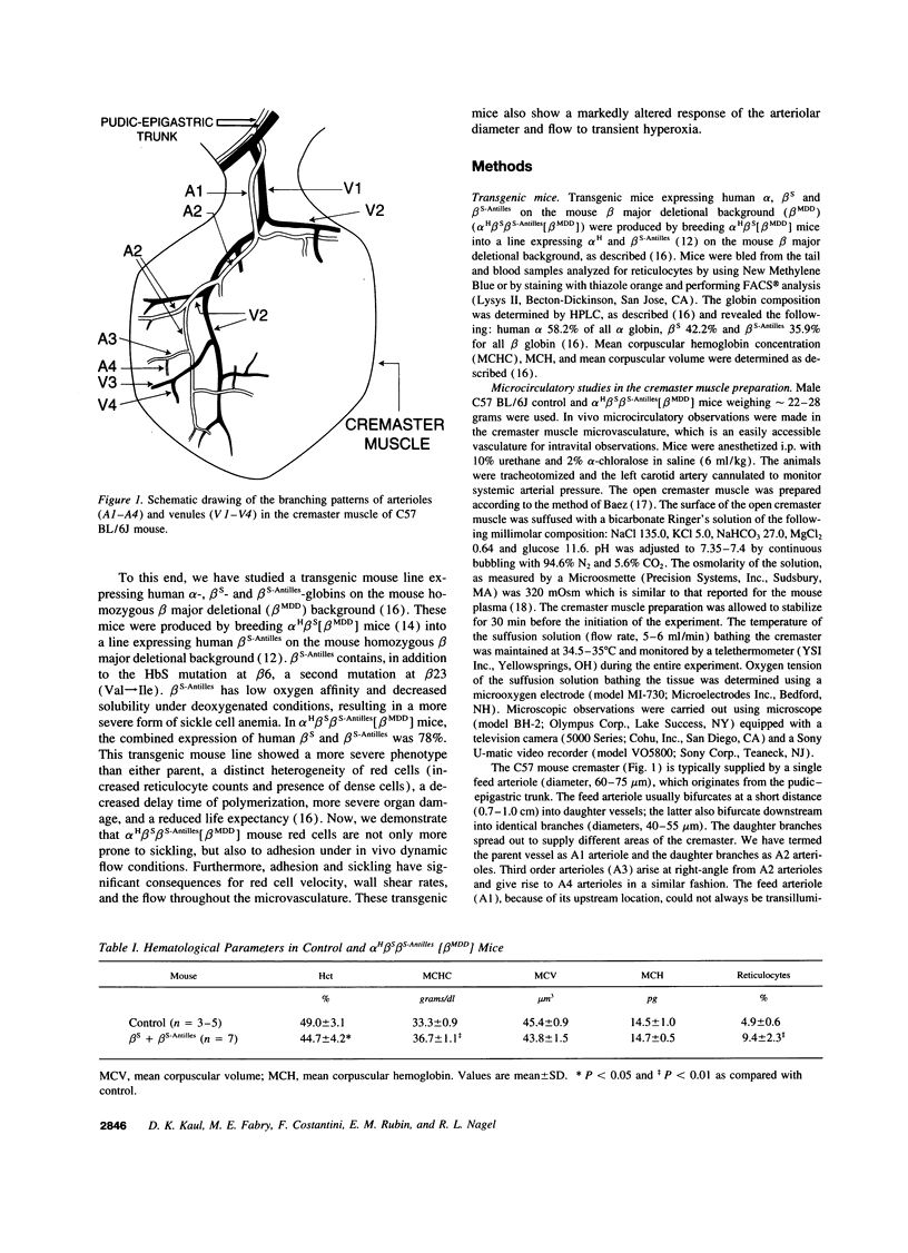
Full text
PDF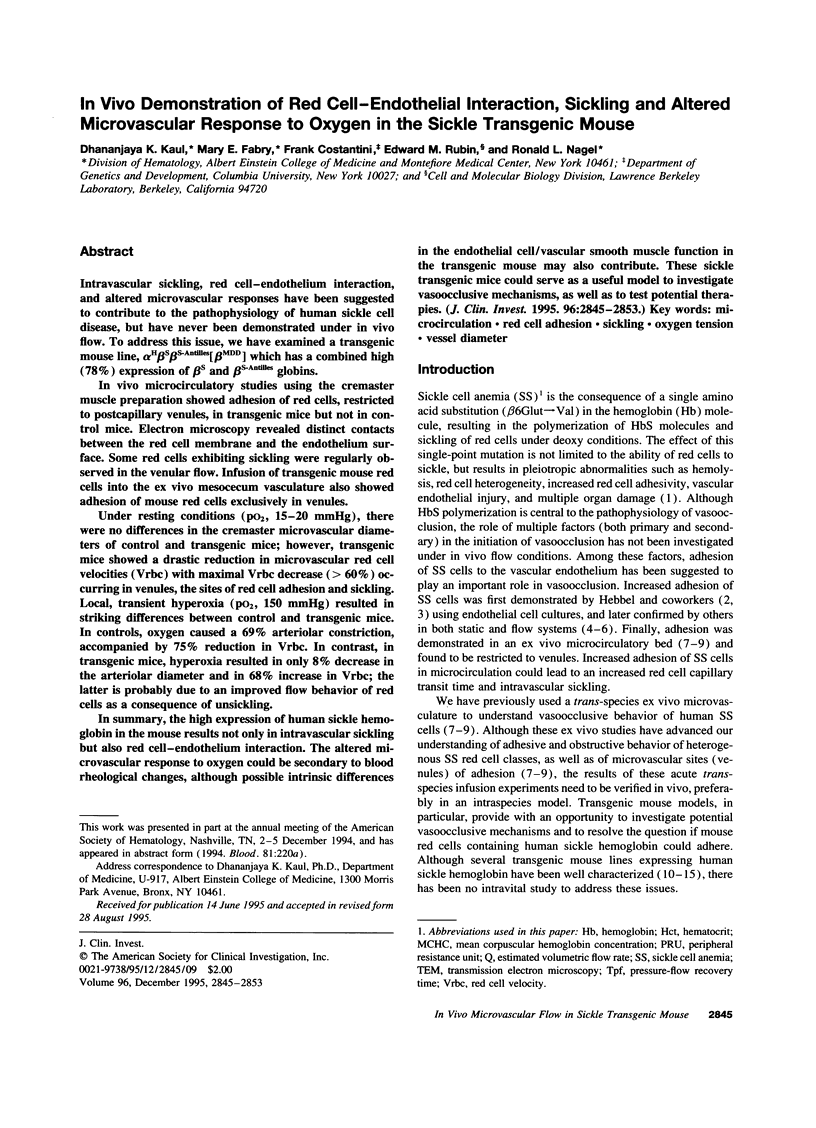
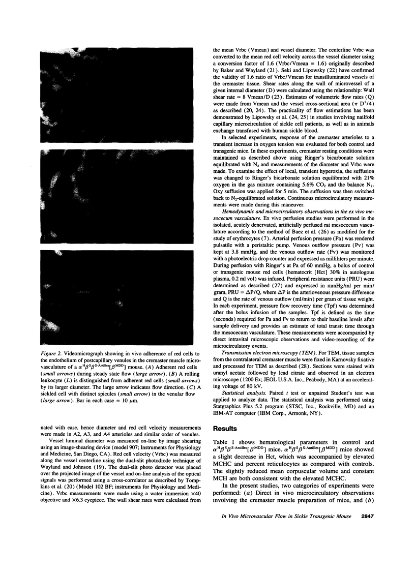
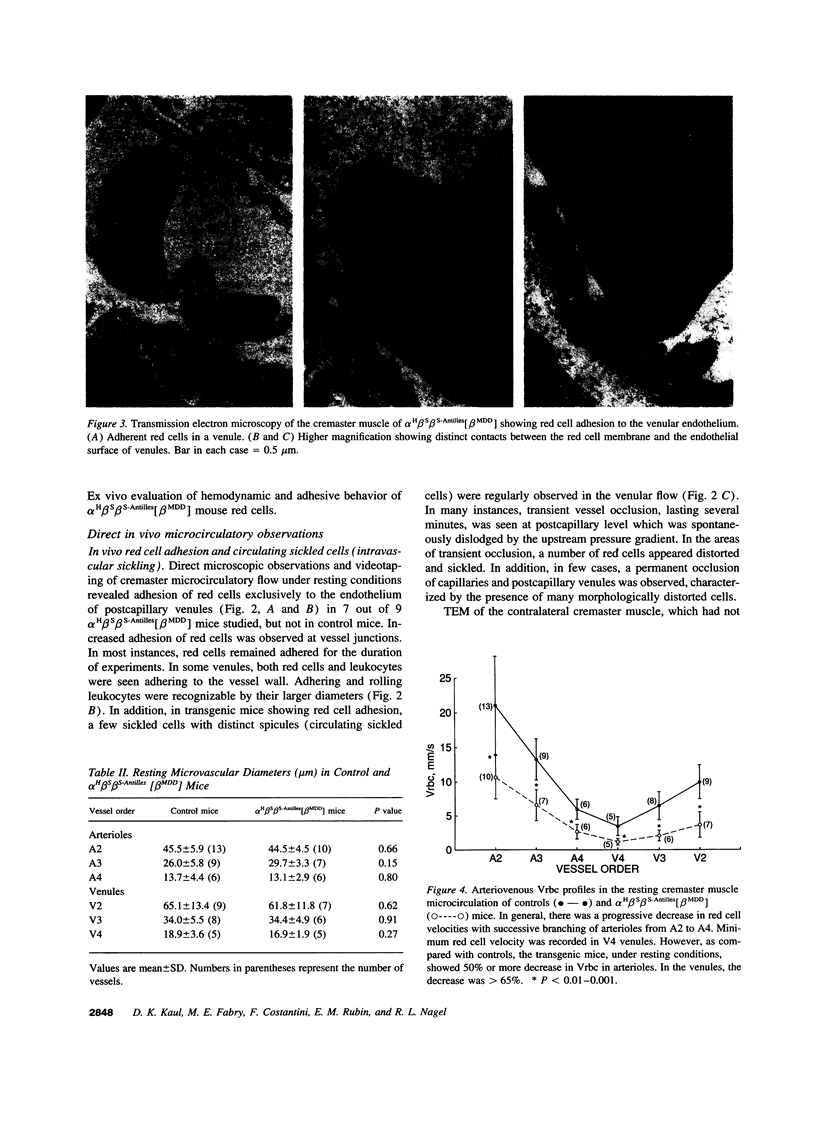
Images in this article
Selected References
These references are in PubMed. This may not be the complete list of references from this article.
- Baez S. An open cremaster muscle preparation for the study of blood vessels by in vivo microscopy. Microvasc Res. 1973 May;5(3):384–394. doi: 10.1016/0026-2862(73)90054-x. [DOI] [PubMed] [Google Scholar]
- Baker M., Wayland H. On-line volume flow rate and velocity profile measurement for blood in microvessels. Microvasc Res. 1974 Jan;7(1):131–143. doi: 10.1016/0026-2862(74)90043-0. [DOI] [PubMed] [Google Scholar]
- Ballard S. T., Hill M. A., Meininger G. A. Effect of vasodilation and vasoconstriction on microvascular pressures in skeletal muscle. Microcirc Endothelium Lymphatics. 1991;7(1-3):109–131. [PubMed] [Google Scholar]
- Barabino G. A., McIntire L. V., Eskin S. G., Sears D. A., Udden M. Endothelial cell interactions with sickle cell, sickle trait, mechanically injured, and normal erythrocytes under controlled flow. Blood. 1987 Jul;70(1):152–157. [PubMed] [Google Scholar]
- Bevan J. A., Joyce E. H., Wellman G. C. Flow-dependent dilation in a resistance artery still occurs after endothelium removal. Circ Res. 1988 Nov;63(5):980–985. doi: 10.1161/01.res.63.5.980. [DOI] [PubMed] [Google Scholar]
- Chappey O., Wautier-Pepin M. P., Wautier J. L. Adhesion of erythrocytes to endothelium in pathological situations: a review article. Nouv Rev Fr Hematol. 1994 Aug;36(4):281–288. [PubMed] [Google Scholar]
- Chen D., Kaul D. K. Rheologic and hemodynamic characteristics of red cells of mouse, rat and human. Biorheology. 1994 Jan-Feb;31(1):103–113. doi: 10.3233/bir-1994-31109. [DOI] [PubMed] [Google Scholar]
- Cooke J. P., Stamler J., Andon N., Davies P. F., McKinley G., Loscalzo J. Flow stimulates endothelial cells to release a nitrovasodilator that is potentiated by reduced thiol. Am J Physiol. 1990 Sep;259(3 Pt 2):H804–H812. doi: 10.1152/ajpheart.1990.259.3.H804. [DOI] [PubMed] [Google Scholar]
- Duling B. R. Microvascular responses to alterations in oxygen tension. Circ Res. 1972 Oct;31(4):481–489. doi: 10.1161/01.res.31.4.481. [DOI] [PubMed] [Google Scholar]
- Fabry M. E., Costantini F., Pachnis A., Suzuka S. M., Bank N., Aynedjian H. S., Factor S. M., Nagel R. L. High expression of human beta S- and alpha-globins in transgenic mice: erythrocyte abnormalities, organ damage, and the effect of hypoxia. Proc Natl Acad Sci U S A. 1992 Dec 15;89(24):12155–12159. doi: 10.1073/pnas.89.24.12155. [DOI] [PMC free article] [PubMed] [Google Scholar]
- Fabry M. E., Nagel R. L., Pachnis A., Suzuka S. M., Costantini F. High expression of human beta S- and alpha-globins in transgenic mice: hemoglobin composition and hematological consequences. Proc Natl Acad Sci U S A. 1992 Dec 15;89(24):12150–12154. doi: 10.1073/pnas.89.24.12150. [DOI] [PMC free article] [PubMed] [Google Scholar]
- Fiebig E., Ley K., Arfors K. E. Rapid leukocyte accumulation by "spontaneous" rolling and adhesion in the exteriorized rabbit mesentery. Int J Microcirc Clin Exp. 1991 May;10(2):127–144. [PubMed] [Google Scholar]
- Fujii K., Heistad D. D., Faraci F. M. Flow-mediated dilatation of the basilar artery in vivo. Circ Res. 1991 Sep;69(3):697–705. doi: 10.1161/01.res.69.3.697. [DOI] [PubMed] [Google Scholar]
- Greaves D. R., Fraser P., Vidal M. A., Hedges M. J., Ropers D., Luzzatto L., Grosveld F. A transgenic mouse model of sickle cell disorder. Nature. 1990 Jan 11;343(6254):183–185. doi: 10.1038/343183a0. [DOI] [PubMed] [Google Scholar]
- Hebbel R. P., Moldow C. F., Steinberg M. H. Modulation of erythrocyte-endothelial interactions and the vasocclusive severity of sickling disorders. Blood. 1981 Nov;58(5):947–952. [PubMed] [Google Scholar]
- Hebbel R. P., Yamada O., Moldow C. F., Jacob H. S., White J. G., Eaton J. W. Abnormal adherence of sickle erythrocytes to cultured vascular endothelium: possible mechanism for microvascular occlusion in sickle cell disease. J Clin Invest. 1980 Jan;65(1):154–160. doi: 10.1172/JCI109646. [DOI] [PMC free article] [PubMed] [Google Scholar]
- Hoover R., Rubin R., Wise G., Warren R. Adhesion of normal and sickle erythrocytes to endothelial monolayer cultures. Blood. 1979 Oct;54(4):872–876. [PubMed] [Google Scholar]
- Hutchins P. M., Bond R. F., Green H. D. Participation of oxygen in the local control of skeletal muscle microvasculature. Circ Res. 1974 Jan;34(1):85–93. doi: 10.1161/01.res.40.4.85. [DOI] [PubMed] [Google Scholar]
- Kaul D. K., Chen D., Zhan J. Adhesion of sickle cells to vascular endothelium is critically dependent on changes in density and shape of the cells. Blood. 1994 May 15;83(10):3006–3017. [PubMed] [Google Scholar]
- Kaul D. K., Fabry M. E., Nagel R. L. Microvascular sites and characteristics of sickle cell adhesion to vascular endothelium in shear flow conditions: pathophysiological implications. Proc Natl Acad Sci U S A. 1989 May;86(9):3356–3360. doi: 10.1073/pnas.86.9.3356. [DOI] [PMC free article] [PubMed] [Google Scholar]
- Kaul D. K., Fabry M. E., Windisch P., Baez S., Nagel R. L. Erythrocytes in sickle cell anemia are heterogeneous in their rheological and hemodynamic characteristics. J Clin Invest. 1983 Jul;72(1):22–31. doi: 10.1172/JCI110960. [DOI] [PMC free article] [PubMed] [Google Scholar]
- Kaul D. K., Nagel R. L., Llena J. F., Shear H. L. Cerebral malaria in mice: demonstration of cytoadherence of infected red blood cells and microrheologic correlates. Am J Trop Med Hyg. 1994 Apr;50(4):512–521. doi: 10.4269/ajtmh.1994.50.512. [DOI] [PubMed] [Google Scholar]
- Kaul D. K., Nagel R. L. Sickle cell vasoocclusion: many issues and some answers. Experientia. 1993 Jan 15;49(1):5–15. doi: 10.1007/BF01928783. [DOI] [PubMed] [Google Scholar]
- Lipowsky H. H., Sheikh N. U., Katz D. M. Intravital microscopy of capillary hemodynamics in sickle cell disease. J Clin Invest. 1987 Jul;80(1):117–127. doi: 10.1172/JCI113036. [DOI] [PMC free article] [PubMed] [Google Scholar]
- Lipowsky H. H., Usami S., Chien S. Human SS red cell rheological behavior in the microcirculation of cremaster muscle. Blood Cells. 1982;8(1):113–126. [PubMed] [Google Scholar]
- Lipowsky H. H., Usami S., Chien S. In vivo measurements of "apparent viscosity" and microvessel hematocrit in the mesentery of the cat. Microvasc Res. 1980 May;19(3):297–319. doi: 10.1016/0026-2862(80)90050-3. [DOI] [PubMed] [Google Scholar]
- Messina E. J., Sun D., Koller A., Wolin M. S., Kaley G. Increases in oxygen tension evoke arteriolar constriction by inhibiting endothelial prostaglandin synthesis. Microvasc Res. 1994 Sep;48(2):151–160. doi: 10.1006/mvre.1994.1046. [DOI] [PubMed] [Google Scholar]
- Mohandas N., Evans E. Sickle erythrocyte adherence to vascular endothelium. Morphologic correlates and the requirement for divalent cations and collagen-binding plasma proteins. J Clin Invest. 1985 Oct;76(4):1605–1612. doi: 10.1172/JCI112144. [DOI] [PMC free article] [PubMed] [Google Scholar]
- Nash G. B., Johnson C. S., Meiselman H. J. Influence of oxygen tension on the viscoelastic behavior of red blood cells in sickle cell disease. Blood. 1986 Jan;67(1):110–118. [PubMed] [Google Scholar]
- Prewitt R. L., Johnson P. C. The effect of oxygen on arteriolar red cell velocity and capillary density in the rat cremaster muscle. Microvasc Res. 1976 Jul;12(1):59–70. doi: 10.1016/0026-2862(76)90007-8. [DOI] [PubMed] [Google Scholar]
- Rodgers G. P., Schechter A. N., Noguchi C. T., Klein H. G., Nienhuis A. W., Bonner R. F. Periodic microcirculatory flow in patients with sickle-cell disease. N Engl J Med. 1984 Dec 13;311(24):1534–1538. doi: 10.1056/NEJM198412133112403. [DOI] [PubMed] [Google Scholar]
- Rubin E. M., Witkowska H. E., Spangler E., Curtin P., Lubin B. H., Mohandas N., Clift S. M. Hypoxia-induced in vivo sickling of transgenic mouse red cells. J Clin Invest. 1991 Feb;87(2):639–647. doi: 10.1172/JCI115041. [DOI] [PMC free article] [PubMed] [Google Scholar]
- Ryan T. M., Townes T. M., Reilly M. P., Asakura T., Palmiter R. D., Brinster R. L., Behringer R. R. Human sickle hemoglobin in transgenic mice. Science. 1990 Feb 2;247(4942):566–568. doi: 10.1126/science.2154033. [DOI] [PubMed] [Google Scholar]
- Seki J., Lipowsky H. H. In vivo and in vitro measurements of red cell velocity under epifluorescence microscopy. Microvasc Res. 1989 Jul;38(1):110–124. doi: 10.1016/0026-2862(89)90020-4. [DOI] [PubMed] [Google Scholar]
- Trudel M., De Paepe M. E., Chrétien N., Saadane N., Jacmain J., Sorette M., Hoang T., Beuzard Y. Sickle cell disease of transgenic SAD mice. Blood. 1994 Nov 1;84(9):3189–3197. [PubMed] [Google Scholar]
- Wayland H., Johnson P. C. Erythrocyte velocity measurement in microvessels by a two-slit photometric method. J Appl Physiol. 1967 Feb;22(2):333–337. doi: 10.1152/jappl.1967.22.2.333. [DOI] [PubMed] [Google Scholar]