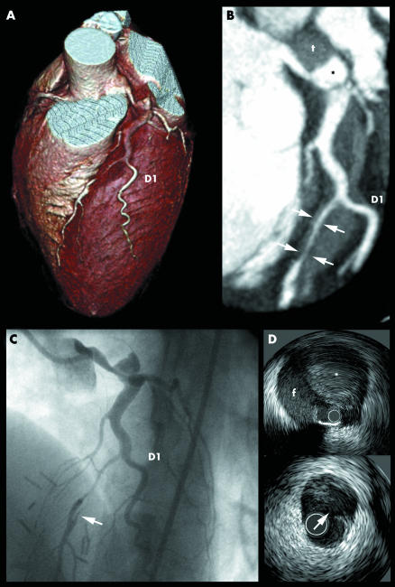
A 33 year old woman, with hereditary haemorrhagic telangiectasia (Osler‐Weber‐Rendu disease), presented to the hospital 48 hours after a prolonged episode of chest pain. The ECG, showing pathological Q waves in leads V1–V3 and inverted T waves in leads V1–V6, and increased cardiac enzymes confirmed the diagnosis of a semi‐recent anterior myocardial infarction. Cardiac 64 row multislice computed tomography (panels A and B) revealed a dissection originating from the left main (LM) artery with antegrade extension along the course of the left anterior descending coronary artery (LAD) and re‐entry distal to the first diagonal branch (D1). These findings were subsequently confirmed by coronary angiography and intravascular ultrasound (panels C and D). Both the LM and mid LAD were successfully treated with a bare metal stent.
Spontaneous coronary artery dissection is a rare condition, the aetiology of which remains unclear. In this case it might be related to the fragility of the vessel wall, which characterises Osler‐Weber‐Rendu disease.
Three dimensional (A) and multiplanar reconstructed (B) multislice computed tomography image showing the false lumen (asterisk) with thrombus (t) within the left main (LM) artery. The dissection is accompanied by a large haematoma along the course of the left anterior descending (LAD) artery that compresses the lumen (arrows) distal to the origin of first diagonal branch (D1). (C) Coronary angiography confirms the LM dissection and critical narrowing of the mid LAD. (D) Intravascular ultrasound shows the false lumen (f) with thrombus (asterisk) and an intimal tear (arrow) corresponding to the distal re‐entry point of the dissection. Angiographically the tear is visible as a “flap” within the lumen.


