

Abstract
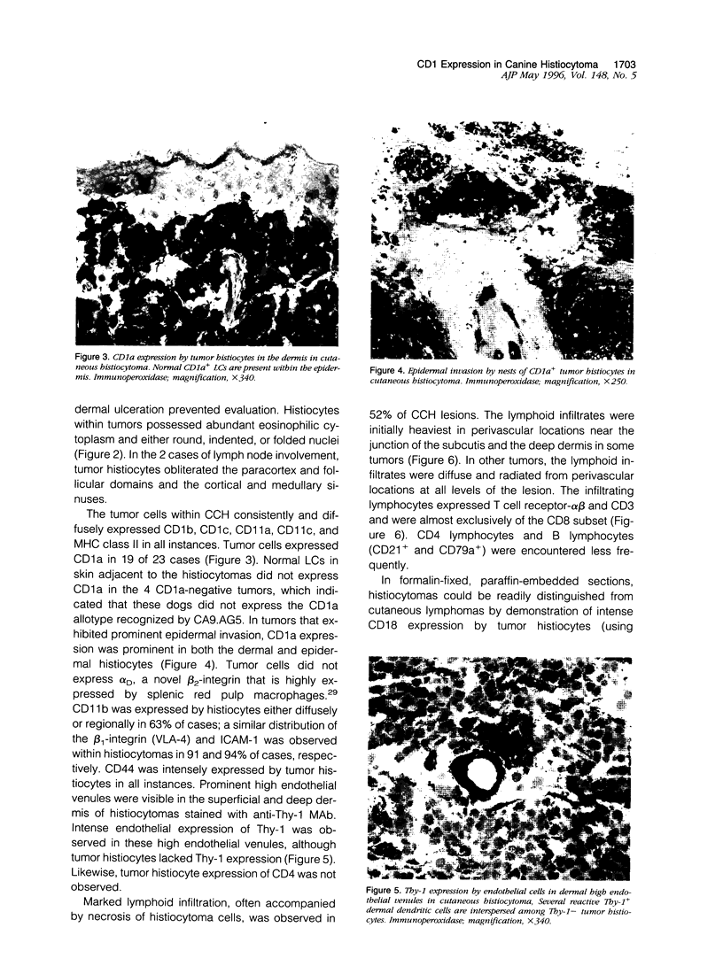
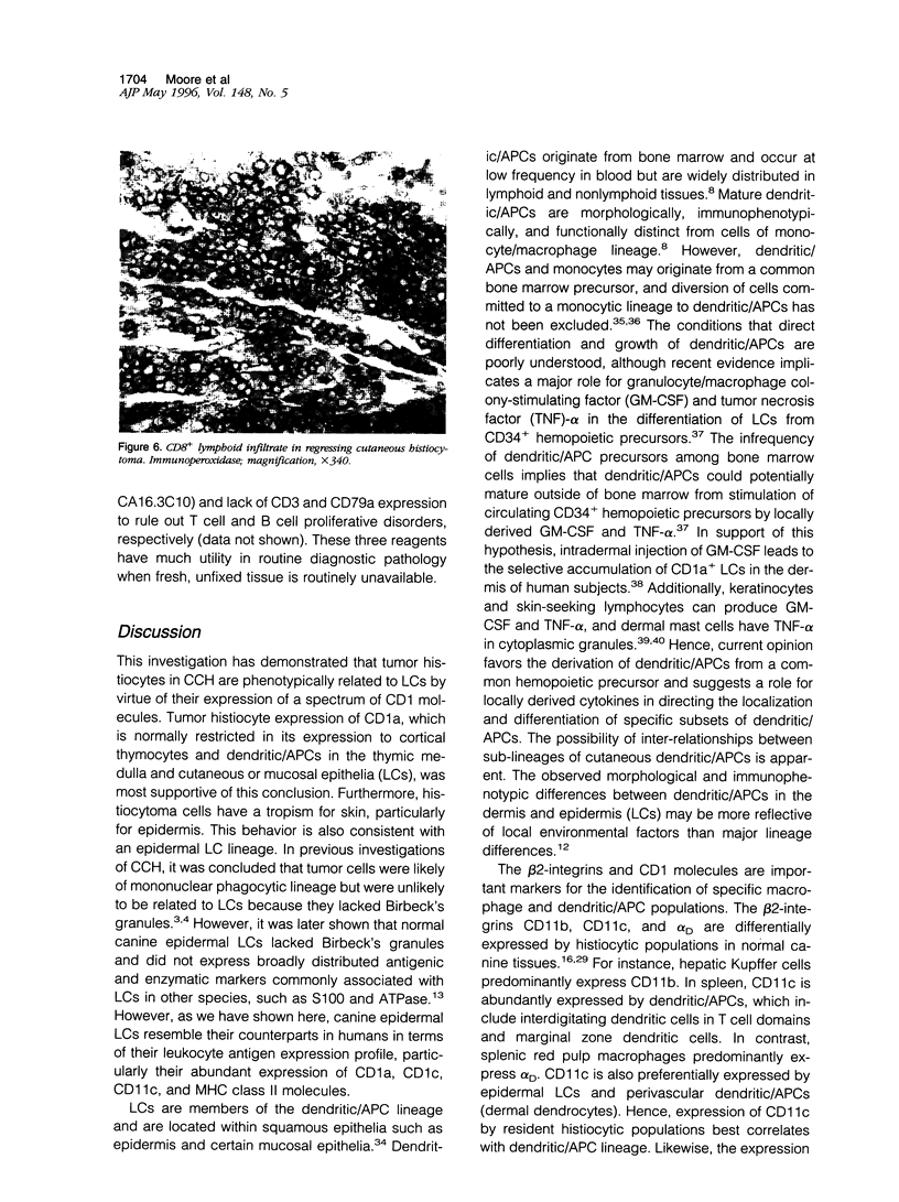
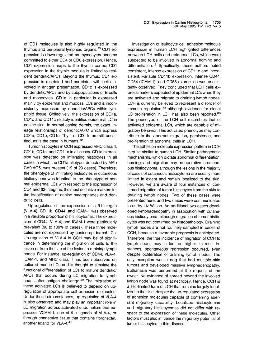
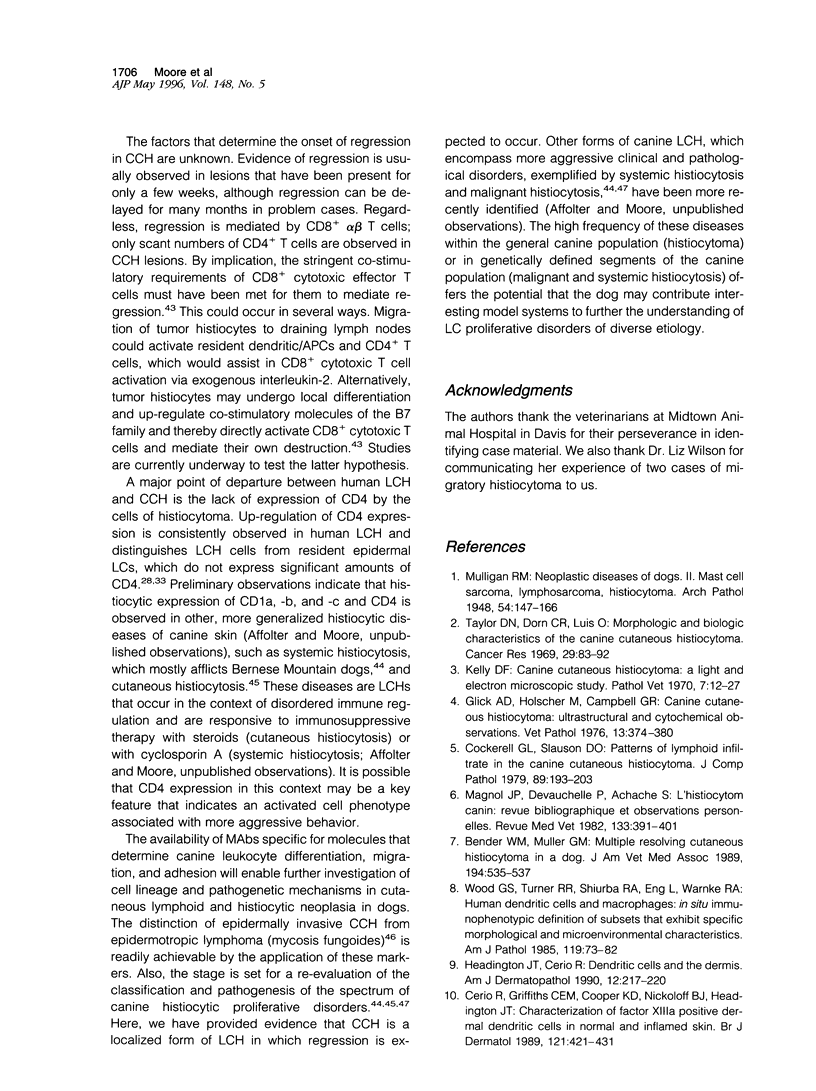
Canine cutaneous histiocytoma (CCH) is a common, benign neoplasm of the dog. Histiocytomas most commonly occur as solitary lesions that undergo spontaneous regression. The age-specific incidence rate for histiocytomas drops precipitously after 3 years, although histiocytomas occur in dogs of all ages. Langerhans cells (LCs) in humans and dogs express abundant major histocompatibility complex class II molecules and a variety of leukocyte antigens characteristic of dendritic cell differentiation including CD1a, CD1b, CD1c, and CD11c. The immunophenotype of CCH resembled that of cutaneous LCs by virtue of the expression of CD1 molecules (CD1a, -b, and -c), CD11c, and major histocompatibility complex class II. Furthermore, histiocytoma cells had a tropism for epidermis, which was also consistent with an epidermal LC lineage. The expression of adhesion molecules such as CD11b (variable), CD44, CD54 (ICAM-1), and CD49d (VLA-4) in CCH indicated that the infiltrating cells had some of the characteristics of activated LCs, as these molecules are not expressed by normal, resting canine epidermal LCs. CCH did not express Thy-1 or CD4. Thy-1 expression is a characteristic of human and canine dermal dendrocytes, which are perivascular dendritic antigen-presenting cells closely related to epidermal LCs. CD4 expression is prevalent in human LC histiocytosis, and in this respect CCH differed from human LC histiocytosis. Here we demonstrate that CCH is a localized form of self-limiting LC histiocytosis, which predominantly expresses an epidermal LC phenotype. CCH occurs as solitary or, less commonly, as multiple cutaneous nodules or plaques, which rarely may extend beyond the skin to local lymph nodes. Regression of CCH occurs spontaneously in the vast majority of cases in primary and secondary sites, and is mediated by CD8+ alpha beta T cells. The high frequency of CCH within the general canine population offers the potential that the dog may provide an interesting model system to further the understanding of LC proliferative disorders, particularly the self-limiting, cutaneous form of human LC histiocytosis.
Full text
PDF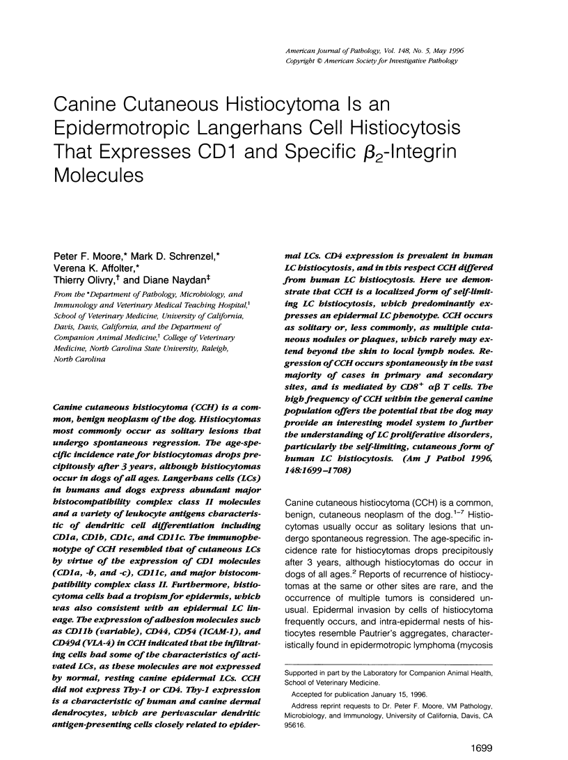
Images in this article
Selected References
These references are in PubMed. This may not be the complete list of references from this article.
- Aiba S., Nakagawa S., Ozawa H., Miyake K., Yagita H., Tagami H. Up-regulation of alpha 4 integrin on activated Langerhans cells: analysis of adhesion molecules on Langerhans cells relating to their migration from skin to draining lymph nodes. J Invest Dermatol. 1993 Feb;100(2):143–147. doi: 10.1111/1523-1747.ep12462783. [DOI] [PubMed] [Google Scholar]
- Bender W. M., Muller G. H. Multiple, resolving, cutaneous histiocytoma in a dog. J Am Vet Med Assoc. 1989 Feb 15;194(4):535–537. [PubMed] [Google Scholar]
- Bieber T., Jürgens M., Wollenberg A., Sander E., Hanau D., de la Salle H. Characterization of the protein tyrosine phosphatase CD45 on human epidermal Langerhans cells. Eur J Immunol. 1995 Feb;25(2):317–321. doi: 10.1002/eji.1830250202. [DOI] [PubMed] [Google Scholar]
- Bos J. D., Kapsenberg M. L. The skin immune system: progress in cutaneous biology. Immunol Today. 1993 Feb;14(2):75–78. doi: 10.1016/0167-5699(93)90062-P. [DOI] [PubMed] [Google Scholar]
- Calabi F., Bradbury A. The CD1 system. Tissue Antigens. 1991 Jan;37(1):1–9. doi: 10.1111/j.1399-0039.1991.tb01836.x. [DOI] [PubMed] [Google Scholar]
- Caux C., Dezutter-Dambuyant C., Schmitt D., Banchereau J. GM-CSF and TNF-alpha cooperate in the generation of dendritic Langerhans cells. Nature. 1992 Nov 19;360(6401):258–261. doi: 10.1038/360258a0. [DOI] [PubMed] [Google Scholar]
- Cerio R., Griffiths C. E., Cooper K. D., Nickoloff B. J., Headington J. T. Characterization of factor XIIIa positive dermal dendritic cells in normal and inflamed skin. Br J Dermatol. 1989 Oct;121(4):421–431. doi: 10.1111/j.1365-2133.1989.tb15509.x. [DOI] [PubMed] [Google Scholar]
- Chu T., Jaffe R. The normal Langerhans cell and the LCH cell. Br J Cancer Suppl. 1994 Sep;23:S4–10. [PMC free article] [PubMed] [Google Scholar]
- Cobbold S., Metcalfe S. Monoclonal antibodies that define canine homologues of human CD antigens: summary of the First International Canine Leukocyte Antigen Workshop (CLAW). Tissue Antigens. 1994 Mar;43(3):137–154. doi: 10.1111/j.1399-0039.1994.tb02315.x. [DOI] [PubMed] [Google Scholar]
- Cockerell G. L., Slauson D. O. Patterns of lymphoid infiltrate in the canine cutaneous histiocytoma. J Comp Pathol. 1979 Apr;89(2):193–203. doi: 10.1016/0021-9975(79)90058-6. [DOI] [PubMed] [Google Scholar]
- Danilenko D. M., Moore P. F., Rossitto P. V. Canine leukocyte cell adhesion molecules (LeuCAMs): characterization of the CD11/CD18 family. Tissue Antigens. 1992 Jul;40(1):13–21. doi: 10.1111/j.1399-0039.1992.tb01952.x. [DOI] [PubMed] [Google Scholar]
- Danilenko D. M., Rossitto P. V., Van der Vieren M., Le Trong H., McDonough S. P., Affolter V. K., Moore P. F. A novel canine leukointegrin, alpha d beta 2, is expressed by specific macrophage subpopulations in tissue and a minor CD8+ lymphocyte subpopulation in peripheral blood. J Immunol. 1995 Jul 1;155(1):35–44. [PubMed] [Google Scholar]
- Emile J. F., Fraitag S., Leborgne M., de Prost Y., Brousse N. Langerhans' cell histiocytosis cells are activated Langerhans' cells. J Pathol. 1994 Oct;174(2):71–76. doi: 10.1002/path.1711740202. [DOI] [PubMed] [Google Scholar]
- Favara B. E. Langerhans' cell histiocytosis pathobiology and pathogenesis. Semin Oncol. 1991 Feb;18(1):3–7. [PubMed] [Google Scholar]
- Fivenson D. P., Beck E. R., Dunstan R. W., Nickoloff B. J., Moore P. F. Dermal dendrocytes and T-cells in canine mycosis fungoides. Support for an animal model of human cutaneous T-cell lymphoma. Cancer. 1992 Oct 15;70(8):2091–2098. doi: 10.1002/1097-0142(19921015)70:8<2091::aid-cncr2820700814>3.0.co;2-2. [DOI] [PubMed] [Google Scholar]
- Furue M., Nindl M., Kawabe K., Nakamura K., Ishibashi Y., Sagawa K. Epitopes for CD1a, CD1b, and CD1c antigens are differentially mapped on Langerhans cells, dermal dendritic cells, keratinocytes, and basement membrane zone in human skin. J Am Acad Dermatol. 1992 Sep;27(3):419–426. doi: 10.1016/0190-9622(92)70211-w. [DOI] [PubMed] [Google Scholar]
- Glick A. D., Holscher M., Campbell G. R. Canine cutaneous histiocytoma: ultrastructural and cytochemical observations. Vet Pathol. 1976;13(5):374–380. doi: 10.1177/030098587601300507. [DOI] [PubMed] [Google Scholar]
- Headington J. T., Cerio R. Dendritic cells and the dermis: 1990. Am J Dermatopathol. 1990 Jun;12(3):217–220. doi: 10.1097/00000372-199006000-00001. [DOI] [PubMed] [Google Scholar]
- Howard C. J., Sopp P., Bembridge G., Young J., Parsons K. R. Comparison of CD1 monoclonal antibodies on bovine cells and tissues. Vet Immunol Immunopathol. 1993 Nov;39(1-3):77–83. doi: 10.1016/0165-2427(93)90166-2. [DOI] [PubMed] [Google Scholar]
- Jones M., Cordell J. L., Beyers A. D., Tse A. G., Mason D. Y. Detection of T and B cells in many animal species using cross-reactive anti-peptide antibodies. J Immunol. 1993 Jun 15;150(12):5429–5435. [PubMed] [Google Scholar]
- Kaplan G., Walsh G., Guido L. S., Meyn P., Burkhardt R. A., Abalos R. M., Barker J., Frindt P. A., Fajardo T. T., Celona R. Novel responses of human skin to intradermal recombinant granulocyte/macrophage-colony-stimulating factor: Langerhans cell recruitment, keratinocyte growth, and enhanced wound healing. J Exp Med. 1992 Jun 1;175(6):1717–1728. doi: 10.1084/jem.175.6.1717. [DOI] [PMC free article] [PubMed] [Google Scholar]
- Kasinrerk W., Baumruker T., Majdic O., Knapp W., Stockinger H. CD1 molecule expression on human monocytes induced by granulocyte-macrophage colony-stimulating factor. J Immunol. 1993 Jan 15;150(2):579–584. [PubMed] [Google Scholar]
- Kelly D. F. Canine cutaneous histiocytoma. A light and electron microscopic study. Pathol Vet. 1970;7(1):12–27. doi: 10.1177/030098587000700103. [DOI] [PubMed] [Google Scholar]
- Le Varlet B., Dezutter-Dambuyant C., Staquet M. J., Delorme P., Schmitt D. Human epidermal Langerhans cells express integrins of the beta 1 subfamily. J Invest Dermatol. 1991 Apr;96(4):518–522. doi: 10.1111/1523-1747.ep12470229. [DOI] [PubMed] [Google Scholar]
- Mason D. Y., Cordell J., Brown M., Pallesen G., Ralfkiaer E., Rothbard J., Crumpton M., Gatter K. C. Detection of T cells in paraffin wax embedded tissue using antibodies against a peptide sequence from the CD3 antigen. J Clin Pathol. 1989 Nov;42(11):1194–1200. doi: 10.1136/jcp.42.11.1194. [DOI] [PMC free article] [PubMed] [Google Scholar]
- Mays M. B., Bergeron J. A. Cutaneous histiocytosis in dogs. J Am Vet Med Assoc. 1986 Feb 15;188(4):377–381. [PubMed] [Google Scholar]
- Meunier L., Gonzalez-Ramos A., Cooper K. D. Heterogeneous populations of class II MHC+ cells in human dermal cell suspensions. Identification of a small subset responsible for potent dermal antigen-presenting cell activity with features analogous to Langerhans cells. J Immunol. 1993 Oct 15;151(8):4067–4080. [PubMed] [Google Scholar]
- Moore P. F., Olivry T., Naydan D. Canine cutaneous epitheliotropic lymphoma (mycosis fungoides) is a proliferative disorder of CD8+ T cells. Am J Pathol. 1994 Feb;144(2):421–429. [PMC free article] [PubMed] [Google Scholar]
- Moore P. F., Rosin A. Malignant histiocytosis of Bernese mountain dogs. Vet Pathol. 1986 Jan;23(1):1–10. doi: 10.1177/030098588602300101. [DOI] [PubMed] [Google Scholar]
- Moore P. F., Rossitto P. V., Danilenko D. M. Canine leukocyte integrins: characterization of a CD18 homologue. Tissue Antigens. 1990 Nov;36(5):211–220. doi: 10.1111/j.1399-0039.1990.tb01831.x. [DOI] [PubMed] [Google Scholar]
- Moore P. F., Rossitto P. V., Danilenko D. M., Wielenga J. J., Raff R. F., Severns E. Monoclonal antibodies specific for canine CD4 and CD8 define functional T-lymphocyte subsets and high-density expression of CD4 by canine neutrophils. Tissue Antigens. 1992 Aug;40(2):75–85. doi: 10.1111/j.1399-0039.1992.tb01963.x. [DOI] [PubMed] [Google Scholar]
- Moore P. F. Systemic histiocytosis of Bernese mountain dogs. Vet Pathol. 1984 Nov;21(6):554–563. doi: 10.1177/030098588402100602. [DOI] [PubMed] [Google Scholar]
- Murphy G. F. Cell membrane glycoproteins and Langerhans cells. Hum Pathol. 1985 Feb;16(2):103–112. doi: 10.1016/s0046-8177(85)80057-5. [DOI] [PubMed] [Google Scholar]
- Nash R. A., Scherf U., Storb R. Molecular cloning of the CD3 epsilon subunit of the T-cell receptor/CD3 complex in dog. Immunogenetics. 1991;33(5-6):396–398. doi: 10.1007/BF00216700. [DOI] [PubMed] [Google Scholar]
- Rossi G., Heveker N., Thiele B., Gelderblom H., Steinbach F. Development of a Langerhans cell phenotype from peripheral blood monocytes. Immunol Lett. 1992 Feb;31(2):189–197. doi: 10.1016/0165-2478(92)90145-e. [DOI] [PubMed] [Google Scholar]
- Sandmaier B. M., Storb R., Appelbaum F. R., Gallatin W. M. An antibody that facilitates hematopoietic engraftment recognizes CD44. Blood. 1990 Aug 1;76(3):630–635. [PubMed] [Google Scholar]
- Schwartz R. H. Costimulation of T lymphocytes: the role of CD28, CTLA-4, and B7/BB1 in interleukin-2 production and immunotherapy. Cell. 1992 Dec 24;71(7):1065–1068. doi: 10.1016/s0092-8674(05)80055-8. [DOI] [PubMed] [Google Scholar]
- Sepulveda-Merrill C., Mayall S., Hamblin A. S., Breathnach S. M. Antigen-presenting capacity in normal human dermis is mainly subserved by CD1a+ cells. Br J Dermatol. 1994 Jul;131(1):15–22. doi: 10.1111/j.1365-2133.1994.tb08451.x. [DOI] [PubMed] [Google Scholar]
- Smith C. W., Entman M. L., Lane C. L., Beaudet A. L., Ty T. I., Youker K., Hawkins H. K., Anderson D. C. Adherence of neutrophils to canine cardiac myocytes in vitro is dependent on intercellular adhesion molecule-1. J Clin Invest. 1991 Oct;88(4):1216–1223. doi: 10.1172/JCI115424. [DOI] [PMC free article] [PubMed] [Google Scholar]
- Springer T. A. Traffic signals for lymphocyte recirculation and leukocyte emigration: the multistep paradigm. Cell. 1994 Jan 28;76(2):301–314. doi: 10.1016/0092-8674(94)90337-9. [DOI] [PubMed] [Google Scholar]
- Stingl G., Tamaki K., Katz S. I. Origin and function of epidermal Langerhans cells. Immunol Rev. 1980;53:149–174. doi: 10.1111/j.1600-065x.1980.tb01043.x. [DOI] [PubMed] [Google Scholar]
- Taylor D. O., Dorn C. R., Luis O. H. Morphologic and biologic characteristics of the canine cutaneous histiocytoma. Cancer Res. 1969 Jan;29(1):83–92. [PubMed] [Google Scholar]
- Teunissen M. B., Wormmeester J., Krieg S. R., Peters P. J., Vogels I. M., Kapsenberg M. L., Bos J. D. Human epidermal Langerhans cells undergo profound morphologic and phenotypical changes during in vitro culture. J Invest Dermatol. 1990 Feb;94(2):166–173. doi: 10.1111/1523-1747.ep12874439. [DOI] [PubMed] [Google Scholar]
- Vedel J., Vincendeau P., Bézian J. H., Taïeb A. Flow cytometry analysis of adhesion molecules on human Langerhans cells. Clin Exp Dermatol. 1992 Jul;17(4):240–245. doi: 10.1111/j.1365-2230.1992.tb02157.x. [DOI] [PubMed] [Google Scholar]
- Wood G. S., Turner R. R., Shiurba R. A., Eng L., Warnke R. A. Human dendritic cells and macrophages. In situ immunophenotypic definition of subsets that exhibit specific morphologic and microenvironmental characteristics. Am J Pathol. 1985 Apr;119(1):73–82. [PMC free article] [PubMed] [Google Scholar]
- Wood G. S., Warner N. L., Warnke R. A. Anti-Leu-3/T4 antibodies react with cells of monocyte/macrophage and Langerhans lineage. J Immunol. 1983 Jul;131(1):212–216. [PubMed] [Google Scholar]
- de Graaf J. H., Tamminga R. Y., Kamps W. A., Timens W. Langerhans' cell histiocytosis: expression of leukocyte cellular adhesion molecules suggests abnormal homing and differentiation. Am J Pathol. 1994 Mar;144(3):466–472. [PMC free article] [PubMed] [Google Scholar]