Abstract
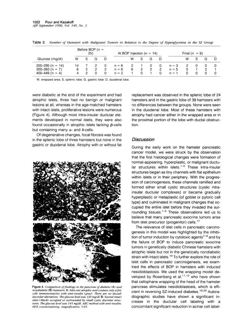
Previous studies have shown that some N-nitrosobis (2-oxopropyl)amine (BOP)-induced ductal/ductular pancreatic cancers in the hamster model develop within islets and that streptozotocin (SZ) pretreatment that caused islet degeneration and atrophy inhibits pancreatic cancer induction. Hence, it appears that in this model islets play a significant role in exocrine pancreatic carcinogenesis. To examine whether stimulation of islet cell proliferation (nesidioblastosis) enhances pancreatic exocrine cancer development, we tested the effect of the pancreatic carcinogen BOP in hamsters after induction of nesidioblastosis by cellophane wrapping. Before wrapping, hamsters were treated with SZ to inhibit pancreatic tumor induction in the unwrapped pancreatic tissues. Control groups with a wrapped pancreas did not receive SZ. Six weeks after SZ treatment, all hamsters were treated with BOP (10 mg/kg body weight) weekly for 10 weeks and the experiment was terminated 38 weeks after the last BOP treatment. Many animals recovered from their diabetes at the time when BOP was injected and many more after BOP treatment. Only nine hamsters remained diabetic until the end of the experiment. Both SZ-treated and control groups developed proliferative and malignant pancreatic ductal-type lesions primarily in the wrapped area (47%) but less frequently in the larger segments of the pancreas, including the splenic lobe (34%), gastric lobe (13%), and duodenal lobe (6%). Only a few lesions developed in the unwrapped pancreatic region of nine diabetic hamsters with atrophic islets, whereas seven of these hamsters had tumors in the wrapped area. Histologically, most tumors appeared to originate from islets, many invasive carcinomas had foci of islets, and some tumor cells showed reactivity with anti-insulin. The results show that, in the BOP hamster model, islets are the site of formation of the major fraction of exocrine pancreatic cancer and that induction of nesidioblastosis enhances pancreatic carcinogenesis.
Full text
PDF


Images in this article
Selected References
These references are in PubMed. This may not be the complete list of references from this article.
- BENCOSME S. A. The histogenesis and cytology of the pancreatic islets in the rabbit. Am J Anat. 1955 Jan;96(1):103–151. doi: 10.1002/aja.1000960105. [DOI] [PubMed] [Google Scholar]
- Bell R. H., Jr, Pour P. M. Pancreatic carcinogenicity of N-nitrosobis(2-oxopropyl)-amine in diabetic and non-diabetic Chinese hamsters. Cancer Lett. 1987 Feb;34(2):221–230. doi: 10.1016/0304-3835(87)90014-0. [DOI] [PubMed] [Google Scholar]
- Bell R. H., Jr, Sayers H. J., Pour P. M., Ray M. B., McCullough P. J. Importance of diabetes in inhibition of pancreatic cancer by streptozotocin. J Surg Res. 1989 May;46(5):515–519. doi: 10.1016/0022-4804(89)90170-4. [DOI] [PubMed] [Google Scholar]
- Bell R. H., Jr, Strayer D. S. Streptozotocin prevents development of nitrosamine-induced pancreatic cancer in the Syrian hamster. J Surg Oncol. 1983 Dec;24(4):258–262. doi: 10.1002/jso.2930240404. [DOI] [PubMed] [Google Scholar]
- Laidlaw G. F. Nesidioblastoma, the islet tumor of the pancreas. Am J Pathol. 1938 Mar;14(2):125–134.5. [PMC free article] [PubMed] [Google Scholar]
- Pour P. M., Donnelly K., Stepan K. Modification of pancreatic carcinogenesis in the hamster model. 3. Inhibitory effect of alloxan. Am J Pathol. 1983 Mar;110(3):310–314. [PMC free article] [PubMed] [Google Scholar]
- Pour P. M., Duckworth W., Carlson K., Kazakoff K. Insulin therapy prevents spontaneous recovery from streptozotocin-induced diabetes in Syrian hamsters. An autoradiographic and immunohistochemical study. Virchows Arch A Pathol Anat Histopathol. 1990;417(4):333–341. doi: 10.1007/BF01605785. [DOI] [PubMed] [Google Scholar]
- Pour P. M. Histogenesis of exocrine pancreatic cancer in the hamster model. Environ Health Perspect. 1984 Jun;56:229–243. doi: 10.1289/ehp.8456229. [DOI] [PMC free article] [PubMed] [Google Scholar]
- Pour P. M., Kazakoff K., Carlson K. Inhibition of streptozotocin-induced islet cell tumors and N-nitrosobis(2-oxopropyl)amine-induced pancreatic exocrine tumors in Syrian hamsters by exogenous insulin. Cancer Res. 1990 Mar 1;50(5):1634–1639. [PubMed] [Google Scholar]
- Pour P. M., Kazakoff K., Dulaney K. A new multilabeling technique for simultaneous demonstration of different islet cells in permanent slides. Int J Pancreatol. 1993 Apr;13(2):139–142. doi: 10.1007/BF02786082. [DOI] [PubMed] [Google Scholar]
- Pour P. M. Mechanism of pseudoductular (tubular) formation during pancreatic carcinogenesis in the hamster model. An electron-microscopic and immunohistochemical study. Am J Pathol. 1988 Feb;130(2):335–344. [PMC free article] [PubMed] [Google Scholar]
- Pour P. M., Takahashi M., Donnelly T., Stepan K. Modification of pancreatic carcinogenesis in the hamster model. IX. Effect of pancreatitis. J Natl Cancer Inst. 1983 Sep;71(3):607–613. [PubMed] [Google Scholar]
- Pour P., Althoff J., Takahashi M. Early lesions of pancreatic ductal carcinoma in the hamster model. Am J Pathol. 1977 Aug;88(2):291–308. [PMC free article] [PubMed] [Google Scholar]
- Pour P. Islet cells as a component of pancreatic ductal neoplasms. I. Experimental study: ductular cells, including islet cell precursors, as primary progenitor cells of tumors. Am J Pathol. 1978 Feb;90(2):295–316. [PMC free article] [PubMed] [Google Scholar]
- Pour P., Salmasi S. Z., Runge R. G. Ductular origin of pancreatic cancer and its multiplicity in man comparable to experimentally induced tumors. A preliminary study. Cancer Lett. 1979 Feb;6(2):89–97. doi: 10.1016/s0304-3835(79)80006-3. [DOI] [PubMed] [Google Scholar]
- Rafaeloff R., Rosenberg L., Vinik A. I. Expression of growth factors in a pancreatic islet regeneration model. Adv Exp Med Biol. 1992;321:133–141. doi: 10.1007/978-1-4615-3448-8_14. [DOI] [PubMed] [Google Scholar]
- Rosenberg L., Brown R. A., Duguid W. P. A new approach to the induction of duct epithelial hyperplasia and nesidioblastosis by cellophane wrapping of the hamster pancreas. J Surg Res. 1983 Jul;35(1):63–72. doi: 10.1016/0022-4804(83)90127-0. [DOI] [PubMed] [Google Scholar]
- Rosenberg L., Duguid W. P., Brown R. A., Vinik A. I. Induction of nesidioblastosis will reverse diabetes in Syrian golden hamster. Diabetes. 1988 Mar;37(3):334–341. doi: 10.2337/diab.37.3.334. [DOI] [PubMed] [Google Scholar]
- Rosenberg L., Duguid W. P., Vinik A. I. The effect of cellophane wrapping of the pancreas in the Syrian golden hamster: autoradiographic observations. Pancreas. 1989;4(1):31–37. doi: 10.1097/00006676-198902000-00005. [DOI] [PubMed] [Google Scholar]
- Rosenberg L., Vinik A. I. In vitro stimulation of hamster pancreatic duct growth by an extract derived from the "wrapped" pancreas. Pancreas. 1993 Mar;8(2):255–260. doi: 10.1097/00006676-199303000-00018. [DOI] [PubMed] [Google Scholar]
- Rosenberg L., Vinik A. I. Induction of endocrine cell differentiation: a new approach to management of diabetes. J Lab Clin Med. 1989 Jul;114(1):75–83. [PubMed] [Google Scholar]
- Tomioka T., Fujii H., Hirota M., Ueno K., Pour P. M. The patterns of beta-cell regeneration in untreated diabetic and insulin-treated diabetic Syrian hamsters after streptozotocin treatment. Int J Pancreatol. 1991 May;8(4):355–366. doi: 10.1007/BF02952727. [DOI] [PubMed] [Google Scholar]
- Tomioka T., Toshkov I., Kazakoff K., Andrén-Sandberg A., Takahashi T., Büchler M., Friess H., Vaughn R., Pour P. M. Cellular and subcellular localization of transforming growth factor-alpha and epidermal growth factor receptor in normal and diseased human and hamster pancreas. Teratog Carcinog Mutagen. 1995;15(5):231–250. doi: 10.1002/tcm.1770150503. [DOI] [PubMed] [Google Scholar]


