Abstract
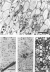
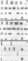
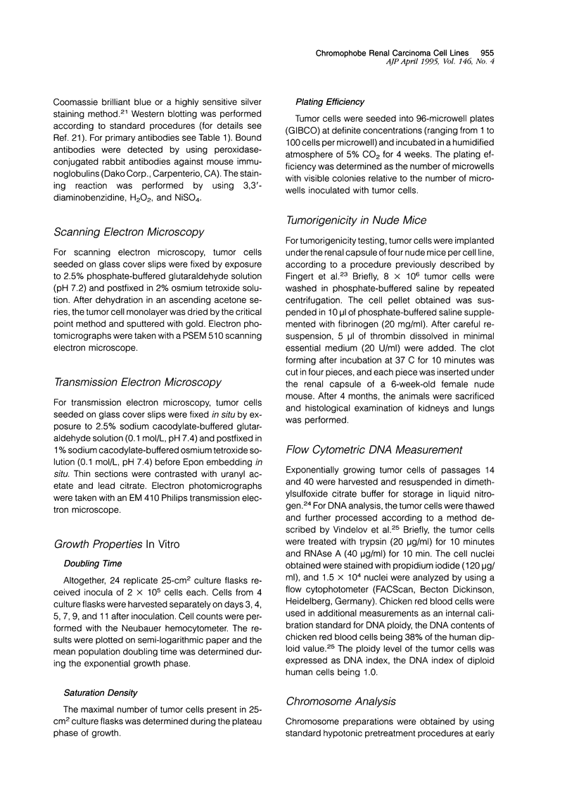
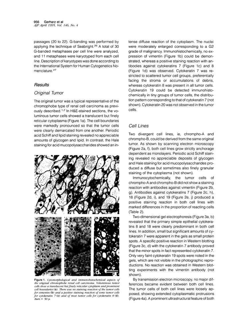
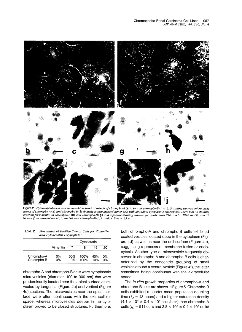
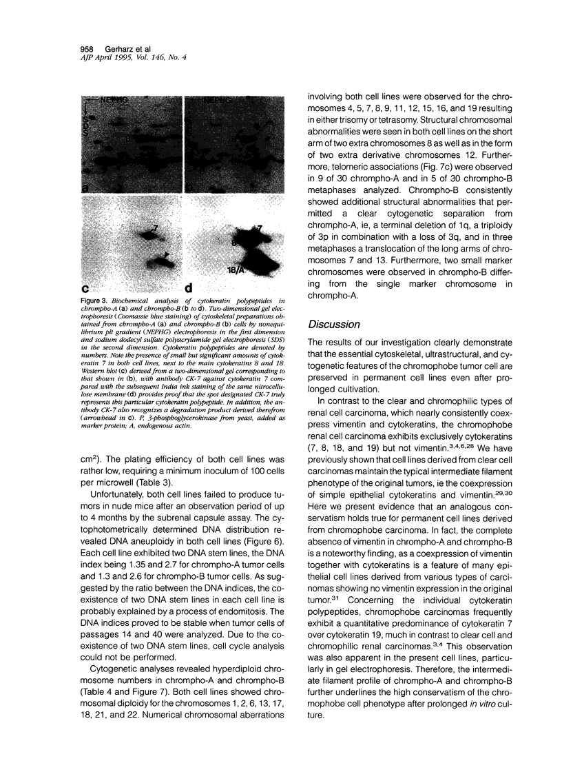
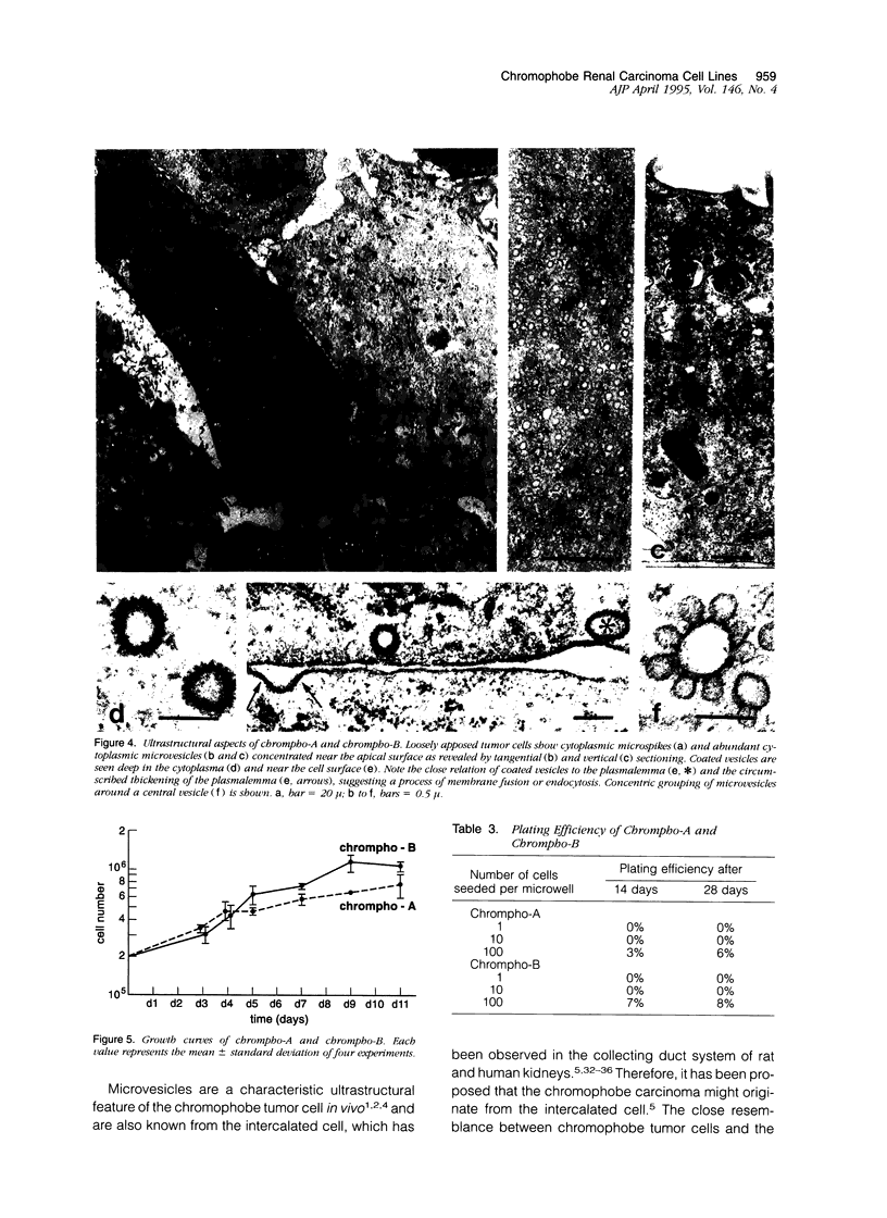
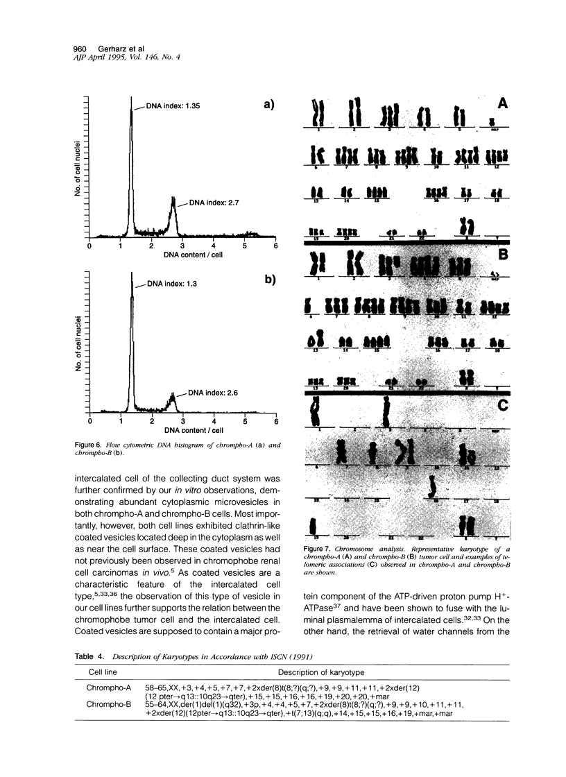
The chromophobe renal cell carcinoma is a distinct type of renal cancer presumably derived from the intercalated cell of the collecting duct system and exhibiting a better prognosis than other types of renal cell carcinoma. Chromophobe carcinomas can be separated from other types of renal cell carcinoma by their characteristic cytomorphology, ultrastructural appearance, cytoskeletal architecture, and cytogenetic aberrations. As no permanent cell line of the chromophobe tumor type has previously been described, we are the first to report on the successful establishment and characterization of two divergent permanent cell lines, ie, chrompho-A and chrompho-B, derived from the same chromophobe renal cell carcinoma. With immunocytochemistry, two-dimensional gel electrophoresis, and Western blot, chrompho-A and chrompho-B exclusively exhibited cytokeratins (Nos. 7, 8, 18, and 19) but not vimentin. Ultrastructural studies revealed numerous cytoplasmic microvesicles as well as coated vesicles that are known to be characteristic features of the intercalated cell. Chrompho-B cells exhibited a shorter mean population doubling time (tD = 43 hours) than chrompho-A cells (tD = 51 hours). Both cell lines failed to produce tumors in nude mice with the subrenal capsule assay. Cytogenetic analyses revealed hyperdiploid chromosome numbers in both cell lines with telomeric associations as well as numeric aberrations known from chromophobe renal cell carcinomas in vivo.
Full text
PDF
Images in this article
Selected References
These references are in PubMed. This may not be the complete list of references from this article.
- Achtstaetter T., Hatzfeld M., Quinlan R. A., Parmelee D. C., Franke W. W. Separation of cytokeratin polypeptides by gel electrophoretic and chromatographic techniques and their identification by immunoblotting. Methods Enzymol. 1986;134:355–371. doi: 10.1016/0076-6879(86)34102-8. [DOI] [PubMed] [Google Scholar]
- Anglard P., Trahan E., Liu S., Latif F., Merino M. J., Lerman M. I., Zbar B., Linehan W. M. Molecular and cellular characterization of human renal cell carcinoma cell lines. Cancer Res. 1992 Jan 15;52(2):348–356. [PubMed] [Google Scholar]
- Bachmann S., Kriz W., Kuhn C., Franke W. W. Differentiation of cell types in the mammalian kidney by immunofluorescence microscopy using antibodies to intermediate filament proteins and desmoplakins. Histochemistry. 1983;77(3):365–394. doi: 10.1007/BF00490899. [DOI] [PubMed] [Google Scholar]
- Beham A., Ratschek M., Zatloukal K., Schmid C., Denk H. Distribution of cytokeratins, vimentin and desmoplakins in normal renal tissue, renal cell carcinomas and oncocytoma as revealed by immunofluorescence microscopy. Virchows Arch A Pathol Anat Histopathol. 1992;421(3):209–215. doi: 10.1007/BF01611177. [DOI] [PubMed] [Google Scholar]
- Bonsib S. M., Lager D. J. Chromophobe cell carcinoma: analysis of five cases. Am J Surg Pathol. 1990 Mar;14(3):260–267. doi: 10.1097/00000478-199003000-00007. [DOI] [PubMed] [Google Scholar]
- Brown D., Gluck S., Hartwig J. Structure of the novel membrane-coating material in proton-secreting epithelial cells and identification as an H+ATPase. J Cell Biol. 1987 Oct;105(4):1637–1648. doi: 10.1083/jcb.105.4.1637. [DOI] [PMC free article] [PubMed] [Google Scholar]
- Crotty T. B., Lawrence K. M., Moertel C. A., Bartelt D. H., Jr, Batts K. P., Dewald G. W., Farrow G. M., Jenkins R. B. Cytogenetic analysis of six renal oncocytomas and a chromophobe cell renal carcinoma. Evidence that -Y, -1 may be a characteristic anomaly in renal oncocytomas. Cancer Genet Cytogenet. 1992 Jul 1;61(1):61–66. doi: 10.1016/0165-4608(92)90372-f. [DOI] [PubMed] [Google Scholar]
- Dørup J. Ultrastructure of distal nephron cells in rat renal cortex. J Ultrastruct Res. 1985 Jul-Aug;92(1-2):101–118. doi: 10.1016/0889-1605(85)90132-6. [DOI] [PubMed] [Google Scholar]
- Ebert T., Bander N. H., Finstad C. L., Ramsawak R. D., Old L. J. Establishment and characterization of human renal cancer and normal kidney cell lines. Cancer Res. 1990 Sep 1;50(17):5531–5536. [PubMed] [Google Scholar]
- Erlandson R. A., Reuter V. E. Renal tumor in a 62-year-old male. Ultrastruct Pathol. 1988 Sep-Oct;12(5):561–567. doi: 10.3109/01913128809032240. [DOI] [PubMed] [Google Scholar]
- Fingert H. J., Chen Z., Mizrahi N., Gajewski W. H., Bamberg M. P., Kradin R. L. Rapid growth of human cancer cells in a mouse model with fibrin clot subrenal capsule assay. Cancer Res. 1987 Jul 15;47(14):3824–3829. [PubMed] [Google Scholar]
- Franke W. W., Schmid E., Schiller D. L., Winter S., Jarasch E. D., Moll R., Denk H., Jackson B. W., Illmensee K. Differentiation-related patterns of expression of proteins of intermediate-size filaments in tissues and cultured cells. Cold Spring Harb Symp Quant Biol. 1982;46(Pt 1):431–453. doi: 10.1101/sqb.1982.046.01.041. [DOI] [PubMed] [Google Scholar]
- Gerharz C. D., Moll R., Störkel S., Ramp U., Thoenes W., Gabbert H. E. Ultrastructural appearance and cytoskeletal architecture of the clear, chromophilic, and chromophobe types of human renal cell carcinoma in vitro. Am J Pathol. 1993 Mar;142(3):851–859. [PMC free article] [PubMed] [Google Scholar]
- Gerharz C. D., Ramp U., Olert J., Moll R., Störkel S., Marx N., Gabbert H. E. Cytomorphological, cytogenetic, and molecular biological characterization of four new human renal carcinoma cell lines of the clear cell type. Virchows Arch. 1994;424(4):403–409. doi: 10.1007/BF00190563. [DOI] [PubMed] [Google Scholar]
- Holthöfer H., Miettinen A., Paasivuo R., Lehto V. P., Linder E., Alfthan O., Virtanen I. Cellular origin and differentiation of renal carcinomas. A fluorescence microscopic study with kidney-specific antibodies, antiintermediate filament antibodies, and lectins. Lab Invest. 1983 Sep;49(3):317–326. [PubMed] [Google Scholar]
- Höehn W., Schroeder F. H. Renal cell carcinoma: two new cell lines and a serially transplantable nude mouse tumor (NC 65). Preliminary report. Invest Urol. 1978 Sep;16(2):106–112. [PubMed] [Google Scholar]
- Kovacs G. Molecular differential pathology of renal cell tumours. Histopathology. 1993 Jan;22(1):1–8. doi: 10.1111/j.1365-2559.1993.tb00061.x. [DOI] [PubMed] [Google Scholar]
- Madsen K. M., Tisher C. C. Response of intercalated cells of rat outer medullary collecting duct to chronic metabolic acidosis. Lab Invest. 1984 Sep;51(3):268–276. [PubMed] [Google Scholar]
- Matsuda M., Osafune M., Nakano E., Kotake T., Sonoda T., Watanabe S., Hada T., Okochi T., Higashino K., Yamamura Y. Characterization of an established cell line from human renal carcinoma. Cancer Res. 1979 Nov;39(11):4694–4699. [PubMed] [Google Scholar]
- Moll R., Dhouailly D., Sun T. T. Expression of keratin 5 as a distinctive feature of epithelial and biphasic mesotheliomas. An immunohistochemical study using monoclonal antibody AE14. Virchows Arch B Cell Pathol Incl Mol Pathol. 1989;58(2):129–145. doi: 10.1007/BF02890064. [DOI] [PubMed] [Google Scholar]
- Moll R., Zimbelmann R., Goldschmidt M. D., Keith M., Laufer J., Kasper M., Koch P. J., Franke W. W. The human gene encoding cytokeratin 20 and its expression during fetal development and in gastrointestinal carcinomas. Differentiation. 1993 Jun;53(2):75–93. doi: 10.1111/j.1432-0436.1993.tb00648.x. [DOI] [PubMed] [Google Scholar]
- Myers C. E., Bulger R. E., Tisher C. C., Trump B. F. Human ultrastructure. IV. Collecting duct of healthy individuals. Lab Invest. 1966 Dec;15(12):1921–1950. [PubMed] [Google Scholar]
- Oosterwijk E., Van Muijen G. N., Oosterwijk-Wakka J. C., Warnaar S. O. Expression of intermediate-sized filaments in developing and adult human kidney and in renal cell carcinoma. J Histochem Cytochem. 1990 Mar;38(3):385–392. doi: 10.1177/38.3.1689337. [DOI] [PubMed] [Google Scholar]
- Pitz S., Moll R., Störkel S., Thoenes W. Expression of intermediate filament proteins in subtypes of renal cell carcinomas and in renal oncocytomas. Distinction of two classes of renal cell tumors. Lab Invest. 1987 Jun;56(6):642–653. [PubMed] [Google Scholar]
- Seabright M. A rapid banding technique for human chromosomes. Lancet. 1971 Oct 30;2(7731):971–972. doi: 10.1016/s0140-6736(71)90287-x. [DOI] [PubMed] [Google Scholar]
- Stetson D. L., Wade J. B., Giebisch G. Morphologic alterations in the rat medullary collecting duct following potassium depletion. Kidney Int. 1980 Jan;17(1):45–56. doi: 10.1038/ki.1980.6. [DOI] [PubMed] [Google Scholar]
- Strange K., Willingham M. C., Handler J. S., Harris H. W., Jr Apical membrane endocytosis via coated pits is stimulated by removal of antidiuretic hormone from isolated, perfused rabbit cortical collecting tubule. J Membr Biol. 1988 Jul;103(1):17–28. doi: 10.1007/BF01871929. [DOI] [PubMed] [Google Scholar]
- Störkel S., Steart P. V., Drenckhahn D., Thoenes W. The human chromophobe cell renal carcinoma: its probable relation to intercalated cells of the collecting duct. Virchows Arch B Cell Pathol Incl Mol Pathol. 1989;56(4):237–245. doi: 10.1007/BF02890022. [DOI] [PubMed] [Google Scholar]
- Störkel S., Thoenes W., Jacobi G. H., Lippold R. Prognostic parameters in renal cell carcinoma--a new approach. Eur Urol. 1989;16(6):416–422. doi: 10.1159/000471633. [DOI] [PubMed] [Google Scholar]
- Thoenes W., Störkel S., Rumpelt H. J. Histopathology and classification of renal cell tumors (adenomas, oncocytomas and carcinomas). The basic cytological and histopathological elements and their use for diagnostics. Pathol Res Pract. 1986 May;181(2):125–143. doi: 10.1016/S0344-0338(86)80001-2. [DOI] [PubMed] [Google Scholar]
- Thoenes W., Störkel S., Rumpelt H. J. Human chromophobe cell renal carcinoma. Virchows Arch B Cell Pathol Incl Mol Pathol. 1985;48(3):207–217. doi: 10.1007/BF02890129. [DOI] [PubMed] [Google Scholar]
- Thoenes W., Störkel S., Rumpelt H. J., Moll R., Baum H. P., Werner S. Chromophobe cell renal carcinoma and its variants--a report on 32 cases. J Pathol. 1988 Aug;155(4):277–287. doi: 10.1002/path.1711550402. [DOI] [PubMed] [Google Scholar]
- Vindeløv L. L., Christensen I. J., Nissen N. I. A detergent-trypsin method for the preparation of nuclei for flow cytometric DNA analysis. Cytometry. 1983 Mar;3(5):323–327. doi: 10.1002/cyto.990030503. [DOI] [PubMed] [Google Scholar]
- Vindeløv L. L., Christensen I. J., Nissen N. I. Standardization of high-resolution flow cytometric DNA analysis by the simultaneous use of chicken and trout red blood cells as internal reference standards. Cytometry. 1983 Mar;3(5):328–331. doi: 10.1002/cyto.990030504. [DOI] [PubMed] [Google Scholar]
- van den Berg E., van der Hout A. H., Oosterhuis J. W., Störkel S., Dijkhuizen T., Dam A., Zweers H. M., Mensink H. J., Buys C. H., de Jong B. Cytogenetic analysis of epithelial renal-cell tumors: relationship with a new histopathological classification. Int J Cancer. 1993 Sep 9;55(2):223–227. doi: 10.1002/ijc.2910550210. [DOI] [PubMed] [Google Scholar]


