Abstract
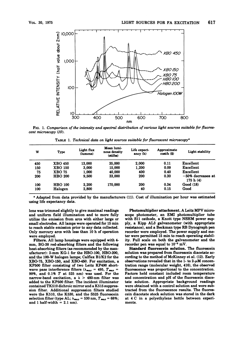
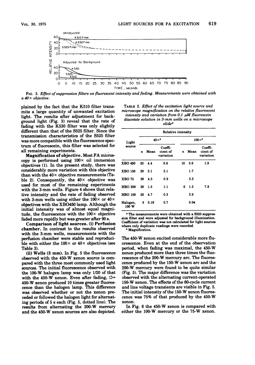
A variety of fluorescent excitation light sources were compared using a standard fluorescein solution or a bacterial conjugate with immunofluorescent microscopy. Quantitative data were obtained with microscope photometric apparatus. Both the quantitative data and comparative conjugate titering suggest that the 450-W xenon arc excited significantly more fluorescence than did the more commonly used 250-W mercury arc or the 100-W halogen lamp. The conjugate could be diluted 4 to 32 times more using the 450-W xenon. Additional advantages of 450-W xenon excitation include sufficient energy of wavelengths between 470 to 490 mm, thus permitting narrow-band excitation resulting in less autofluorescence and the ability to perform fluorescent-antibody procedures without the darkening of ambient room light.
Full text
PDF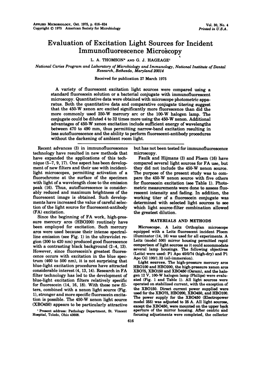
Selected References
These references are in PubMed. This may not be the complete list of references from this article.
- Faulk W. P., Hijmans W. Recent developments in immunofluorescence. Prog Allergy. 1972;16:9–39. doi: 10.1159/000393067. [DOI] [PubMed] [Google Scholar]
- Hijmans W., Schuit H. R. Immunofluorescence studies on immunoglobulins in the lymphoid cells of human peripheral blood. Clin Exp Immunol. 1972 Aug;11(4):483–494. [PMC free article] [PubMed] [Google Scholar]
- Hijmans W., Schuit H. R., Teiko Y., Schechter I. An immunofluorescence study on the specificity of antibodies synthesized in separate cells after the administration of an immunogen with double specificity. Eur J Immunol. 1972 Feb;2(1):1–4. doi: 10.1002/eji.1830020102. [DOI] [PubMed] [Google Scholar]
- Jongsma A. P., Hijmans W., Ploem J. S. Quantitative immunofluorescence. Standardization and calibration in microfluorometry. Histochemie. 1971;25(4):329–343. doi: 10.1007/BF00278226. [DOI] [PubMed] [Google Scholar]
- Ploem J. S. A study of filters and light sources in immunofluorescence microscopy. Ann N Y Acad Sci. 1971 Jun 21;177:414–429. doi: 10.1111/j.1749-6632.1971.tb35070.x. [DOI] [PubMed] [Google Scholar]
- Ploem J. S. The use of a vertical illuminator with interchangeable dichroic mirrors for fluorescence microscopy with incidental light. Z Wiss Mikrosk. 1967 Nov;68(3):129–142. [PubMed] [Google Scholar]
- van Boxtel C. J., Engelfriet C. P., Feltkamp T. E. Immunofluorescence microphotometry for the detection of platelet antibodies. I. Standardization of the method. Scand J Immunol. 1973;2(3):217–229. doi: 10.1111/j.1365-3083.1973.tb02032.x. [DOI] [PubMed] [Google Scholar]
- van der Ploeg M., Ploem J. S. Filter combinations and light sources for fluorescence microscopy of quinacrine mustard or quinacrine stained chromosomes. Histochemie. 1973;33(1):61–70. doi: 10.1007/BF00304227. [DOI] [PubMed] [Google Scholar]


