Abstract
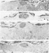
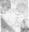
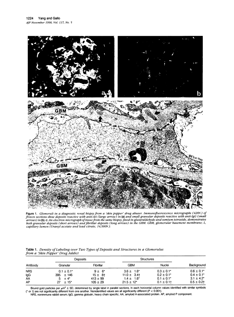
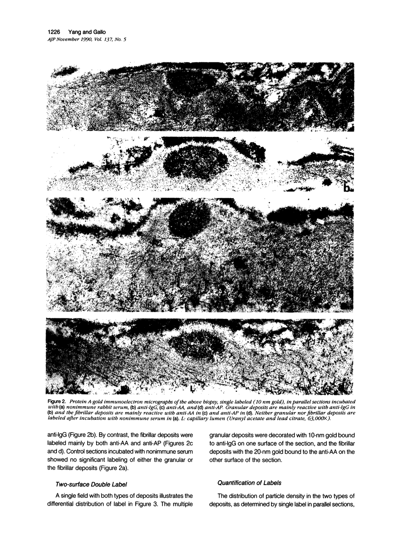
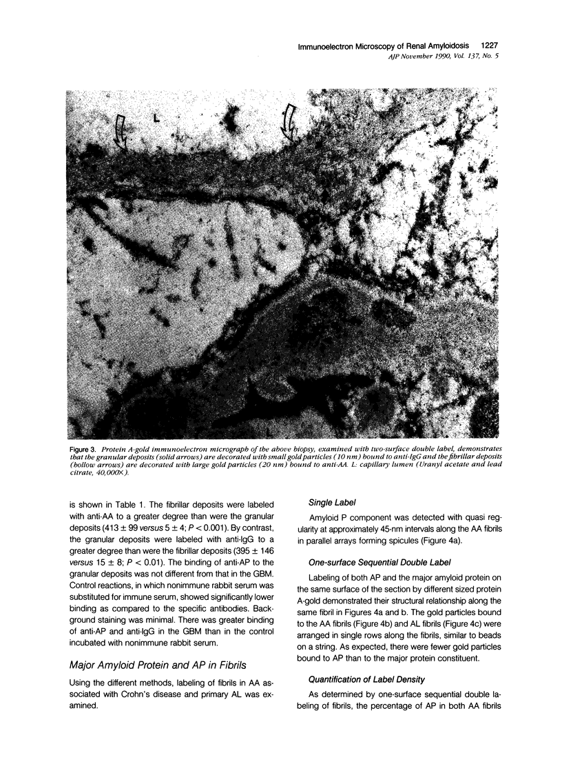
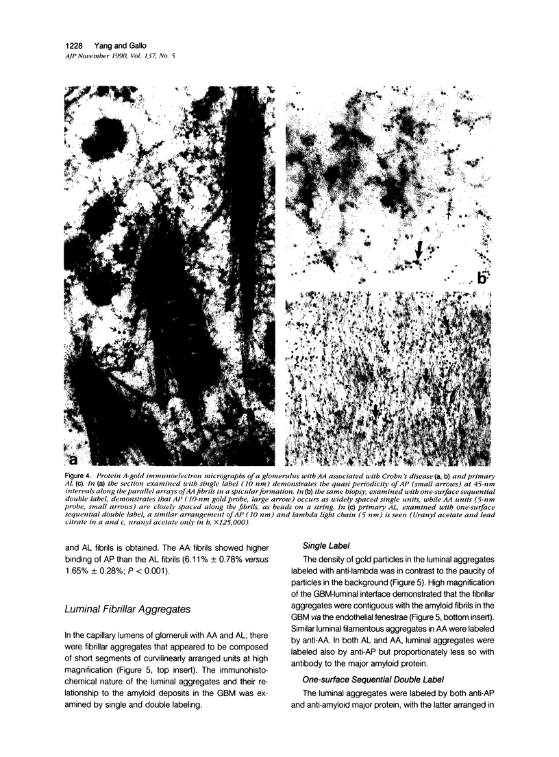
Glomeruli of archival renal biopsies, stored frozen at -70 degrees C, from three patients with amyloid were examined by protein A-gold immunoelectron microscopy. In one with both fibrillar and granular deposits from a 'skin popper' drug abuser, the granular deposits were labeled with anti-IgG, while the fibrillar deposits were labeled with anti-amyloid-A (AA) protein and amyloid P component (AP), suggesting coexisting immune complex disease and AA due to different, but possibly related, pathogenesis. In studies using double-label immunostaining of primary amyloidosis-lambda light chain type (AL) and AA associated with Crohn's disease, AP occurred as widely separated single units along the amyloid fibrils and represented 1.5% and 6.5% of the total gold label in AL and AA, respectively, while the major fibril protein was labeled in single rows, similar to beads on a string. Fibrillar aggregates in the capillary lumens were labeled similarly by antisera to the major protein and AP and appeared to be contiguous with the fibrillar deposits at the glomerular basement membrane (GBM)-luminal interface, suggesting intravascular fibrillogenesis.
Full text
PDF
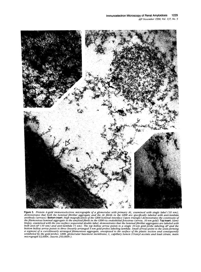
Images in this article
Selected References
These references are in PubMed. This may not be the complete list of references from this article.
- Bendayan M. Double immunocytochemical labeling applying the protein A-gold technique. J Histochem Cytochem. 1982 Jan;30(1):81–85. doi: 10.1177/30.1.6172469. [DOI] [PubMed] [Google Scholar]
- Gallo G. R., Feiner H. D., Chuba J. V., Beneck D., Marion P., Cohen D. H. Characterization of tissue amyloid by immunofluorescence microscopy. Clin Immunol Immunopathol. 1986 Jun;39(3):479–490. doi: 10.1016/0090-1229(86)90175-3. [DOI] [PubMed] [Google Scholar]
- Herrera G. A. Ultrastructural postembedding immunogold labeling: applications to diagnostic pathology. Ultrastruct Pathol. 1989 Sep-Dec;13(5-6):485–499. doi: 10.3109/01913128909074532. [DOI] [PubMed] [Google Scholar]
- Kraehenbuhl J. P., Racine L., Griffiths G. W. Attempts to quantitate immunocytochemistry at the electron microscope level. Histochem J. 1980 May;12(3):317–332. doi: 10.1007/BF01006953. [DOI] [PubMed] [Google Scholar]
- Menchel S., Cohen D., Gross E., Frangione B., Gallo G. AA protein-related renal amyloidosis in drug addicts. Am J Pathol. 1983 Aug;112(2):195–199. [PMC free article] [PubMed] [Google Scholar]
- Roth J., Heitz P. U. Immunolabeling with the protein A-gold technique: an overview. Ultrastruct Pathol. 1989 Sep-Dec;13(5-6):467–484. doi: 10.3109/01913128909074531. [DOI] [PubMed] [Google Scholar]
- Roth J. The preparation of protein A-gold complexes with 3 nm and 15nm gold particles and their use in labelling multiple antigens on ultra-thin sections. Histochem J. 1982 Sep;14(5):791–801. doi: 10.1007/BF01033628. [DOI] [PubMed] [Google Scholar]
- Shirahama T., Cohen A. S. Fine structure of the glomerulus in human and experimental renal amyloidosis. Am J Pathol. 1967 Nov;51(5):869–911. [PMC free article] [PubMed] [Google Scholar]
- Shore I., Moss J. The use of formvar films on both sides of a section to facilitate the selected surface technique for double immunostaining at the electron microscope level. Histochem J. 1988 Mar;20(3):183–184. doi: 10.1007/BF01746682. [DOI] [PubMed] [Google Scholar]
- Skinner M., Shirahama T., Benson M. D., Cohen A. S. Murine amyloid protein AA in casein-induced experimental amyloidosis. Lab Invest. 1977 Apr;36(4):420–427. [PubMed] [Google Scholar]
- Tagliavini F., Giaccone G., Frangione B., Bugiani O. Preamyloid deposits in the cerebral cortex of patients with Alzheimer's disease and nondemented individuals. Neurosci Lett. 1988 Nov 11;93(2-3):191–196. doi: 10.1016/0304-3940(88)90080-8. [DOI] [PubMed] [Google Scholar]
- Verga L., Frangione B., Tagliavini F., Giaccone G., Migheli A., Bugiani O. Alzheimer patients and Down patients: cerebral preamyloid deposits differ ultrastructurally and histochemically from the amyloid of senile plaques. Neurosci Lett. 1989 Nov 6;105(3):294–299. doi: 10.1016/0304-3940(89)90636-8. [DOI] [PubMed] [Google Scholar]
- Yang G. C., Morrison A. B. Wide-field electron microscopy. A rapid method for the study of histologic material that provides a bridge between light and electron microscopy. Am J Clin Pathol. 1975 Nov;64(5):648–654. doi: 10.1093/ajcp/64.5.648. [DOI] [PubMed] [Google Scholar]


