Abstract
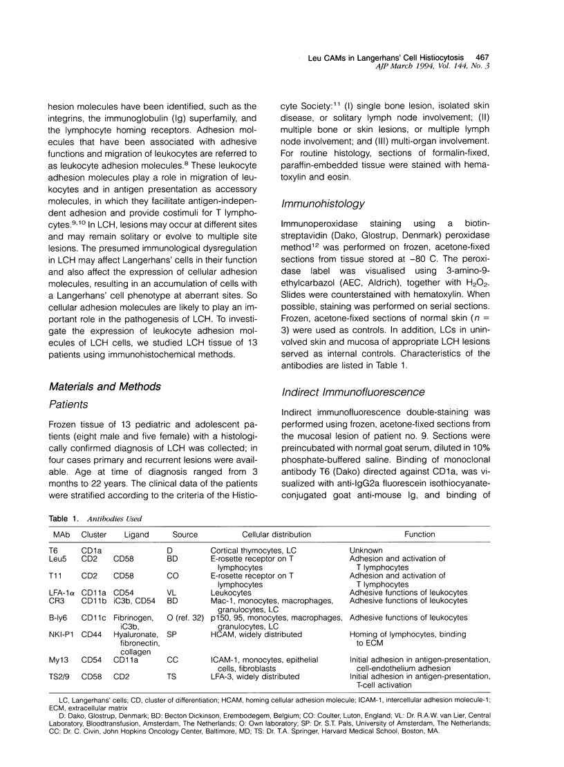
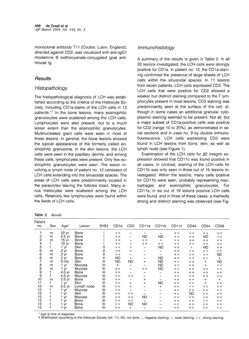
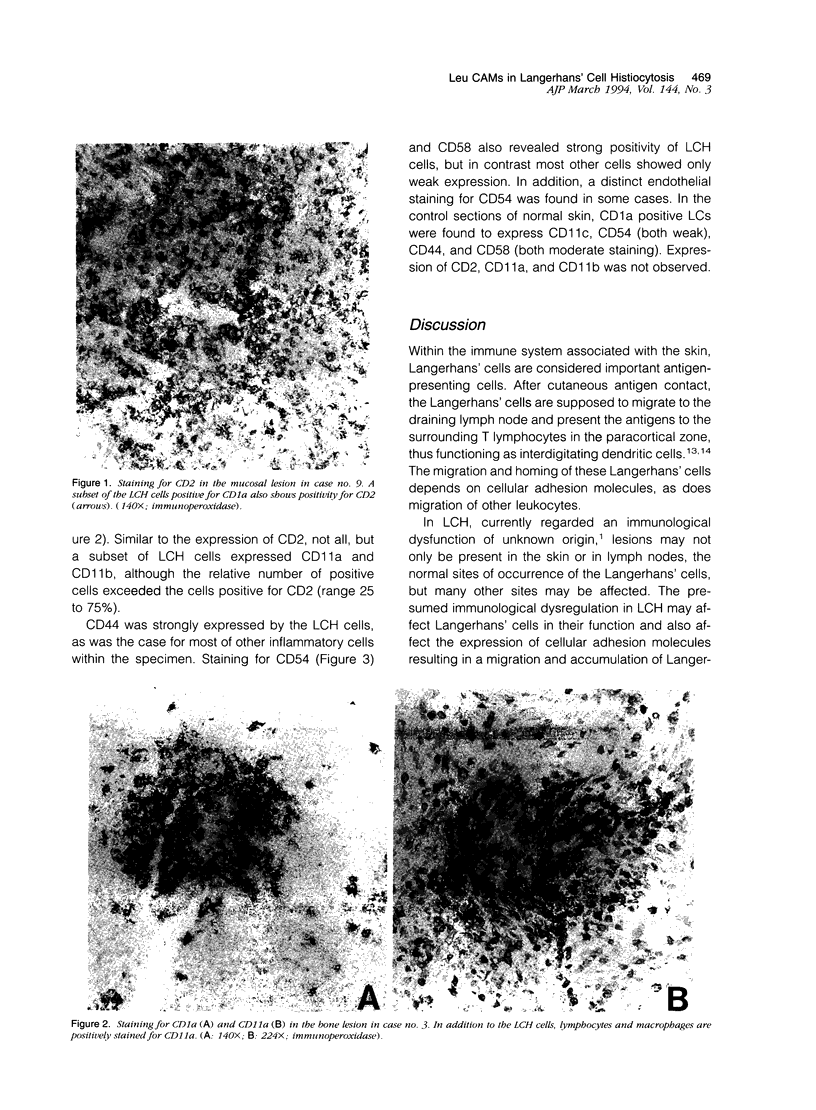
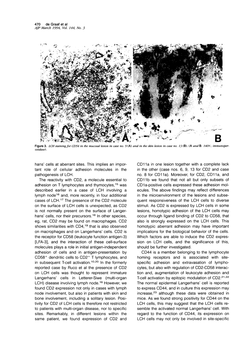
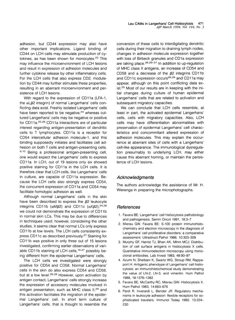
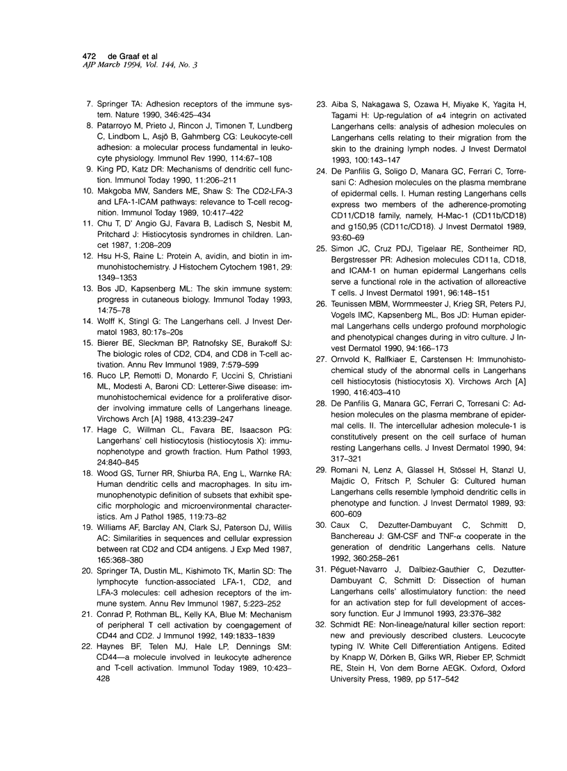
Langerhans' cell histiocytosis (LCH) is characterized by an accumulation of cells with a Langerhans' cell (LC) phenotype. Most patients present with solitary skin or bone lesions, but multi-organ lesions may appear. Twenty-two LCH-tissue sections from 13 children and adolescents, with lesions at different sites, were investigated for the expression of leukocyte cellular adhesion molecules. Surprisingly, the LCH cells showed expression for CD2 in 11 lesions. Staining of LCH cells for CD11a and CD11b was positive in six and three lesions, respectively. Staining for CD11c, CD44, CD54, and CD58 was found consistently positive in all lesions. The strong reactivity for CD54 (intercellular adhesion molecule-1) and CD58 (leukocyte function antigen-3) is in contrast with the epidermal LC. LCs in culture are known to up-regulate the expression of CD54 and CD58. These changes are thought to reflect the in vivo situation during migration of activated LCs from the skin to the draining lymph node. It can be concluded that the abnormal cells in LCH not only share characteristics with the epidermal LC, but have additional characteristics of the activated LC, a cell capable of migration. The presumed immunological dysregulation in LCH may affect the expression of cellular adhesion molecules, reflected by the inconsistent expression of CD11a and CD11b and the unexpected expression of CD2. These features may contribute to migration of LCs to aberrant sites in combination with abnormal persistence and proliferation.
Full text
PDF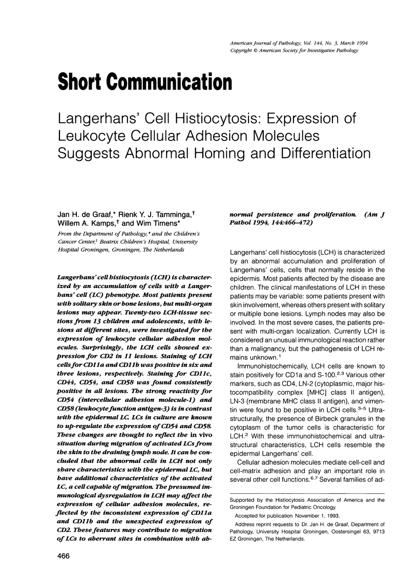
Images in this article
Selected References
These references are in PubMed. This may not be the complete list of references from this article.
- Aiba S., Nakagawa S., Ozawa H., Miyake K., Yagita H., Tagami H. Up-regulation of alpha 4 integrin on activated Langerhans cells: analysis of adhesion molecules on Langerhans cells relating to their migration from skin to draining lymph nodes. J Invest Dermatol. 1993 Feb;100(2):143–147. doi: 10.1111/1523-1747.ep12462783. [DOI] [PubMed] [Google Scholar]
- Azumi N., Sheibani K., Swartz W. G., Stroup R. M., Rappaport H. Antigenic phenotype of Langerhans cell histiocytosis: an immunohistochemical study demonstrating the value of LN-2, LN-3, and vimentin. Hum Pathol. 1988 Dec;19(12):1376–1382. doi: 10.1016/s0046-8177(88)80229-6. [DOI] [PubMed] [Google Scholar]
- Bierer B. E., Sleckman B. P., Ratnofsky S. E., Burakoff S. J. The biologic roles of CD2, CD4, and CD8 in T-cell activation. Annu Rev Immunol. 1989;7:579–599. doi: 10.1146/annurev.iy.07.040189.003051. [DOI] [PubMed] [Google Scholar]
- Bos J. D., Kapsenberg M. L. The skin immune system: progress in cutaneous biology. Immunol Today. 1993 Feb;14(2):75–78. doi: 10.1016/0167-5699(93)90062-P. [DOI] [PubMed] [Google Scholar]
- Caux C., Dezutter-Dambuyant C., Schmitt D., Banchereau J. GM-CSF and TNF-alpha cooperate in the generation of dendritic Langerhans cells. Nature. 1992 Nov 19;360(6401):258–261. doi: 10.1038/360258a0. [DOI] [PubMed] [Google Scholar]
- Conrad P., Rothman B. L., Kelley K. A., Blue M. L. Mechanism of peripheral T cell activation by coengagement of CD44 and CD2. J Immunol. 1992 Sep 15;149(6):1833–1839. [PubMed] [Google Scholar]
- De Panfilis G., Manara G. C., Ferrari C., Torresani C. Adhesion molecules on the plasma membrane of epidermal cells. II. The intercellular adhesion molecule-1 is constitutively present on the cell surface of human resting Langerhans cells. J Invest Dermatol. 1990 Mar;94(3):317–321. doi: 10.1111/1523-1747.ep12874444. [DOI] [PubMed] [Google Scholar]
- De Panfilis G., Soligo D., Manara G. C., Ferrari C., Torresani C. Adhesion molecules on the plasma membrane of epidermal cells. I. Human resting Langerhans cells express two members of the adherence-promoting CD11/CD18 family, namely, H-Mac-1 (CD11b/CD18) and gp 150,95 (CD11c/CD18). J Invest Dermatol. 1989 Jul;93(1):60–69. doi: 10.1111/1523-1747.ep12277352. [DOI] [PubMed] [Google Scholar]
- Favara B. E. Langerhans' cell histiocytosis pathobiology and pathogenesis. Semin Oncol. 1991 Feb;18(1):3–7. [PubMed] [Google Scholar]
- Favara B. E., McCarthy R. C., Mierau G. W. Histiocytosis X. Hum Pathol. 1983 Aug;14(8):663–676. doi: 10.1016/s0046-8177(83)80138-5. [DOI] [PubMed] [Google Scholar]
- Hage C., Willman C. L., Favara B. E., Isaacson P. G. Langerhans' cell histiocytosis (histiocytosis X): immunophenotype and growth fraction. Hum Pathol. 1993 Aug;24(8):840–845. doi: 10.1016/0046-8177(93)90133-2. [DOI] [PubMed] [Google Scholar]
- Haynes B. F., Telen M. J., Hale L. P., Denning S. M. CD44--a molecule involved in leukocyte adherence and T-cell activation. Immunol Today. 1989 Dec;10(12):423–428. doi: 10.1016/0167-5699(89)90040-6. [DOI] [PubMed] [Google Scholar]
- Hsu S. M., Raine L. Protein A, avidin, and biotin in immunohistochemistry. J Histochem Cytochem. 1981 Nov;29(11):1349–1353. doi: 10.1177/29.11.6172466. [DOI] [PubMed] [Google Scholar]
- King P. D., Katz D. R. Mechanisms of dendritic cell function. Immunol Today. 1990 Jun;11(6):206–211. doi: 10.1016/0167-5699(90)90084-m. [DOI] [PubMed] [Google Scholar]
- Makgoba M. W., Sanders M. E., Shaw S. The CD2-LFA-3 and LFA-1-ICAM pathways: relevance to T-cell recognition. Immunol Today. 1989 Dec;10(12):417–422. doi: 10.1016/0167-5699(89)90039-X. [DOI] [PubMed] [Google Scholar]
- Mierau G. W., Favara B. E. S-100 protein immunohistochemistry and electron microscopy in the diagnosis of Langerhans cell proliferative disorders: a comparative assessment. Ultrastruct Pathol. 1986;10(4):303–309. doi: 10.3109/01913128609064194. [DOI] [PubMed] [Google Scholar]
- Murphy G. F., Harrist T. J., Bhan A. K., Mihm M. C., Jr Distribution of cell surface antigens in histiocytosis X cells. Quantitative immunoelectron microscopy using monoclonal antibodies. Lab Invest. 1983 Jan;48(1):90–97. [PubMed] [Google Scholar]
- Ornvold K., Ralfkiaer E., Carstensen H. Immunohistochemical study of the abnormal cells in Langerhans cell histiocytosis (histiocytosis x). Virchows Arch A Pathol Anat Histopathol. 1990;416(5):403–410. doi: 10.1007/BF01605145. [DOI] [PubMed] [Google Scholar]
- Pardi R., Inverardi L., Bender J. R. Regulatory mechanisms in leukocyte adhesion: flexible receptors for sophisticated travelers. Immunol Today. 1992 Jun;13(6):224–230. doi: 10.1016/0167-5699(92)90159-5. [DOI] [PubMed] [Google Scholar]
- Patarroyo M., Prieto J., Rincon J., Timonen T., Lundberg C., Lindbom L., Asjö B., Gahmberg C. G. Leukocyte-cell adhesion: a molecular process fundamental in leukocyte physiology. Immunol Rev. 1990 Apr;114:67–108. doi: 10.1111/j.1600-065x.1990.tb00562.x. [DOI] [PubMed] [Google Scholar]
- Péguet-Navarro J., Dalbiez-Gauthier C., Dezutter-Dambuyant C., Schmitt D. Dissection of human Langerhans cells' allostimulatory function: the need for an activation step for full development of accessory function. Eur J Immunol. 1993 Feb;23(2):376–382. doi: 10.1002/eji.1830230212. [DOI] [PubMed] [Google Scholar]
- Romani N., Lenz A., Glassel H., Stössel H., Stanzl U., Majdic O., Fritsch P., Schuler G. Cultured human Langerhans cells resemble lymphoid dendritic cells in phenotype and function. J Invest Dermatol. 1989 Nov;93(5):600–609. doi: 10.1111/1523-1747.ep12319727. [DOI] [PubMed] [Google Scholar]
- Ruco L. P., Remotti D., Monardo F., Uccini S., Cristiani M. L., Modesti A., Baroni C. D. Letterer-Siwe disease: immunohistochemical evidence for a proliferative disorder involving immature cells of Langerhans lineage. Virchows Arch A Pathol Anat Histopathol. 1988;413(3):239–247. doi: 10.1007/BF00718616. [DOI] [PubMed] [Google Scholar]
- Simon J. C., Cruz P. D., Jr, Tigelaar R. E., Sontheimer R. D., Bergstresser P. R. Adhesion molecules CD11a, CD18, and ICAM-1 on human epidermal Langerhans cells serve a functional role in the activation of alloreactive T cells. J Invest Dermatol. 1991 Jan;96(1):148–151. doi: 10.1111/1523-1747.ep12515946. [DOI] [PubMed] [Google Scholar]
- Springer T. A. Adhesion receptors of the immune system. Nature. 1990 Aug 2;346(6283):425–434. doi: 10.1038/346425a0. [DOI] [PubMed] [Google Scholar]
- Springer T. A., Dustin M. L., Kishimoto T. K., Marlin S. D. The lymphocyte function-associated LFA-1, CD2, and LFA-3 molecules: cell adhesion receptors of the immune system. Annu Rev Immunol. 1987;5:223–252. doi: 10.1146/annurev.iy.05.040187.001255. [DOI] [PubMed] [Google Scholar]
- Teunissen M. B., Wormmeester J., Krieg S. R., Peters P. J., Vogels I. M., Kapsenberg M. L., Bos J. D. Human epidermal Langerhans cells undergo profound morphologic and phenotypical changes during in vitro culture. J Invest Dermatol. 1990 Feb;94(2):166–173. doi: 10.1111/1523-1747.ep12874439. [DOI] [PubMed] [Google Scholar]
- Williams A. F., Barclay A. N., Clark S. J., Paterson D. J., Willis A. C. Similarities in sequences and cellular expression between rat CD2 and CD4 antigens. J Exp Med. 1987 Feb 1;165(2):368–380. doi: 10.1084/jem.165.2.368. [DOI] [PMC free article] [PubMed] [Google Scholar]
- Wolff K., Stingl G. The Langerhans cell. J Invest Dermatol. 1983 Jun;80 (Suppl):17s–21s. [PubMed] [Google Scholar]
- Wood G. S., Turner R. R., Shiurba R. A., Eng L., Warnke R. A. Human dendritic cells and macrophages. In situ immunophenotypic definition of subsets that exhibit specific morphologic and microenvironmental characteristics. Am J Pathol. 1985 Apr;119(1):73–82. [PMC free article] [PubMed] [Google Scholar]


