Abstract
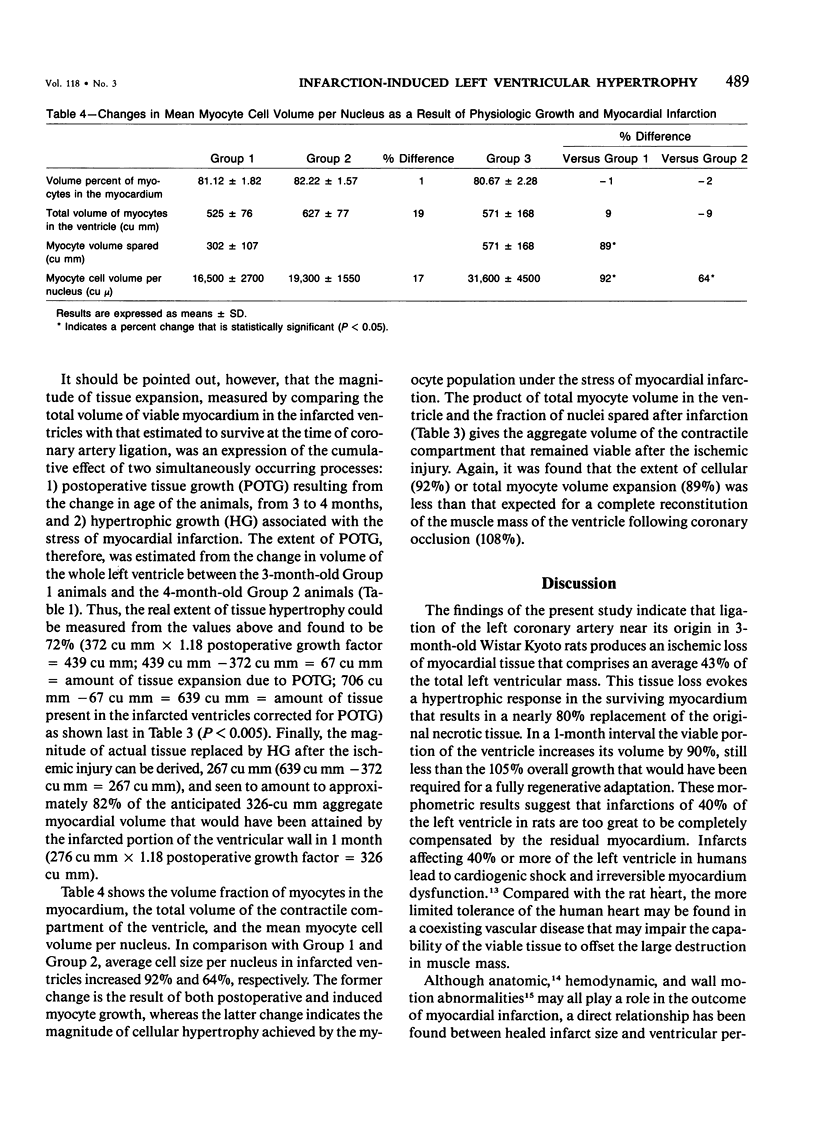
For determination of the effects of myocardial infarction on the recovery potential of muscle mass in the surviving tissue, ligation of the left coronary artery was performed in 3-month-old rats, and the infarcted ventricles were analyzed morphometrically a month after surgery. Comparisons were made with 4-month-old control rats that underwent sham operations and with 3-month-old control rats that were not operated upon for evaluation of the magnitude of infarct size and discrimination of the relative contribution of tissue growth that occurred in the surviving myocardium solely as a result of the change in age, from 3 to 4 months (postoperative tissue growth, or POTG), from the additional growth induced by infarction (hypertrophic growth, or HG). Coronary occlusion induced a 276-cu mm loss of ventricular tissue volume that corresponded to 43% of the total left ventricular mass, 648 cu mm. Over a 30-day period the remaining 372 cu mm of viable tissue expanded by 90% with an overall volume gain of 334 cu mm. This tissue augmentation consisted of 20% POTG, 67 cu mm, and 80% HG, 267 cu mm. Total myocyte volume increased 89%, from 302 cu mm to 571 cu mm, and average myocyte cell volume per nucleus increased 92%, from 16,500 cu mu to 31,600 cu mu. The expansion of the myocyte mass was the result of a 21% POTG and a 79% HG. Corresponding values for the myocyte population were 19% and 81%.
Full text
PDF
Images in this article
Selected References
These references are in PubMed. This may not be the complete list of references from this article.
- Anversa P., Beghi C., Levicky V., McDonald S. L., Kikkawa Y. Morphometry of right ventricular hypertrophy induced by strenuous exercise in rat. Am J Physiol. 1982 Dec;243(6):H856–H861. doi: 10.1152/ajpheart.1982.243.6.H856. [DOI] [PubMed] [Google Scholar]
- Anversa P., Beghi C., McDonald S. L., Levicky V., Kikkawa Y., Olivetti G. Morphometry of right ventricular hypertrophy induced by myocardial infarction in the rat. Am J Pathol. 1984 Sep;116(3):504–513. [PMC free article] [PubMed] [Google Scholar]
- Anversa P., Loud A. V., Giacomelli F., Wiener J. Absolute morphometric study of myocardial hypertrophy in experimental hypertension. II. Ultrastructure of myocytes and interstitium. Lab Invest. 1978 May;38(5):597–609. [PubMed] [Google Scholar]
- Anversa P., Olivetti G., Melissari M., Loud A. V. Stereological measurement of cellular and subcellular hypertrophy and hyperplasia in the papillary muscle of adult rat. J Mol Cell Cardiol. 1980 Aug;12(8):781–795. doi: 10.1016/0022-2828(80)90080-2. [DOI] [PubMed] [Google Scholar]
- Anversa P., Vitali-Mazza L., Visioli O., Marchetti G. Experimental cardiac hypertrophy: a quantitative ultrastructural study in the compensatory stage. J Mol Cell Cardiol. 1971 Dec;3(3):213–227. doi: 10.1016/0022-2828(71)90042-3. [DOI] [PubMed] [Google Scholar]
- Bishop S. P., Melsen L. R. Myocardial necrosis, fibrosis, and DNA synthesis in experimental cardiac hypertrophy induced by sudden pressure overload. Circ Res. 1976 Aug;39(2):238–245. doi: 10.1161/01.res.39.2.238. [DOI] [PubMed] [Google Scholar]
- Bishop S. P., Oparil S., Reynolds R. H., Drummond J. L. Regional myocyte size in normotensive and spontaneously hypertensive rats. Hypertension. 1979 Jul-Aug;1(4):378–383. doi: 10.1161/01.hyp.1.4.378. [DOI] [PubMed] [Google Scholar]
- Cosby R. S., Giddings J. A., See J. R., Mayo M. Late complications of myocardial infarction. Adv Cardiol. 1978;23:73–81. doi: 10.1159/000401075. [DOI] [PubMed] [Google Scholar]
- Feild B. J., Russell R. O., Jr, Dowling J. T., Rackley C. E. Regional left ventricular performance in the year following myocardial infarction. Circulation. 1972 Oct;46(4):679–689. doi: 10.1161/01.cir.46.4.679. [DOI] [PubMed] [Google Scholar]
- Fishbein M. C., Maclean D., Maroko P. R. Experimental myocardial infarction in the rat: qualitative and quantitative changes during pathologic evolution. Am J Pathol. 1978 Jan;90(1):57–70. [PMC free article] [PubMed] [Google Scholar]
- Fletcher P. J., Pfeffer J. M., Pfeffer M. A., Braunwald E. Left ventricular diastolic pressure-volume relations in rats with healed myocardial infarction. Effects on systolic function. Circ Res. 1981 Sep;49(3):618–626. doi: 10.1161/01.res.49.3.618. [DOI] [PubMed] [Google Scholar]
- Grove D., Zak R., Nair K. G., Aschenbrenner V. Biochemical correlates of cardiac hypertrophy. IV. Observations on the cellular organization of growth during myocardial hypertrophy in the rat. Circ Res. 1969 Oct;25(4):473–485. doi: 10.1161/01.res.25.4.473. [DOI] [PubMed] [Google Scholar]
- HORT W., DACANALIS S., JUST H. UNTERSUCHUNGEN BEI CHRONISCHEM EXPERIMENTELLEN HERZINFARKT DER RATTE. Arch Kreislaufforsch. 1964 Oct;44:288–299. doi: 10.1007/BF02119542. [DOI] [PubMed] [Google Scholar]
- Hochman J. S., Bulkley B. H. Expansion of acute myocardial infarction: an experimental study. Circulation. 1982 Jun;65(7):1446–1450. doi: 10.1161/01.cir.65.7.1446. [DOI] [PubMed] [Google Scholar]
- Hochman J. S., Bulkley B. H. Pathogenesis of left ventricular aneurysms: an experimental study in the rat model. Am J Cardiol. 1982 Jul;50(1):83–88. doi: 10.1016/0002-9149(82)90012-1. [DOI] [PubMed] [Google Scholar]
- JENNINGS R. B., SOMMERS H. M., KALTENBACH J. P., WEST J. J. ELECTROLYTE ALTERATIONS IN ACUTE MYOCARDIAL ISCHEMIC INJURY. Circ Res. 1964 Mar;14:260–269. doi: 10.1161/01.res.14.3.260. [DOI] [PubMed] [Google Scholar]
- Korecky B., Rakusan K. Normal and hypertrophic growth of the rat heart: changes in cell dimensions and number. Am J Physiol. 1978 Feb;234(2):H123–H128. doi: 10.1152/ajpheart.1978.234.2.H123. [DOI] [PubMed] [Google Scholar]
- Loud A. V., Anversa P. Morphometric analysis of biologic processes. Lab Invest. 1984 Mar;50(3):250–261. [PubMed] [Google Scholar]
- Morkin E., Ashford T. P. Myocardial DNA synthesis in experimental cardiac hypertrophy. Am J Physiol. 1968 Dec;215(6):1409–1413. doi: 10.1152/ajplegacy.1968.215.6.1409. [DOI] [PubMed] [Google Scholar]
- Oberpriller J. O., Ferrans V. J., Carroll R. J. Changes in DNA content, number of nuclei and cellular dimensions of young rat atrial myocytes in response to left coronary artery ligation. J Mol Cell Cardiol. 1983 Jan;15(1):31–42. doi: 10.1016/0022-2828(83)90305-x. [DOI] [PubMed] [Google Scholar]
- Page D. L., Caulfield J. B., Kastor J. A., DeSanctis R. W., Sanders C. A. Myocardial changes associated with cardiogenic shock. N Engl J Med. 1971 Jul 15;285(3):133–137. doi: 10.1056/NEJM197107152850301. [DOI] [PubMed] [Google Scholar]
- Page E., McCallister L. P. Quantitative electron microscopic description of heart muscle cells. Application to normal, hypertrophied and thyroxin-stimulated hearts. Am J Cardiol. 1973 Feb;31(2):172–181. doi: 10.1016/0002-9149(73)91030-8. [DOI] [PubMed] [Google Scholar]
- Pfeffer M. A., Pfeffer J. M., Fishbein M. C., Fletcher P. J., Spadaro J., Kloner R. A., Braunwald E. Myocardial infarct size and ventricular function in rats. Circ Res. 1979 Apr;44(4):503–512. doi: 10.1161/01.res.44.4.503. [DOI] [PubMed] [Google Scholar]
- Polimeni P. I., Al-Sadir J. Expansion of extracellular space in the nonischemic zone of the infarcted heart and concomitant changes in tissue electrolyte contents in the rat. Circ Res. 1975 Dec;37(6):725–732. doi: 10.1161/01.res.37.6.725. [DOI] [PubMed] [Google Scholar]
- Roberts C. S., Maclean D., Braunwald E., Maroko P. R., Kloner R. A. Topographic changes in the left ventricle after experimentally induced myocardial infarction in the rat. Am J Cardiol. 1983 Mar 1;51(5):872–876. doi: 10.1016/s0002-9149(83)80147-7. [DOI] [PubMed] [Google Scholar]
- Rubin S. A., Fishbein M. C., Swan H. J. Compensatory hypertrophy in the heart after myocardial infarction in the rat. J Am Coll Cardiol. 1983 Jun;1(6):1435–1441. doi: 10.1016/s0735-1097(83)80046-1. [DOI] [PubMed] [Google Scholar]
- Rumyantsev P. P., Kassem A. M. Cumulative indices of DNA synthesizing myocytes in different compartments of the working myocardium and conductive system of the rat's heart muscle following extensive left ventricle infarction. Virchows Arch B Cell Pathol. 1976 May 26;20(4):329–342. doi: 10.1007/BF02890352. [DOI] [PubMed] [Google Scholar]
- SELYE H., BAJUSZ E., GRASSO S., MENDELL P. Simple techniques for the surgical occlusion of coronary vessels in the rat. Angiology. 1960 Oct;11:398–407. doi: 10.1177/000331976001100505. [DOI] [PubMed] [Google Scholar]
- Turek Z., Grandtner M., Kubát K., Ringnalda B. E., Kreuzer F. Arterial blood gases, muscle fiber diameter and intercapillary distance in cardiac hypertrophy of rats with an old myocardial infarction. Pflugers Arch. 1978 Sep 29;376(3):209–215. doi: 10.1007/BF00584952. [DOI] [PubMed] [Google Scholar]
- Vokonas P. S., Pirzada F., Hood W. B., Jr Experimental myocardial infarction: XII. Dynamic changes in segmental mechanical behavior of infarcted and non-infarcted myocardium. Am J Cardiol. 1976 May;37(6):853–859. doi: 10.1016/0002-9149(76)90109-0. [DOI] [PubMed] [Google Scholar]
- Wallenstein S., Zucker C. L., Fleiss J. L. Some statistical methods useful in circulation research. Circ Res. 1980 Jul;47(1):1–9. doi: 10.1161/01.res.47.1.1. [DOI] [PubMed] [Google Scholar]


