

Abstract
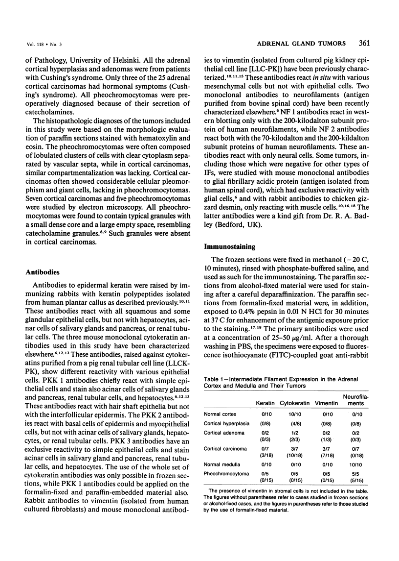
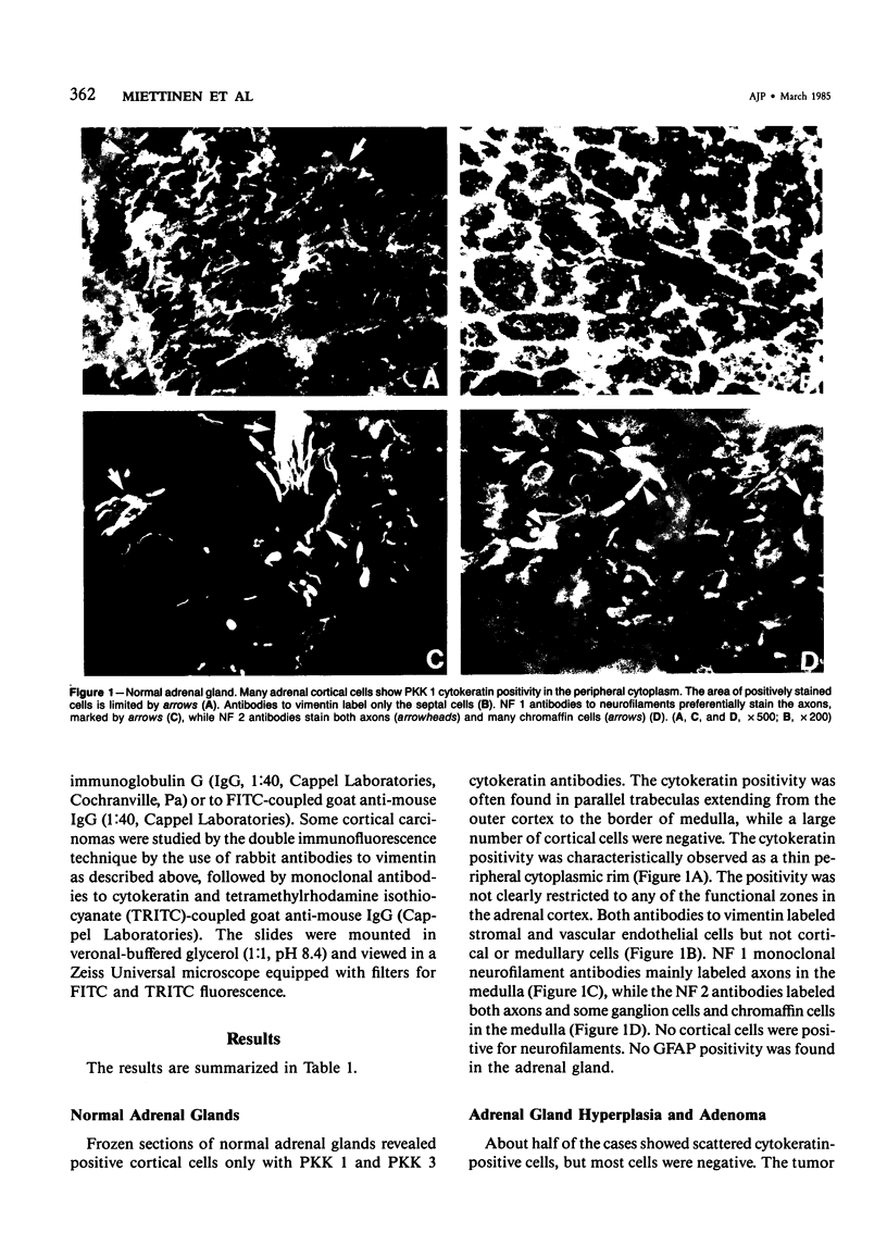
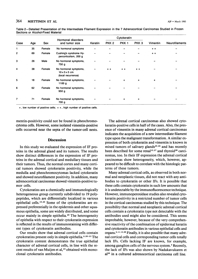
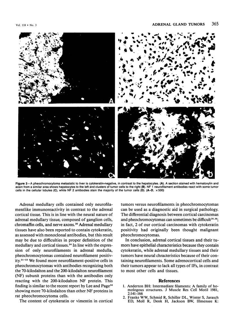
Normal adrenal glands (10 specimens) and adrenal gland tumors (58 cases) were immunohistochemically evaluated for different types of intermediate filament (IF) proteins. Some of the normal cortical cells showed cytokeratin positivity, and no positivity was seen for epidermal keratin or other types of IF. In the adrenal medulla, neurofilament positivity was seen in nerve axons, some ganglion cells, and chromaffin cells; and cytokeratin-positive cells could not be detected. Only the vascular and connective tissue elements showed vimentin positivity in both cortical and medullary areas. In half of the cortical carcinomas (13/25), cytokeratin-positive tumor cells were found. Furthermore, vimentin-positive tumor cells were present in 10 of 25 cases, in some of them together with cytokeratin-positive cells. Thus, the results show heterogeneity among the adrenal cortical carcinomas. Interestingly, many benign adrenal cortical tissues and some carcinomas lacked immunoreactivity for all types of IF, suggesting a poorly developed IF system in these tissues. In contrast to adrenal cortical tumors, pheochromocytomas contained neurofilamentlike immunoreactivity. These results reflect the different cellular nature of adrenal cortical and medullary tumors, which apparently can be distinguished from each other with antibodies to intermediate filament proteins.
Full text
PDF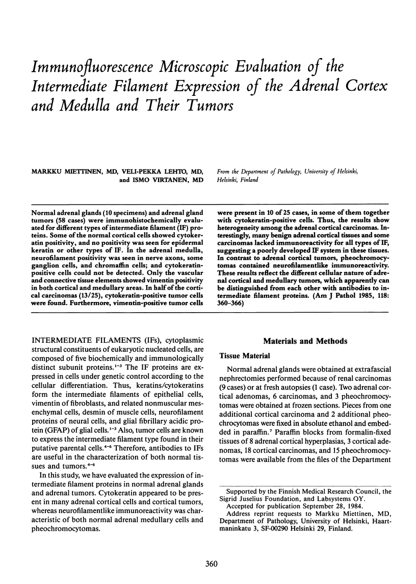
Images in this article
Selected References
These references are in PubMed. This may not be the complete list of references from this article.
- Altmannsberger M., Osborn M., Schauer A., Weber K. Antibodies to different intermediate filament proteins. Cell type-specific markers on paraffin-embedded human tissues. Lab Invest. 1981 Nov;45(5):427–434. [PubMed] [Google Scholar]
- Anderton B. H. Intermediate filaments: a family of homologous structures. J Muscle Res Cell Motil. 1981 Jun;2(2):141–166. doi: 10.1007/BF00711866. [DOI] [PubMed] [Google Scholar]
- Badley R. A., Woods A., Carruthers L., Rees D. A. Cytoskeleton changes in fibroblast adhesion and detachment. J Cell Sci. 1980 Jun;43:379–390. doi: 10.1242/jcs.43.1.379. [DOI] [PubMed] [Google Scholar]
- Brozman M. Immunohistochemical analysis of formaldehyde- and trypsin- or pepsin-treated material. Acta Histochem. 1978;63(2):251–260. doi: 10.1016/S0065-1281(78)80032-4. [DOI] [PubMed] [Google Scholar]
- Franke W. W., Appelhans B., Schmid E., Freudenstein C., Osborn M., Weber K. Identification and characterization of epithelial cells in mammalian tissues by immunofluorescence microscopy using antibodies to prekeratin. Differentiation. 1979;15(1):7–25. doi: 10.1111/j.1432-0436.1979.tb01030.x. [DOI] [PubMed] [Google Scholar]
- Franke W. W., Denk H., Kalt R., Schmid E. Biochemical and immunological identification of cytokeratin proteins present in hepatocytes of mammalian liver tissue. Exp Cell Res. 1981 Feb;131(2):299–318. doi: 10.1016/0014-4827(81)90234-2. [DOI] [PubMed] [Google Scholar]
- Franke W. W., Schiller D. L., Moll R., Winter S., Schmid E., Engelbrecht I., Denk H., Krepler R., Platzer B. Diversity of cytokeratins. Differentiation specific expression of cytokeratin polypeptides in epithelial cells and tissues. J Mol Biol. 1981 Dec 25;153(4):933–959. doi: 10.1016/0022-2836(81)90460-5. [DOI] [PubMed] [Google Scholar]
- Franke W. W., Schmid E., Schiller D. L., Winter S., Jarasch E. D., Moll R., Denk H., Jackson B. W., Illmensee K. Differentiation-related patterns of expression of proteins of intermediate-size filaments in tissues and cultured cells. Cold Spring Harb Symp Quant Biol. 1982;46(Pt 1):431–453. doi: 10.1101/sqb.1982.046.01.041. [DOI] [PubMed] [Google Scholar]
- Herman C. J., Moesker O., Kant A., Huysmans A., Vooijs G. P., Ramaekers F. C. Is renal cell (Grawitz) tumor a carcinosarcoma? Evidence from analysis of intermediate filament types. Virchows Arch B Cell Pathol Incl Mol Pathol. 1983;44(1):73–83. doi: 10.1007/BF02890161. [DOI] [PubMed] [Google Scholar]
- Holthöfer H., Miettinen A., Paasivuo R., Lehto V. P., Linder E., Alfthan O., Virtanen I. Cellular origin and differentiation of renal carcinomas. A fluorescence microscopic study with kidney-specific antibodies, antiintermediate filament antibodies, and lectins. Lab Invest. 1983 Sep;49(3):317–326. [PubMed] [Google Scholar]
- Kay S. Hyperplasia and neoplasia of the adrenal gland. Pathol Annu. 1976;11:103–139. [PubMed] [Google Scholar]
- Krepler R., Denk H., Artlieb U., Moll R. Immunocytochemistry of intermediate filament proteins present in pleomorphic adenomas of the human parotid gland: characterization of different cell types in the same tumor. Differentiation. 1982 May;21(3):191–199. doi: 10.1111/j.1432-0436.1982.tb01213.x. [DOI] [PubMed] [Google Scholar]
- Lazarides E. Intermediate filaments: a chemically heterogeneous, developmentally regulated class of proteins. Annu Rev Biochem. 1982;51:219–250. doi: 10.1146/annurev.bi.51.070182.001251. [DOI] [PubMed] [Google Scholar]
- Lee V. M., Page C. The dynamics of nerve growth factor-induced neurofilament and vimentin filament expression and organization in PC12 cells. J Neurosci. 1984 Jul;4(7):1705–1714. doi: 10.1523/JNEUROSCI.04-07-01705.1984. [DOI] [PMC free article] [PubMed] [Google Scholar]
- Lehto V. P., Virtanen I., Miettinen M., Dahl D., Kahri A. Neurofilaments in adrenal and extra-adrenal pheochromocytoma. Demonstration using immunofluorescence microscopy. Arch Pathol Lab Med. 1983 Sep;107(9):492–494. [PubMed] [Google Scholar]
- Lehtonen E., Lehto V. P., Paasivuo R., Virtanen I. Parietal and visceral endoderm differ in their expression of intermediate filaments. EMBO J. 1983;2(7):1023–1028. doi: 10.1002/j.1460-2075.1983.tb01540.x. [DOI] [PMC free article] [PubMed] [Google Scholar]
- Miettinen M., Lehto V. P., Badley R. A., Virtanen I. Alveolar rhabdomyosarcoma. Demonstration of the muscle type of intermediate filament protein, desmin, as a diagnostic aid. Am J Pathol. 1982 Aug;108(2):246–251. [PMC free article] [PubMed] [Google Scholar]
- Moll R., Franke W. W., Schiller D. L., Geiger B., Krepler R. The catalog of human cytokeratins: patterns of expression in normal epithelia, tumors and cultured cells. Cell. 1982 Nov;31(1):11–24. doi: 10.1016/0092-8674(82)90400-7. [DOI] [PubMed] [Google Scholar]
- O'Hare M. J., Monaghan P., Neville A. M. The pathology of adrenocortical neoplasia: a correlated structural and functional approach to the diagnosis of malignant disease. Hum Pathol. 1979 Mar;10(2):137–154. doi: 10.1016/s0046-8177(79)80004-0. [DOI] [PubMed] [Google Scholar]
- Osborn M., Altmannsberger M., Shaw G., Schauer A., Weber K. Various sympathetic derived human tumors differ in neurofilament expression. Use in diagnosis of neuroblastoma, ganglioneuroblastoma and pheochromocytoma. Virchows Arch B Cell Pathol Incl Mol Pathol. 1982 Aug;40(2):141–156. doi: 10.1007/BF02932859. [DOI] [PubMed] [Google Scholar]
- Osborn M., Weber K. Tumor diagnosis by intermediate filament typing: a novel tool for surgical pathology. Lab Invest. 1983 Apr;48(4):372–394. [PubMed] [Google Scholar]
- Ramaekers F. C., Puts J. J., Moesker O., Kant A., Huysmans A., Haag D., Jap P. H., Herman C. J., Vooijs G. P. Antibodies to intermediate filament proteins in the immunohistochemical identification of human tumours: an overview. Histochem J. 1983 Jul;15(7):691–713. doi: 10.1007/BF01002988. [DOI] [PubMed] [Google Scholar]
- Ramaekers F., Huysmans A., Moesker O., Kant A., Jap P., Herman C., Vooijs P. Monoclonal antibody to keratin filaments, specific for glandular epithelia and their tumors. Use in surgical pathology. Lab Invest. 1983 Sep;49(3):353–361. [PubMed] [Google Scholar]
- Sun T. T., Shih C., Green H. Keratin cytoskeletons in epithelial cells of internal organs. Proc Natl Acad Sci U S A. 1979 Jun;76(6):2813–2817. doi: 10.1073/pnas.76.6.2813. [DOI] [PMC free article] [PubMed] [Google Scholar]
- Trojanowski J. Q., Lee V. M., Schlaepfer W. W. An immunohistochemical study of human central and peripheral nervous system tumors, using monoclonal antibodies against neurofilaments and glial filaments. Hum Pathol. 1984 Mar;15(3):248–257. doi: 10.1016/s0046-8177(84)80188-4. [DOI] [PubMed] [Google Scholar]
- Virtanen I., Lehto V. P., Lehtonen E., Vartio T., Stenman S., Kurki P., Wager O., Small J. V., Dahl D., Badley R. A. Expression of intermediate filaments in cultured cells. J Cell Sci. 1981 Aug;50:45–63. doi: 10.1242/jcs.50.1.45. [DOI] [PubMed] [Google Scholar]
- Virtanen I., von Koskull H., Lehto V. P., Vartio T., Aula P. Cultured human amniotic fluid cells characterized with antibodies against intermediate filaments in indirect immunofluorescence microscopy. J Clin Invest. 1981 Nov;68(5):1348–1355. doi: 10.1172/JCI110382. [DOI] [PMC free article] [PubMed] [Google Scholar]
- van Muijen G. N., Ruiter D. J., Ponec M., Huiskens-van der, Mey C., Warnaar S. O. Monoclonal antibodies with different specificities against cytokeratins. An immunohistochemical study of normal tissues and tumors. Am J Pathol. 1984 Jan;114(1):9–17. [PMC free article] [PubMed] [Google Scholar]