Abstract
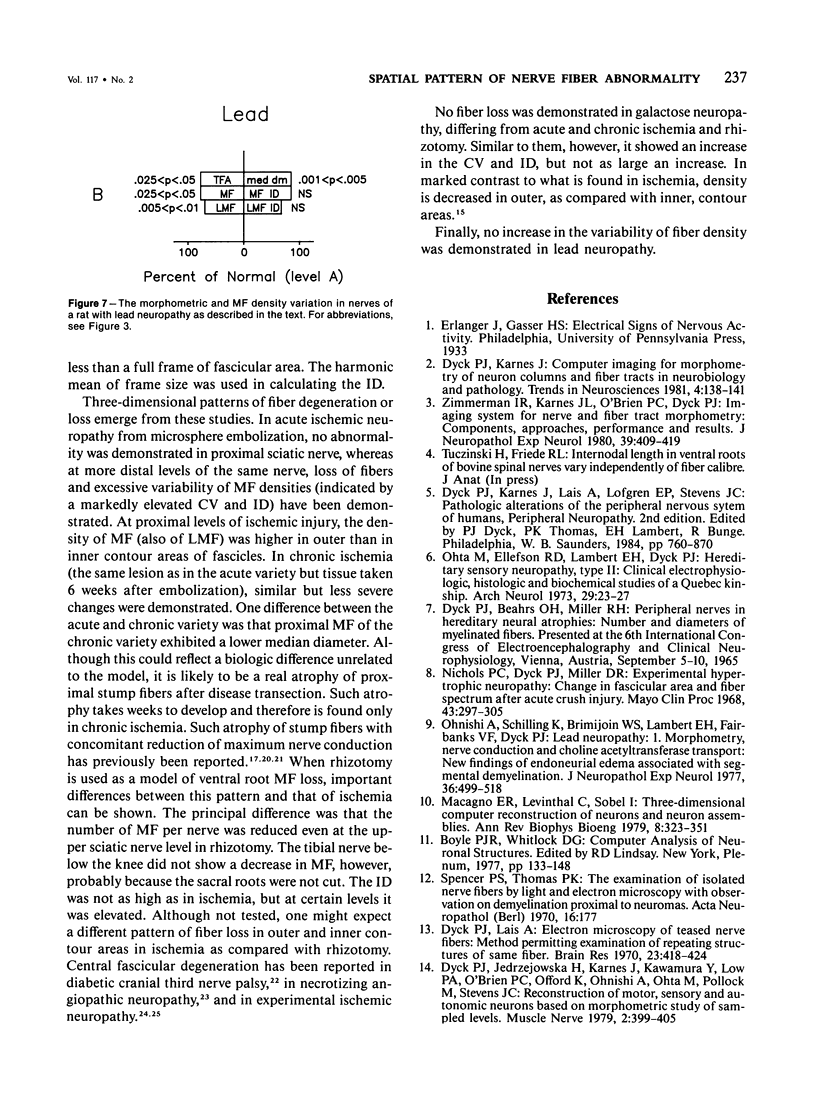
Estimates of the number, density, and size distribution of myelinated fibers at selected levels of roots, spinal tracts, and sampled levels of peripheral nerves may be used in the detection and characterization of alterations of motor, sensory, and autonomic neurons and their axons with development, aging and disease. Use of imaging techniques, now available, increases the reliability, versatility, and speed of such analysis. In this study, the authors evaluated the spatial pattern of fibers in sampled frames and contour areas of transverse sections of nerve fascicles, utilizing, the coefficient of variation and index of dispersion (ID), the latter extensively employed by plant ecologists. The ID was used for recognization of increased, normal, or decreased variability of density within fascicles, between fascicles, and between nerves in health and in various experimental neuropathies. In addition, various morphometric measurements were made in transverse sections at defined levels along the hind limb nerves of rats in acute and chronic ischemia, after rhizotomy and in galactose neuropathy. These stereomorphometric studies, emphasizing the number, size, shape, and spatial pattern of fibers, revealed differences among experimental neuropathies and may be found to be helpful in the characterization and prediction of pathologic mechanisms in neuropathies of unknown cause. Specifically, these approaches could be used for study of whether fiber loss in human diabetic neuropathy is multifocal and determination of the levels of such losses.
Full text
PDF
Selected References
These references are in PubMed. This may not be the complete list of references from this article.
- CRAGG B. G., THOMAS P. K. Changes in conduction velocity and fibre size proximal to peripheral nerve lesions. J Physiol. 1961 Jul;157:315–327. doi: 10.1113/jphysiol.1961.sp006724. [DOI] [PMC free article] [PubMed] [Google Scholar]
- DREYFUS P. M., HAKIM S., ADAMS R. D. Diabetic ophthalmoplegia; report of case, with postmortem study and comments on vascular supply of human oculomotor nerve. AMA Arch Neurol Psychiatry. 1957 Apr;77(4):337–349. [PubMed] [Google Scholar]
- Dyck P. J., Conn D. L., Okazaki H. Necrotizing angiopathic neuropathy. Three-dimensional morphology of fiber degeneration related to sites of occluded vessels. Mayo Clin Proc. 1972 Jul;47(7):461–475. [PubMed] [Google Scholar]
- Dyck P. J., Jedrzejowska H., Karnes J., Kawamura Y., Low P. A., O'Brien P. C., Offord K., Ohnishi A., Ohta M., Pollock M. Reconstruction of motor, sensory, and autonomic neurons based on morphometric study of sampled levels. Muscle Nerve. 1979 Sep-Oct;2(5):399–405. doi: 10.1002/mus.880020513. [DOI] [PubMed] [Google Scholar]
- Dyck P. J., Lais A. C. Electron microscopy of teased nerve fibers: method permitting examination of repeating structures of same fiber. Brain Res. 1970 Oct 28;23(3):418–424. doi: 10.1016/0006-8993(70)90067-3. [DOI] [PubMed] [Google Scholar]
- Gutmann E., Sanders F. K. Recovery of fibre numbers and diameters in the regeneration of peripheral nerves. J Physiol. 1943 Mar 25;101(4):489–518. doi: 10.1113/jphysiol.1943.sp004002. [DOI] [PMC free article] [PubMed] [Google Scholar]
- Korthals J. K., Korthals M. A., Wisniewski H. M. Peripheral nerve ischemia: Part 2. Accumulation of organelles. Ann Neurol. 1978 Dec;4(6):487–498. doi: 10.1002/ana.410040603. [DOI] [PubMed] [Google Scholar]
- Korthals J. K., Wiśniewski H. M. Peripheral nerve ischemia. Part 1. Experimental model. J Neurol Sci. 1975 Jan;24(1):65–76. doi: 10.1016/0022-510x(75)90009-x. [DOI] [PubMed] [Google Scholar]
- Macagno E. R., Levinthal C., Sobel I. Three-dimensional computer reconstruction of neurons and neuronal assemblies. Annu Rev Biophys Bioeng. 1979;8:323–351. doi: 10.1146/annurev.bb.08.060179.001543. [DOI] [PubMed] [Google Scholar]
- Nichols P. C., Dyck P. J., Miller D. R. Experimental hypertrophic neuropathy: change in fascicular area and fiber spectrum after acute crush injury. Mayo Clin Proc. 1968 Apr;43(4):297–305. [PubMed] [Google Scholar]
- Ohnishi A., Schilling K., Brimijoin W. S., Lambert E. H., Fairbanks V. F., Dyck P. J. Lead neuropathy. 1) Morphometry, nerve conduction, and choline acetyltransferase transport: new finding of endoneurial edema associated with segmental demyelination. J Neuropathol Exp Neurol. 1977 May;36(3):499–518. [PubMed] [Google Scholar]
- Ota M., Ellefson R. D., Lambert E. H., Dyck P. J. Hereditary sensory neuropathy, type II. Clinical, electrophysiologic, histologic, and biochemical studies of a Quebec kinship. Arch Neurol. 1973 Jul;29(1):23–37. doi: 10.1001/archneur.1973.00490250041005. [DOI] [PubMed] [Google Scholar]
- Spencer P. S., Thomas P. K. The examination of isolated nerve fibres by light and electron microscopy, with observations on demyelination proximal to neuromas. Acta Neuropathol. 1970;16(3):177–186. doi: 10.1007/BF00687357. [DOI] [PubMed] [Google Scholar]
- Zimmerman I. R., Karnes J. L., O'Brien P. C., Dyck P. J. Imaging system for nerve and fiber tract morphometry: components, approaches, performance, and results. J Neuropathol Exp Neurol. 1980 Jul;39(4):409–419. doi: 10.1097/00005072-198007000-00002. [DOI] [PubMed] [Google Scholar]


