Abstract
The ultrastructure of mucoid "onionskin" intimal thickening in the intrarenal arteries was studied in 12 cases of malignant hypertension. The thickened areas were found to contain proliferating myointimal cells, basement membrane lamellas, and ruthenium-red--positive proteoglycans. The proteoglycans consisted of granules 15--35 nm in diameter and thin filaments about 3 nm thick. The filaments connected the granules to each other and to the basement membranes of the concentric lamellas, to the basement membranes of the endothelial and myointimal cells, and also to the cell surfaces. This arrangement imparted a loose meshwork pattern to the mucoid layer. The granulofilamentous material is considered to be a structural component of the pathologic lesion distinct from plasma insudation. The relationship between the intercellular substances and the myointimal cells is briefly discussed.
Full text
PDF


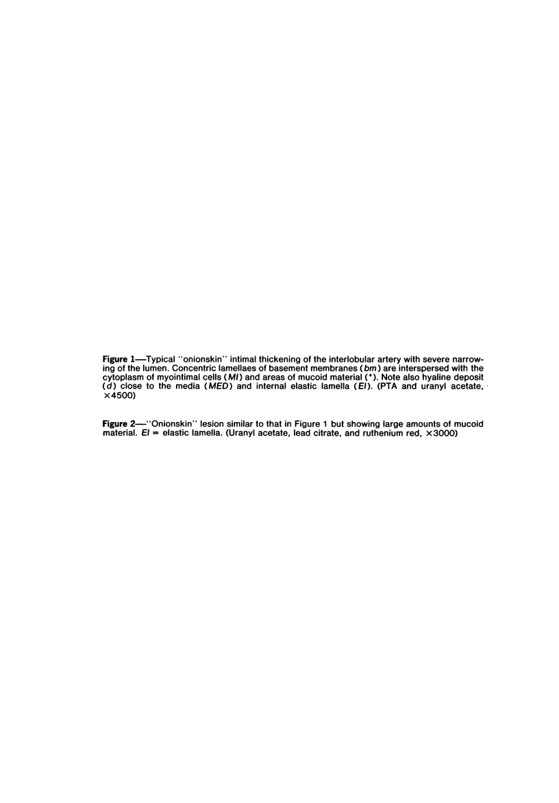
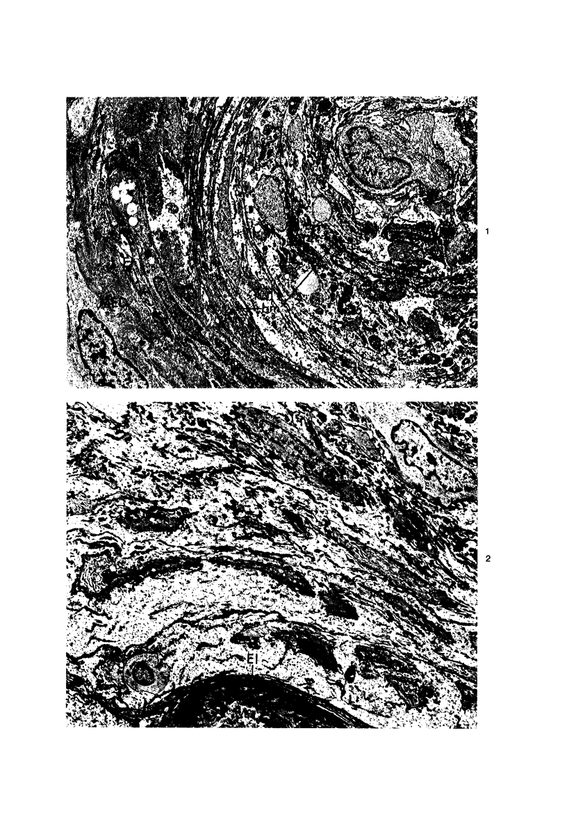


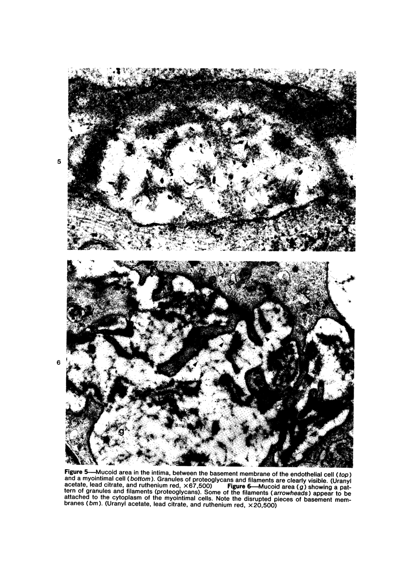

Images in this article
Selected References
These references are in PubMed. This may not be the complete list of references from this article.
- Burkholder P. M. Malignant nephrosclerosis. Arch Pathol. 1965 Dec;80(6):583–589. [PubMed] [Google Scholar]
- De Wolf F., Robertson W. B., Brosens I. The ultrastructure of acute atherosis in hypertensive pregnancy. Am J Obstet Gynecol. 1975 Sep 15;123(2):164–174. doi: 10.1016/0002-9378(75)90522-0. [DOI] [PubMed] [Google Scholar]
- Frederickson R. G., Morse D. E., Low F. N. High-voltage electron microscopy of extracellular fibrillogenesis. Am J Anat. 1977 Sep;150(1):1–33. doi: 10.1002/aja.1001500102. [DOI] [PubMed] [Google Scholar]
- HAUST M. D., MORE R. H., MOVAT H. Z. The role of smooth muscle cells in the fibrogenesis of arteriosclerosis. Am J Pathol. 1960 Oct;37:377–389. [PMC free article] [PubMed] [Google Scholar]
- Hsu H. C., Lee C. J., Lee C. S. The glomerular and arterial changes in chronic rejection of human renal allograft. Taiwan Yi Xue Hui Za Zhi. 1976 Nov;75(11):609–625. [PubMed] [Google Scholar]
- Jones D. B. Arterial and glomerular lesions associated with severe hypertension. Light and electron microscopic studies. Lab Invest. 1974 Oct;31(4):303–313. [PubMed] [Google Scholar]
- KARRER H. E. Electron microscope study of developing chick embryo aorta. J Ultrastruct Res. 1960 Dec;4:420–454. doi: 10.1016/s0022-5320(60)80032-9. [DOI] [PubMed] [Google Scholar]
- Kobayasi T., Asboe-Hansen G. Ruthenium red staining of ultrathin sections of human mast-cell granules. J Microsc. 1971 Feb;93(1):55–60. doi: 10.1111/j.1365-2818.1971.tb02264.x. [DOI] [PubMed] [Google Scholar]
- Kádár A., Gardner D. L., Bush V. Glycosaminoglycans in developing chick-embryo aorta revealed by ruthenium red: an electron-microscope study. J Pathol. 1972 Dec;108(4):275–280. doi: 10.1002/path.1711080403. [DOI] [PubMed] [Google Scholar]
- Kádár A., Veress B., Jellinek H. Ultrastructural elements in experimental intimal thickening. II. Study of the development of elastic elements in intimal proliferation. Exp Mol Pathol. 1969 Oct;11(2):212–223. doi: 10.1016/0014-4800(69)90009-4. [DOI] [PubMed] [Google Scholar]
- Lindner J. Injury and repair of arterial tissue: histochemical alterations. Angiology. 1974 Nov;25(10):628–635. doi: 10.1177/000331977402501003. [DOI] [PubMed] [Google Scholar]
- PARONETTO F. IMMUNOCYTOCHEMICAL OBSERVATIONS ON THE VASCULAR NECROSIS AND RENAL GLOMERULAR LESIONS OF MALIGNANT NEPHROSCLEROSIS. Am J Pathol. 1965 Jun;46:901–915. [PMC free article] [PubMed] [Google Scholar]
- Parker F., Odland G. F. A correlative histochemical, biochemical and electron microscopic study of experimental atherosclerosis in the rabbit aorta with special reference to the myo-intimal cell. Am J Pathol. 1966 Feb;48(2):197–239. [PMC free article] [PubMed] [Google Scholar]
- Pitcock J. A., Johnson J. G., Hatch F. E., Acchiardo S., Muirhead E. E., Brown P. S. Malignant hypertension in blacks. Malignant intrarenal arterial disease as observed by light and electron microscopy. Hum Pathol. 1976 May;7(3):333–346. doi: 10.1016/s0046-8177(76)80043-3. [DOI] [PubMed] [Google Scholar]
- SHEEHAN H. L., DAVIS J. C. Renal ischaemia with good reflow. J Pathol Bacteriol. 1959 Oct;78:351–377. doi: 10.1002/path.1700780204. [DOI] [PubMed] [Google Scholar]
- Sanerkin N. G. Vascular lesions of malignant essential hypertension. J Pathol. 1971 Mar;103(3):177–184. doi: 10.1002/path.1711030306. [DOI] [PubMed] [Google Scholar]
- Shigematsu H., Dikman S. H., Churg J., Grishman E. Glomerular injury in malignant nephrosclerosis. Nephron. 1978;22(4-6):399–408. doi: 10.1159/000181482. [DOI] [PubMed] [Google Scholar]
- Sinclair R. A., Antonovych T. T., Mostofi F. K. Renal proliferative arteriopathies and associated glomerular changes: a light and electron microscopic study. Hum Pathol. 1976 Sep;7(5):565–588. doi: 10.1016/s0046-8177(76)80103-7. [DOI] [PubMed] [Google Scholar]
- Wight T. N., Ross R. Proteoglycans in primate arteries. I. Ultrastructural localization and distribution in the intima. J Cell Biol. 1975 Dec;67(3):660–674. doi: 10.1083/jcb.67.3.660. [DOI] [PMC free article] [PubMed] [Google Scholar]
- Wight T. N., Ross R. Proteoglycans in primate arteries. II. Synthesis and secretion of glycosaminoglycans by arterial smooth muscle cells in culture. J Cell Biol. 1975 Dec;67(3):675–686. doi: 10.1083/jcb.67.3.675. [DOI] [PMC free article] [PubMed] [Google Scholar]


