

Abstract
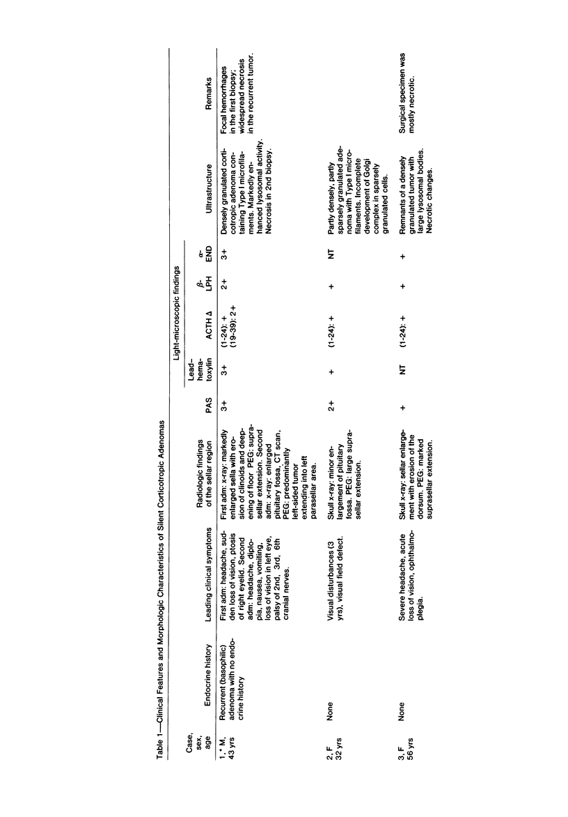
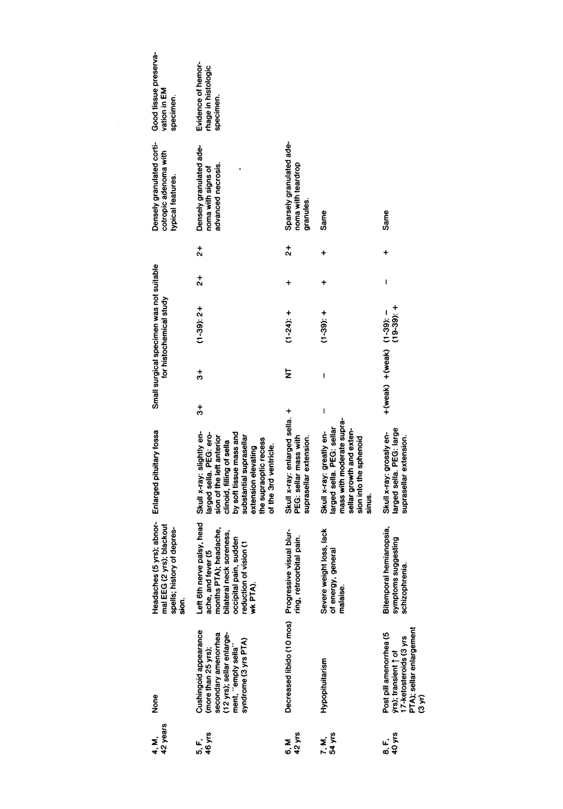
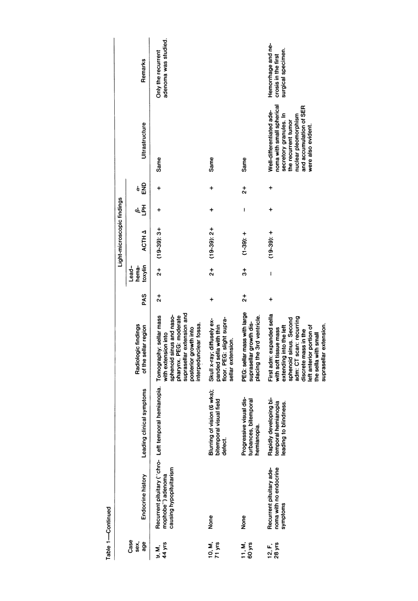
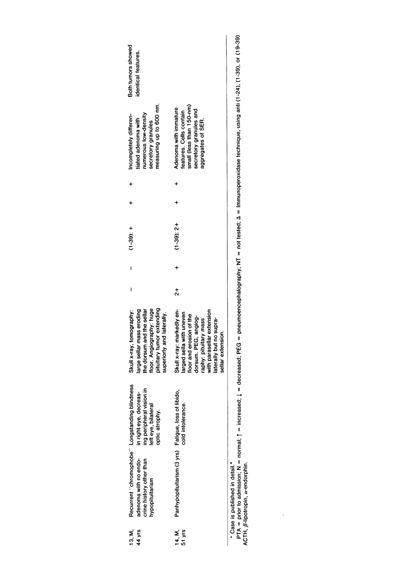
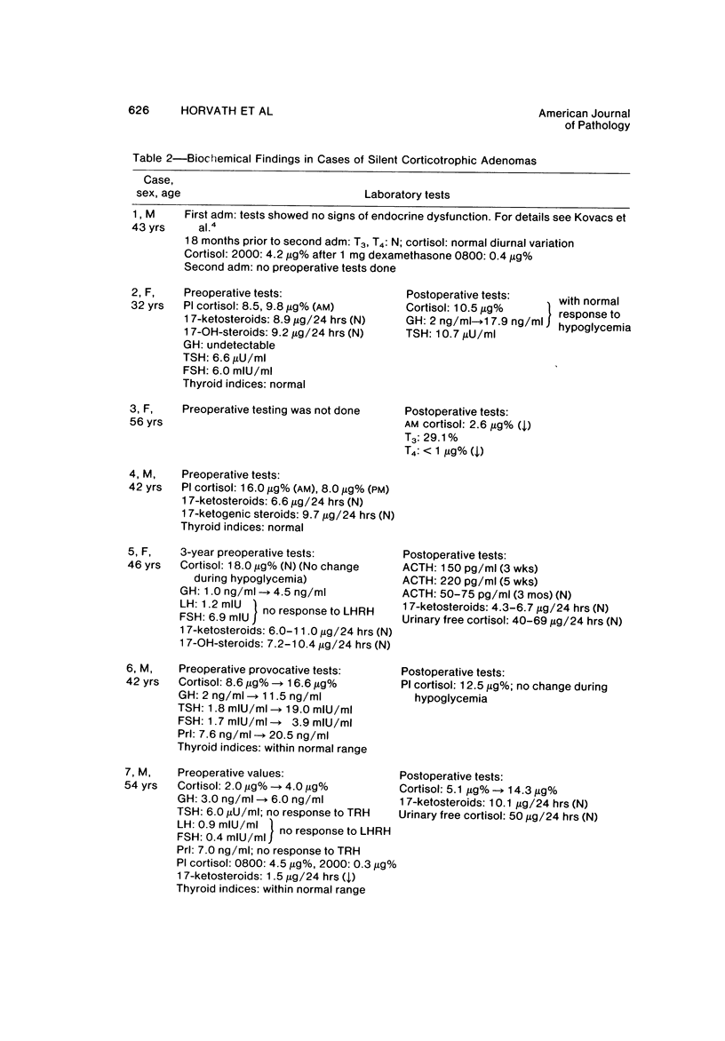
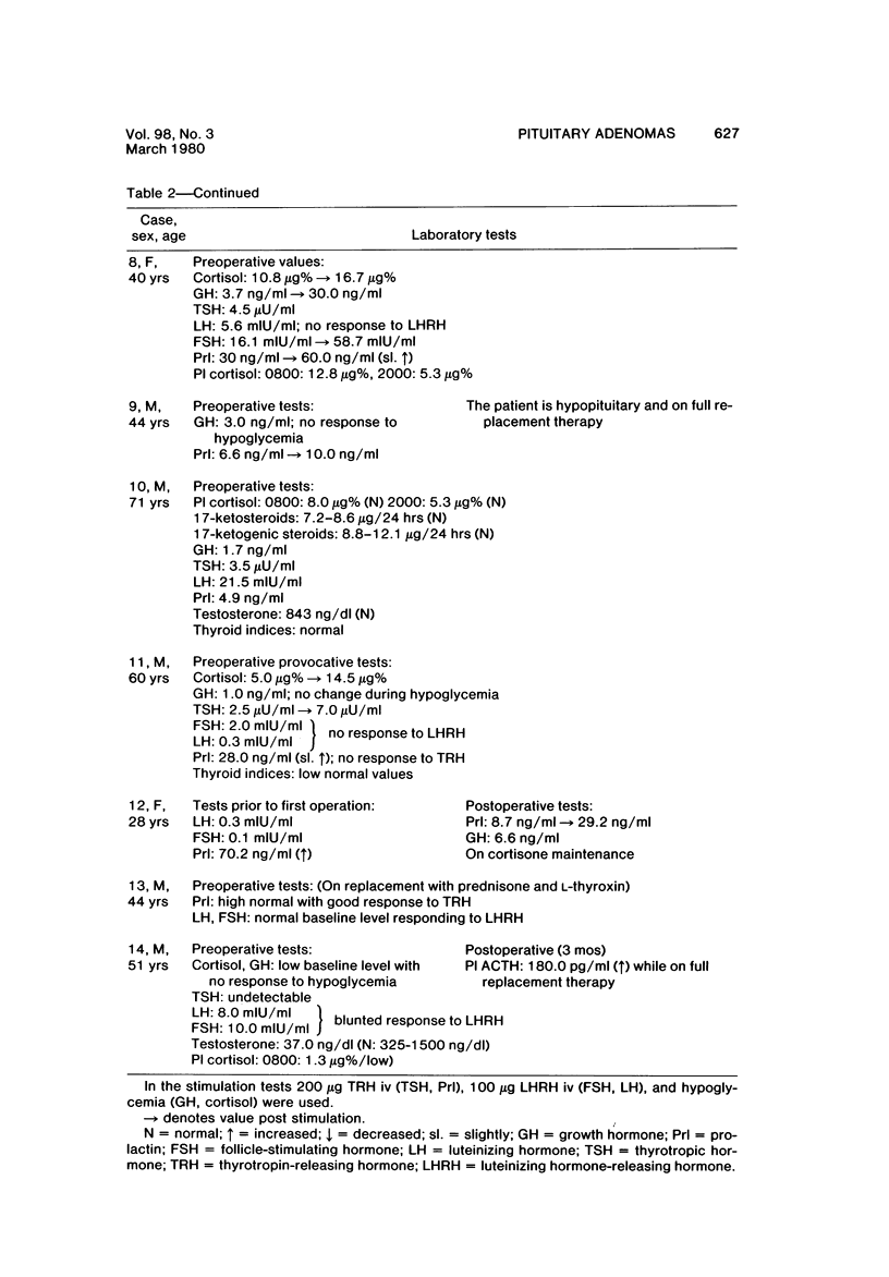
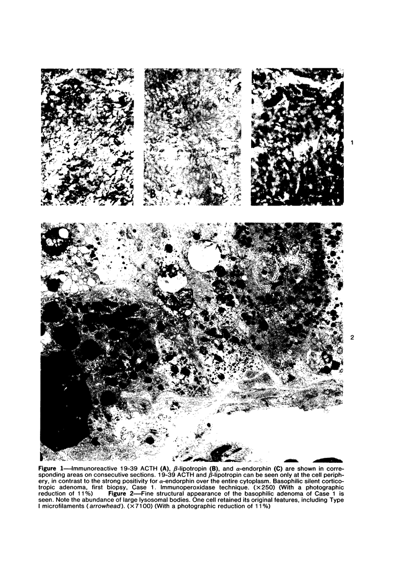
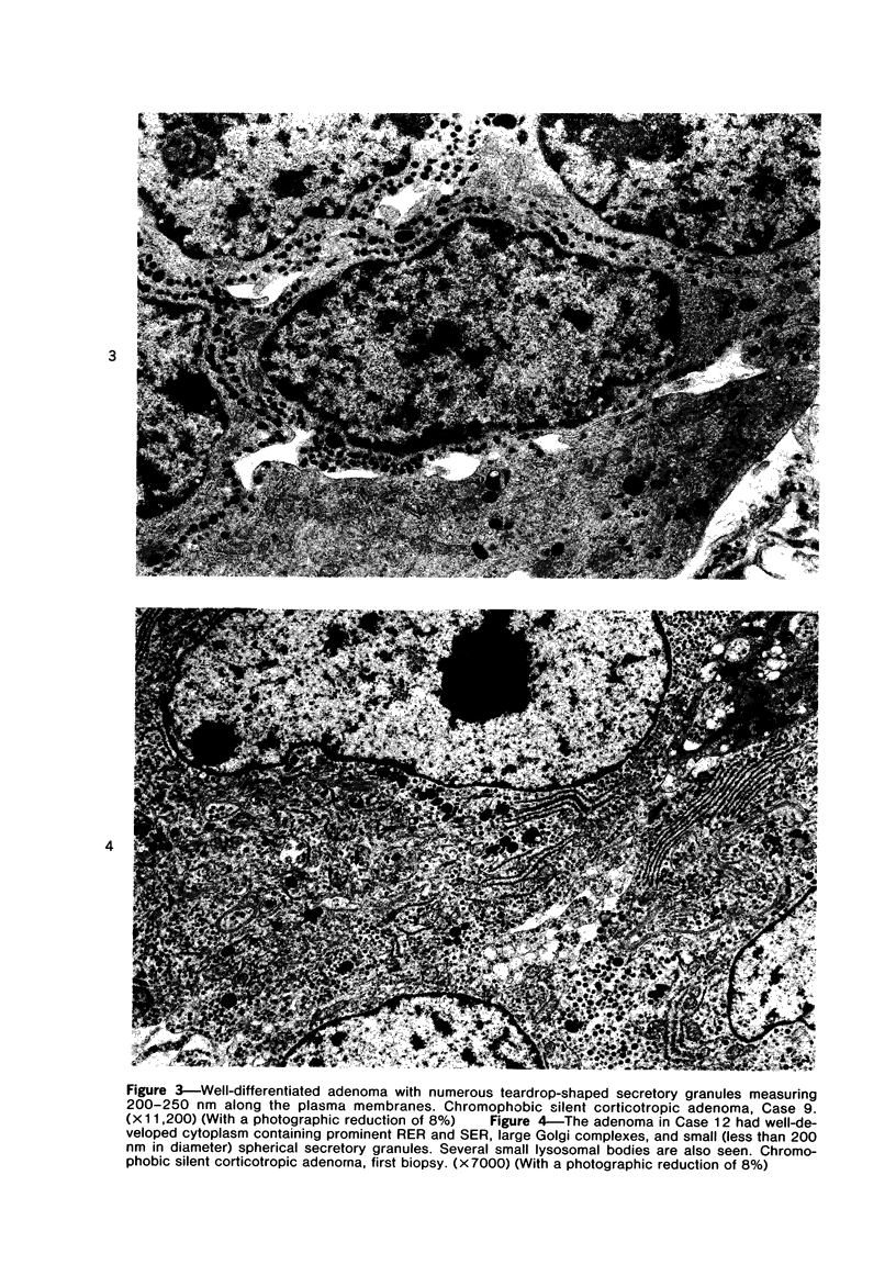
Among 300 surgically removed pituitary adenomas, 17 tumors containing immunoreactive 1-39 adrenocorticotropin (ACTH) and/or 19-39 ACTH, beta-lipotropin, and alpha-endorphin but unassociated with clinical signs of Cushing's disease have been detected. These neoplasms were divided into basophilic adenomas with strong periodic acid-Schiff (PAS) and lead-hematoxylin positivity and chromophobic tumors with moderate or no PAS and lead-hematoxylin positivity. The former were densely granulated tumors with a fine structure strikingly similar to that of functioning corticotropic cell adenomas. The latter were sparsely granulated with varying ultrastructural patterns. The marked morphologic diversity suggests that these adenomas, despite their similar immunocytologic characteristics, represent more than one entity. Clinically, the most common finding was a rapidly progressing visual defect. An unusually high incidence of infarction (5 cases) and recurrence (5 cases) was noted, underlining the importance of correct morphologic diagnosis and careful follow-up.
Full text
PDF


Images in this article


Selected References
These references are in PubMed. This may not be the complete list of references from this article.
- Deftos L. J., Burton D., Catherwood B. D., Bone H. G., Parthemore J. G., Guillemin R., Watkins W. B., Moore R. Y. Demonstration by immunoperoxidase histochemistry of calcitonin in the anterior lobe of the rat pituitary. J Clin Endocrinol Metab. 1978 Aug;47(2):457–460. doi: 10.1210/jcem-47-2-457. [DOI] [PubMed] [Google Scholar]
- Horvath E., Kovacs K. Ultrastructural classification of pituitary adenomas. Can J Neurol Sci. 1976 Feb;3(1):9–21. doi: 10.1017/s0317167100025944. [DOI] [PubMed] [Google Scholar]
- Kovacs K., Corenblum B., Sirek A. M., Penz G., Ezrin C. Localization of prolactin in chromophobe pituitary adenomas: study of human necropsy material by immunoperoxidase technique. J Clin Pathol. 1976 Mar;29(3):250–258. doi: 10.1136/jcp.29.3.250. [DOI] [PMC free article] [PubMed] [Google Scholar]
- Kovacs K., Horvath E., Bayley T. A., Hassaram S. T., Ezrin C. Silent corticotroph cell adenoma with lysosomal accumulation and crinophagy. A distinct clinicopathologic entity. Am J Med. 1978 Mar;64(3):492–499. doi: 10.1016/0002-9343(78)90236-x. [DOI] [PubMed] [Google Scholar]
- Kovacs K., Horvath E., Ezrin C. Pituitary adenomas. Pathol Annu. 1977;12(Pt 2):341–382. [PubMed] [Google Scholar]
- Kovacs K., Horvath E., Kerenyi N. A., Sheppard R. H. Light and electron microscopic features of a pituitary adenoma in Nelson's syndrome. Am J Clin Pathol. 1976 Mar;65(3):337–343. doi: 10.1093/ajcp/65.3.337. [DOI] [PubMed] [Google Scholar]
- Kovacs K., Horvath E. Vascular alterations in adenomas of human pituitary glands. An electron microscopic study. Angiologica. 1973;10(5-6):299–309. doi: 10.1159/000157987. [DOI] [PubMed] [Google Scholar]
- Landolt A. M. Ultrastructure of human sella tumors. Correlations of clinical findings and morphology. Acta Neurochir (Wien) 1975;Suppl 22:1–167. [PubMed] [Google Scholar]
- Mason T. E., Phifer R. F., Spicer S. S., Swallow R. A., Dreskin R. B. An immunoglobulin-enzyme bridge method for localizing tissue antigens. J Histochem Cytochem. 1969 Sep;17(9):563–569. doi: 10.1177/17.9.563. [DOI] [PubMed] [Google Scholar]
- Moriarty G. C. Adenohypophysis: ultrastructural cytochemistry. A review. J Histochem Cytochem. 1973 Oct;21(10):855–894. doi: 10.1177/21.10.855. [DOI] [PubMed] [Google Scholar]
- Rasmussen A. T., Nelson A. A. Pars intermedia basophil adenoma of the hypophysis. Am J Pathol. 1938 May;14(3):297–310.5. [PMC free article] [PubMed] [Google Scholar]
- Sternberger L. A., Hardy P. H., Jr, Cuculis J. J., Meyer H. G. The unlabeled antibody enzyme method of immunohistochemistry: preparation and properties of soluble antigen-antibody complex (horseradish peroxidase-antihorseradish peroxidase) and its use in identification of spirochetes. J Histochem Cytochem. 1970 May;18(5):315–333. doi: 10.1177/18.5.315. [DOI] [PubMed] [Google Scholar]
- Tramu G., Beauvillain J. C., Mazzuca M., Fossati P., Martin-Linquette A., Christiaens J. L. Dissociation des résultants obtenus en immunofluorescence avec des antisérums anti-ACTH dans trois cas d'adénomes "chromophobe" sans hypercorticisme. Etude ultrastructurale. Ann Endocrinol (Paris) 1976 Jan-Feb;37(1):55–56. [PubMed] [Google Scholar]
- Tramu G., Beauvillain J. C., Mazzuca M., Linquette M., Lefebvre J., Fossati P., Christiaens J. L. Adénome hypophysaire à cellules à alpha, 17-39 ACTH et beta, MSH sans hypercorticisme. Ann Endocrinol (Paris) 1978;39(1):51–52. [PubMed] [Google Scholar]