Abstract
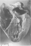
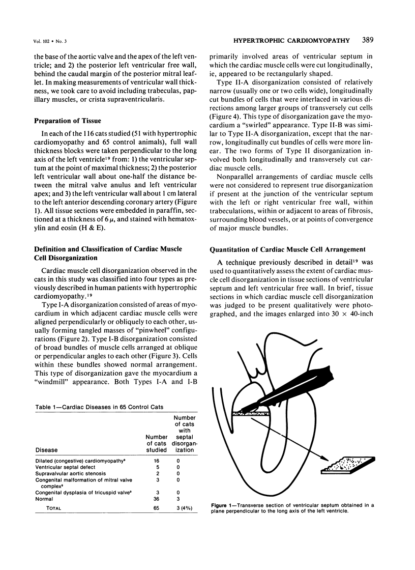
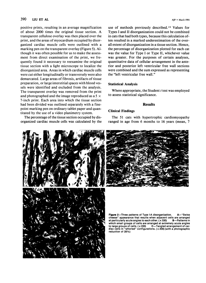
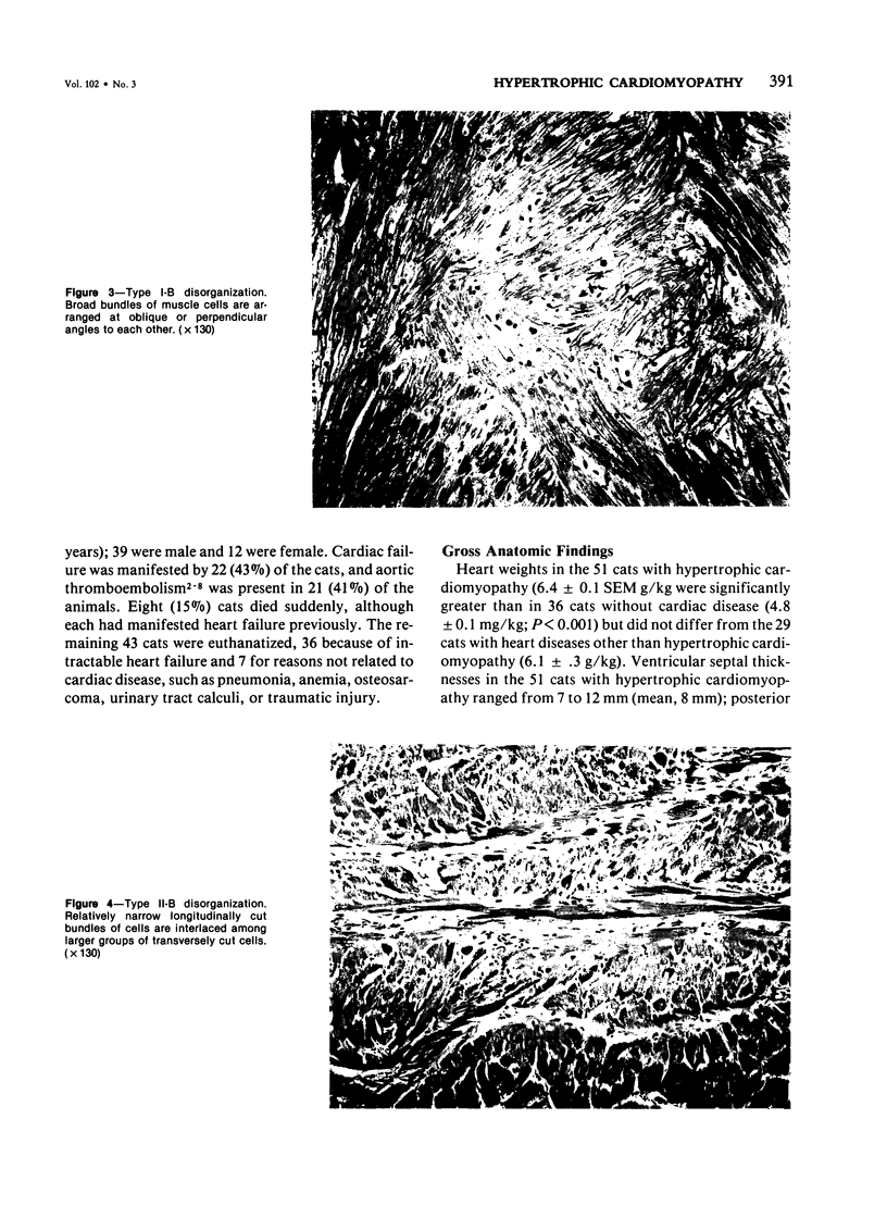
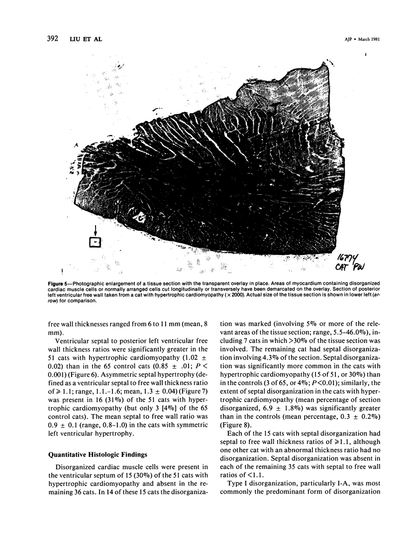
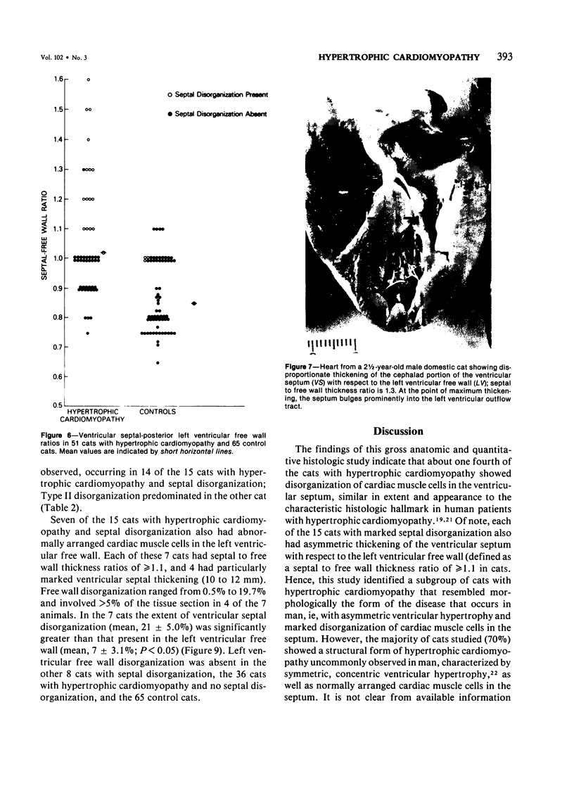
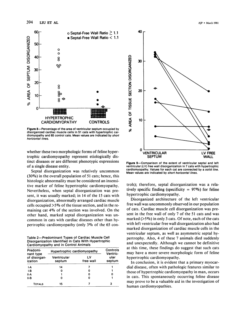
Gross anatomic features and the pattern and extent of cardiac muscle cell disorganization were studied in the hearts of 51 with spontaneously occurring hypertrophic cardiomyopathy. Each cat had a hypertrophied, but nondilated, left ventricle. Ventricular septal disorganization was extensive, involving 5% or more of the relevant areas of the tissue section, in 14 (27%) of the 51 cats. Marked septal disorganization occurred only in those cats with asymmetric septal hypertrophy (ventricular septal to left ventricular free wall thickness ratio of greater than or equal to 1.1) Disorganization of cardiac muscle cells was uncommon and less extensive in the left ventricular free wall of the cats with hypertrophic cardiomyopathy. Disorganization involved the free wall of only 7 cats, each with asymmetric septal hypertrophy, and occupied greater than 5% of the free wall tissue sections in just 3. Hence, about one fourth of this population of cats had hypertrophic cardiomyopathy resembling the human form of this disease, with asymmetric left ventricular hypertrophy and marked disorganization of cardiac muscle cells in the ventricular septum. The majority of cats (about 75%), however, demonstrated a form of hypertrophic cardiomyopathy characterized by symmetric ventricular hypertrophy and normal arrangement of cardiac muscle cells.
Full text
PDF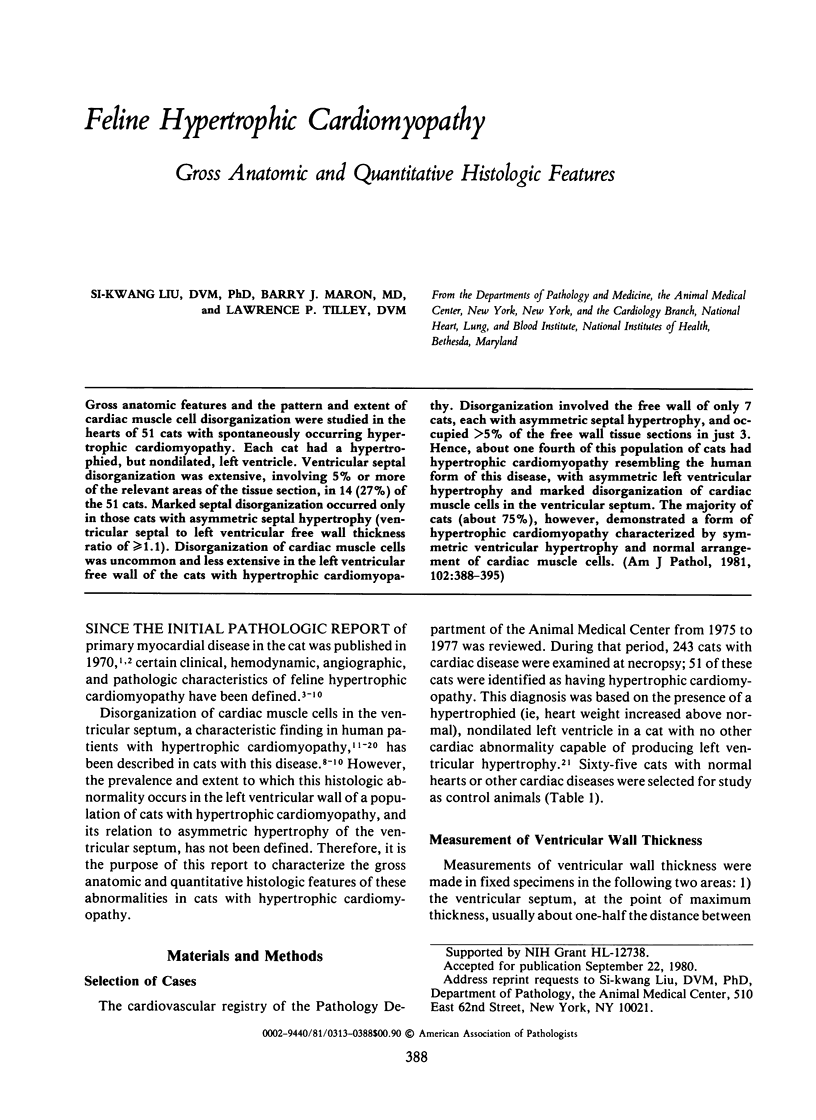
Images in this article
Selected References
These references are in PubMed. This may not be the complete list of references from this article.
- Goodwin J. F. Prospects and predictions for the cardiomyopathies. Circulation. 1974 Aug;50(2):210–219. doi: 10.1161/01.cir.50.2.210. [DOI] [PubMed] [Google Scholar]
- Liu S. K. Acquired cardiac lesions leading to congestive heart failure in the cat. Am J Vet Res. 1970 Nov;31(11):2071–2088. [PubMed] [Google Scholar]
- Liu S. K. Pathology of feline heart diseases. Vet Clin North Am. 1977 May;7(2):323–339. doi: 10.1016/s0091-0279(77)50033-0. [DOI] [PubMed] [Google Scholar]
- Liu S. K., Tashjian R. J., Patnaik A. K. Congestive heart failure in the cat. J Am Vet Med Assoc. 1970 May 1;156(9):1319–1330. [PubMed] [Google Scholar]
- Lord P. F., Wood A., Tilley L. P., Liu S. K. Radiographic and hemodynamic evaluation of cardiomyopathy and thromboembolism in the cat. J Am Vet Med Assoc. 1974 Jan 15;164(2):154–165. [PubMed] [Google Scholar]
- Maron B. J., Edwards J. E., Henry W. L., Clark C. E., Bingle G. J., Epstein S. E. Asymmetric septal hypertrophy (ASH) in infancy. Circulation. 1974 Oct;50(4):809–820. doi: 10.1161/01.cir.50.4.809. [DOI] [PubMed] [Google Scholar]
- Maron B. J., Epstein S. E. Hypertrophic cardiomyopathy: a discussion of nomenclature. Am J Cardiol. 1979 Jun;43(6):1242–1244. doi: 10.1016/0002-9149(79)90160-7. [DOI] [PubMed] [Google Scholar]
- Maron B. J., Ferrans V. J., Henry W. L., Clark C. E., Redwood D. R., Roberts W. C., Morrow A. G., Epstein S. E. Differences in distribution of myocardial abnormalities in patients with obstructive and nonobstructive asymmetric septal hypertrophy (ASH). Light and electron microscopic findings. Circulation. 1974 Sep;50(3):436–446. doi: 10.1161/01.cir.50.3.436. [DOI] [PubMed] [Google Scholar]
- Maron B. J., Gottdiener J. S., Roberts W. C., Henry W. L., Savage D. D., Epstein S. E. Left ventricular outflow tract obstruction due to systolic anterior motion of the anterior mitral leaflet in patients with concentric left ventricular hypertrophy. Circulation. 1978 Mar;57(3):527–533. doi: 10.1161/01.cir.57.3.527. [DOI] [PubMed] [Google Scholar]
- Maron B. J., Henry W. L., Clark C. E., Redwood D. R., Roberts W. C., Epstein S. E. Asymetric septal hypertrophy in childhood. Circulation. 1976 Jan;53(1):9–19. doi: 10.1161/01.cir.53.1.9. [DOI] [PubMed] [Google Scholar]
- Maron B. J., Roberts W. C. Quantitative analysis of cardiac muscle cell disorganization in the ventricular septum of patients with hypertrophic cardiomyopathy. Circulation. 1979 Apr;59(4):689–706. doi: 10.1161/01.cir.59.4.689. [DOI] [PubMed] [Google Scholar]
- Maron B. J., Sato N., Roberts W. C., Edwards J. E., Chandra R. S. Quantitative analysis of cardiac muscle cell disorganization in the ventricular septum. Comparison of fetuses and infants with and without congenital heart disease and patients with hypertrophic cardiomyopathy. Circulation. 1979 Sep;60(3):685–696. doi: 10.1161/01.cir.60.3.685. [DOI] [PubMed] [Google Scholar]
- Roberts W. C., Ferrans V. J. Pathologic anatomy of the cardiomyopathies. Idiopathic dilated and hypertrophic types, infiltrative types, and endomyocardial disease with and without eosinophilia. Hum Pathol. 1975 May;6(3):287–342. [PubMed] [Google Scholar]
- Roberts W. C., Ferrans V. J. Pathological aspects of certain cardiomyopathies. Circ Res. 1974 Aug;35(2):suppl II–II:144. [PubMed] [Google Scholar]
- TEARE D. Asymmetrical hypertrophy of the heart in young adults. Br Heart J. 1958 Jan;20(1):1–8. doi: 10.1136/hrt.20.1.1. [DOI] [PMC free article] [PubMed] [Google Scholar]
- Tilley L. P., Liu S. K., Gilbertson S. R., Wagner B. M., Lord P. F. Primary myocardial disease in the cat. A model for human cardiomyopathy. Am J Pathol. 1977 Mar;86(3):493–522. [PMC free article] [PubMed] [Google Scholar]


