

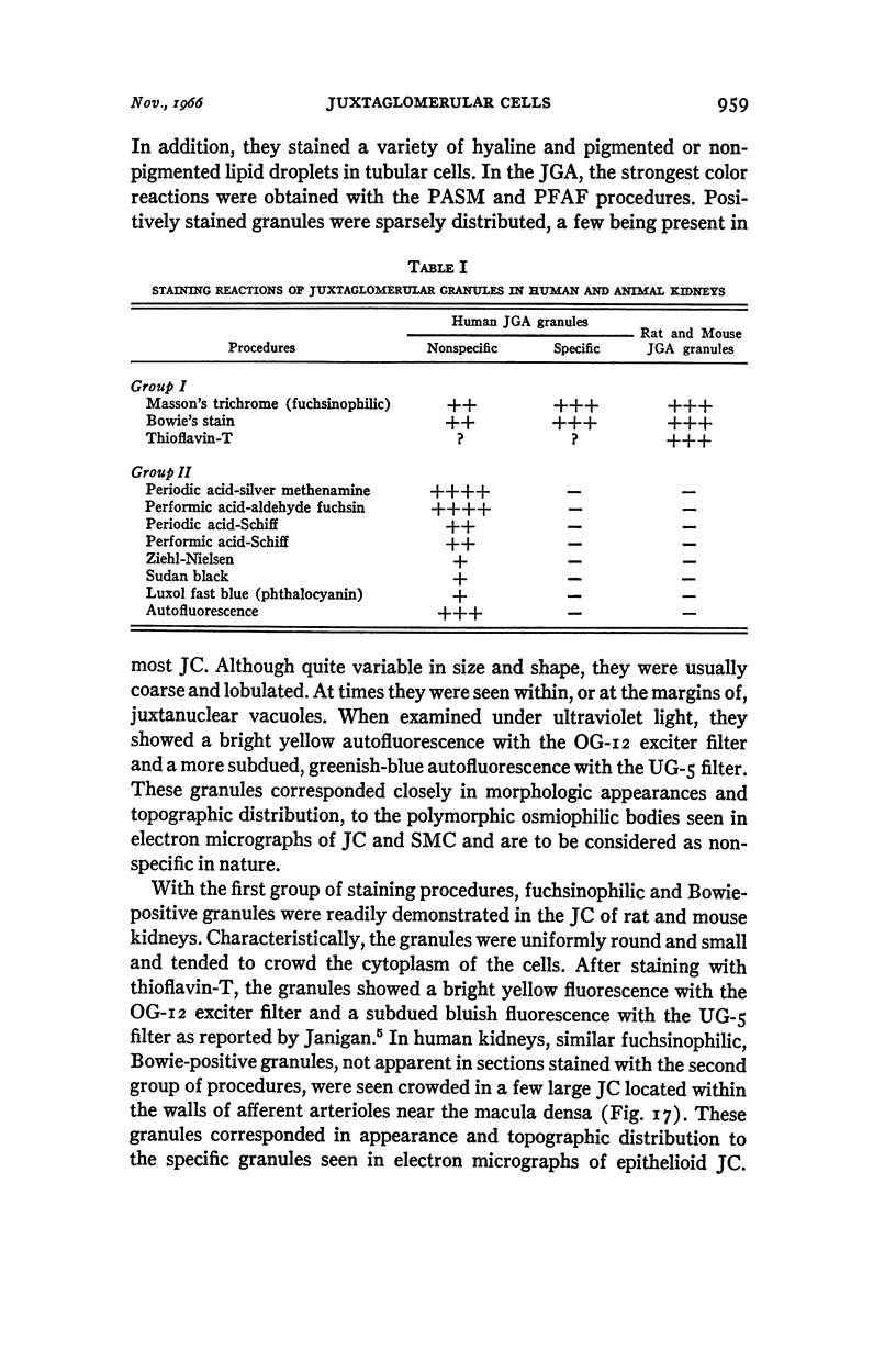
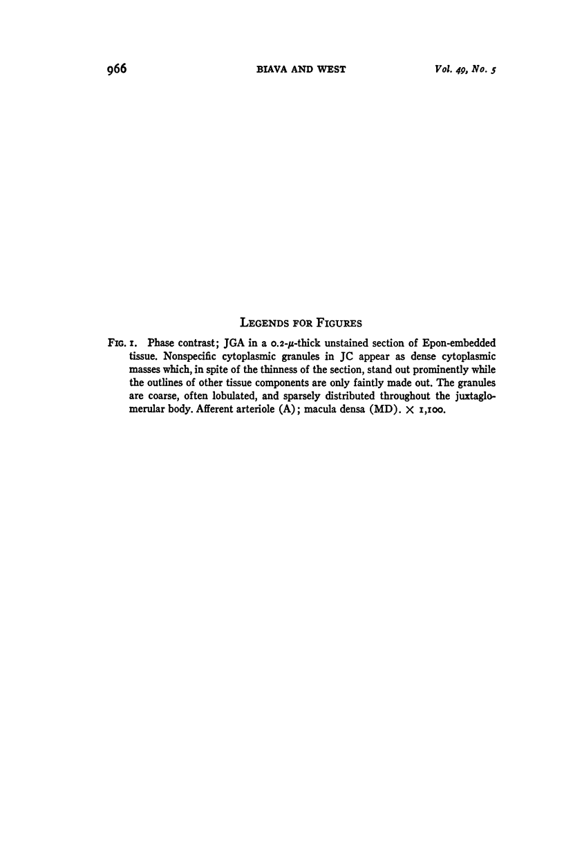
Full text
PDF
Images in this article
Selected References
These references are in PubMed. This may not be the complete list of references from this article.
- BEHNKE O., MOE H. AN ELECTRON MICROSCOPE STUDY OF MATURE AND DIFFERENTIATING PANETH CELLS IN THE RAT, ESPECIALLY OF THEIR ENDOPLASMIC RETICULUM AND LYSOSOMES. J Cell Biol. 1964 Sep;22:633–652. doi: 10.1083/jcb.22.3.633. [DOI] [PMC free article] [PubMed] [Google Scholar]
- BIAVA C., WEST M. LIPOFUSCIN-LIKE GRANULES IN VASCULAR SMOOTH MUSCLE AND JUXTAGLOMERULAR CELLS OF HUMAN KIDNEYS. Am J Pathol. 1965 Aug;47:287–313. [PMC free article] [PubMed] [Google Scholar]
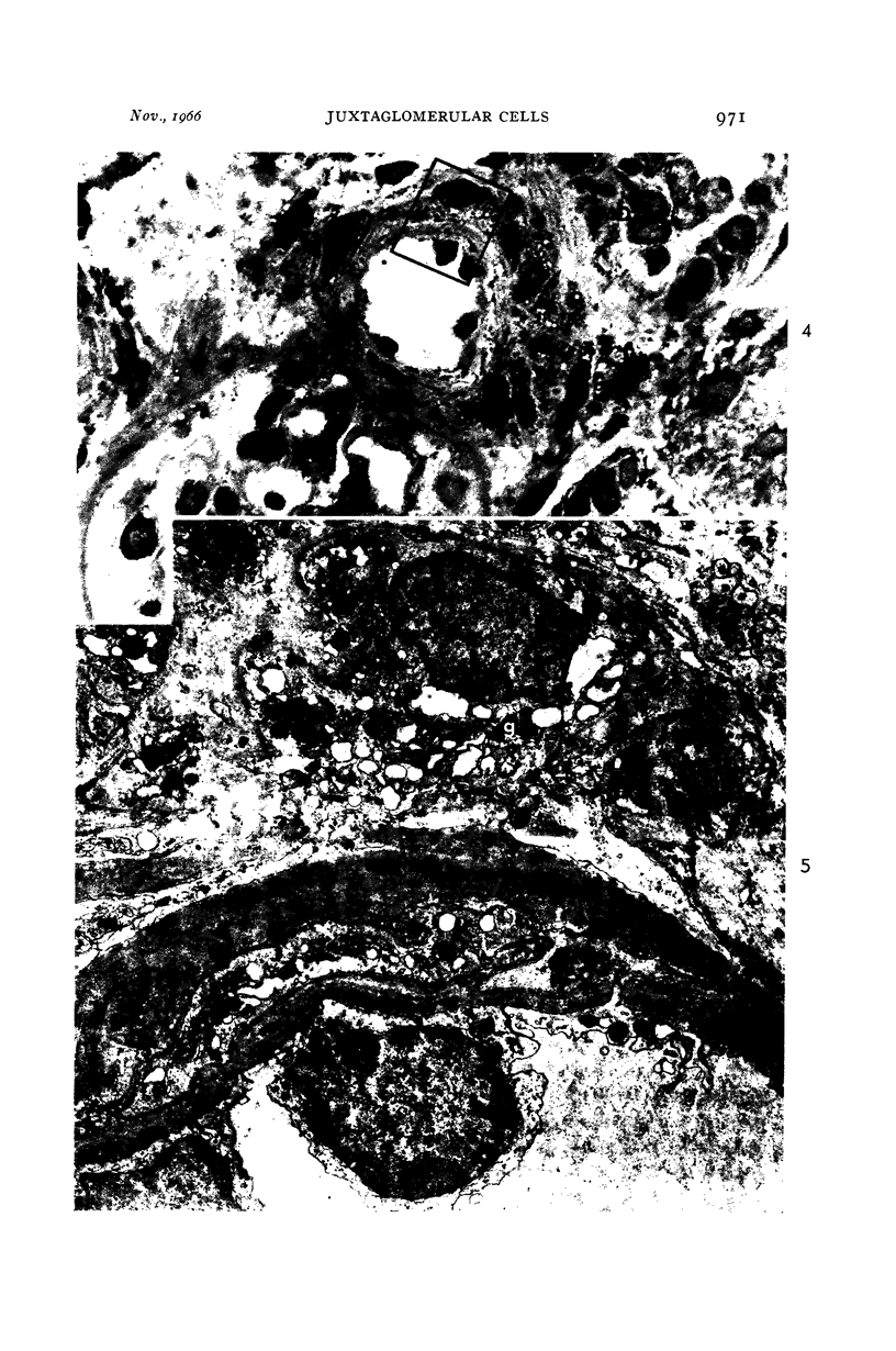
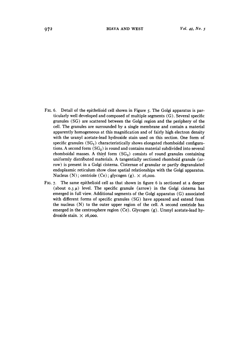
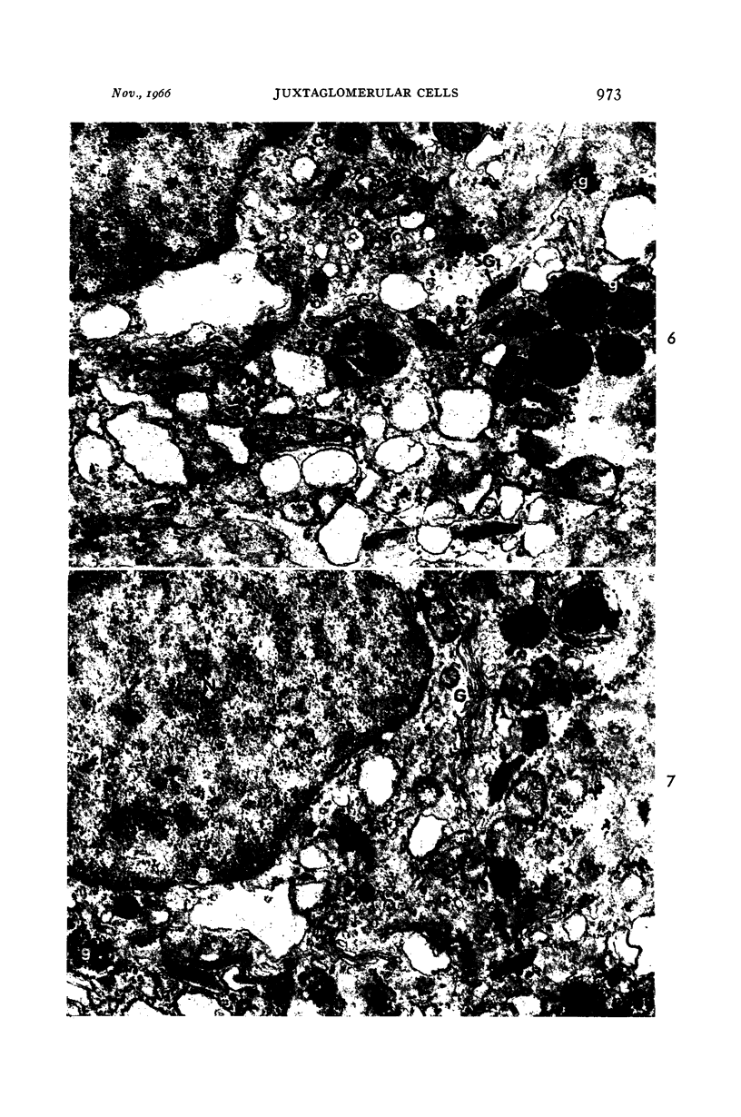
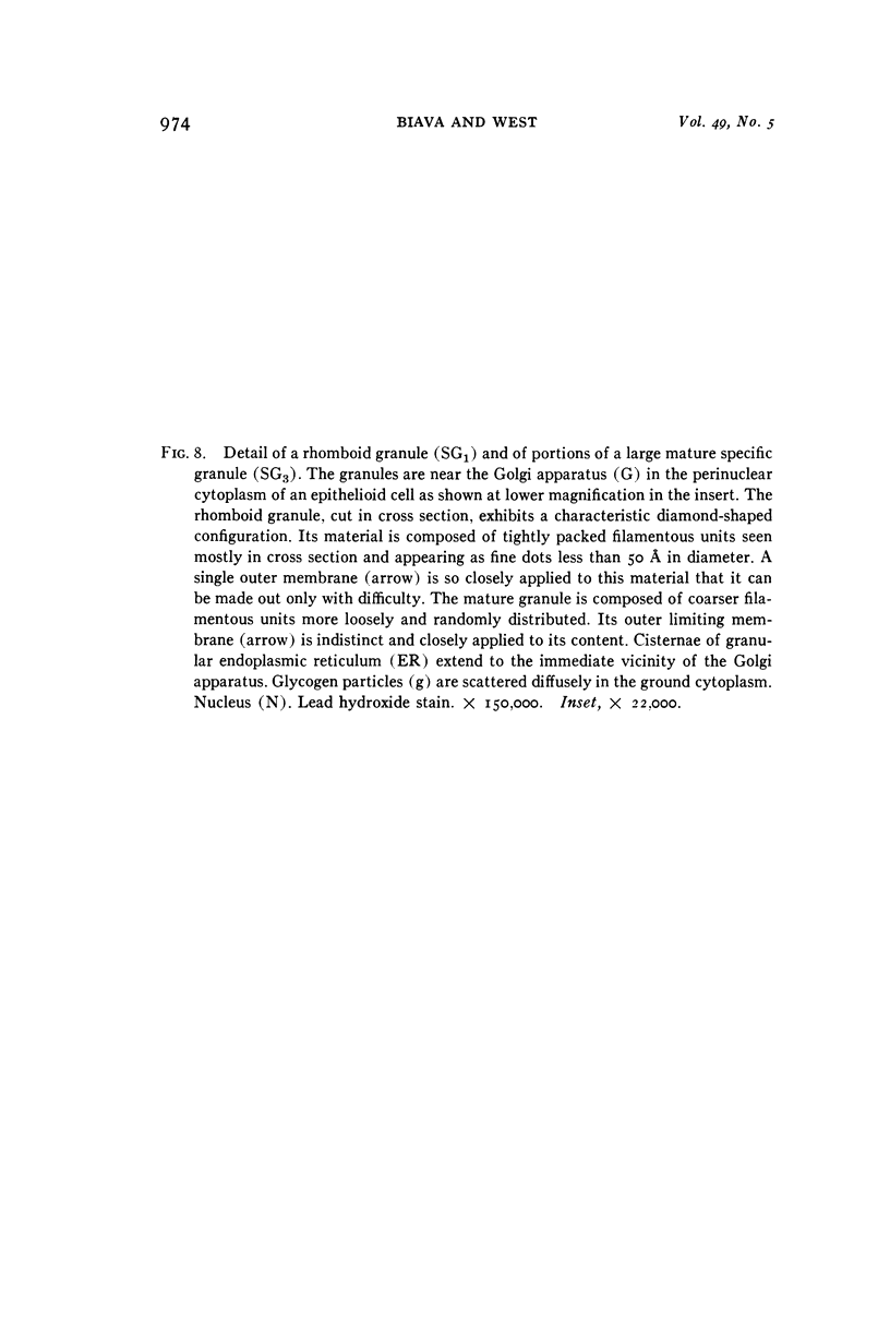
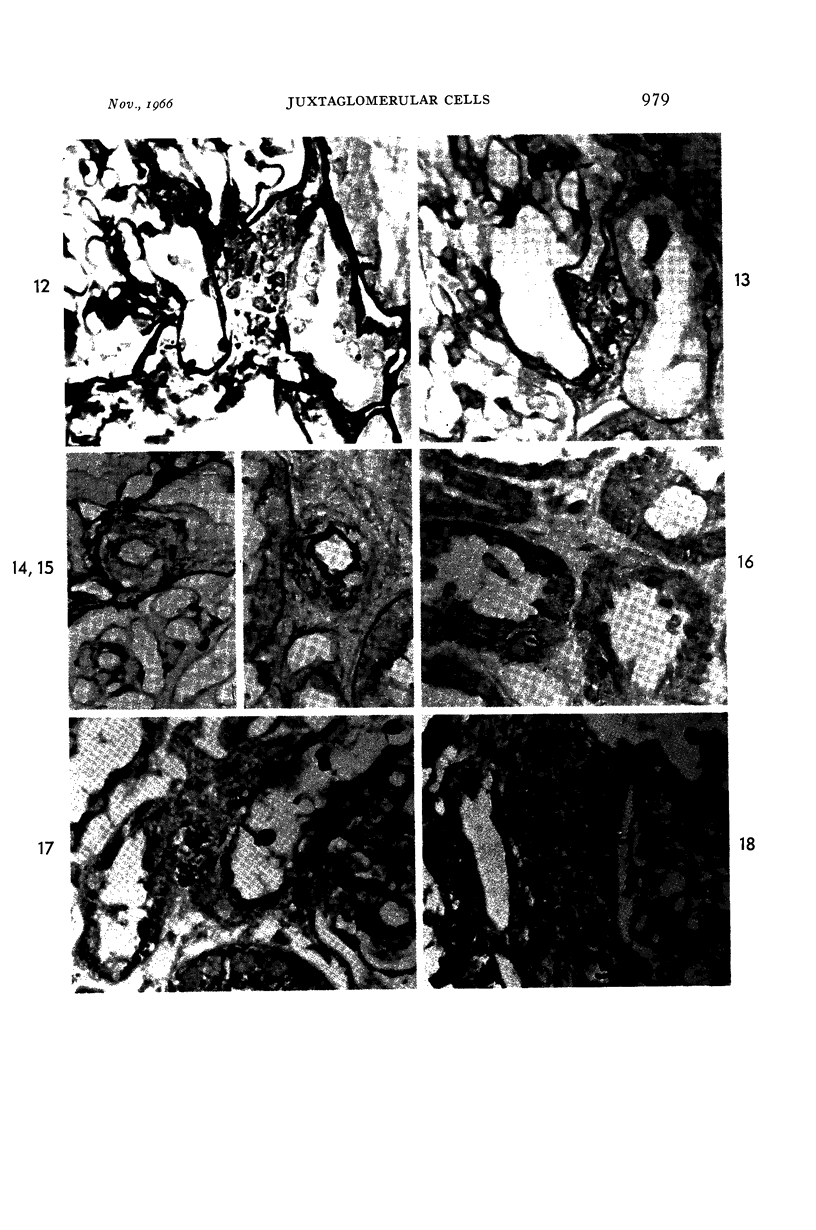
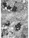
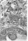
- Biava C., West M. Fine structure of normal human juxtaglomerular cells. I. General structure and intercellular relationships. Am J Pathol. 1966 Oct;49(4):679–721. [PMC free article] [PubMed] [Google Scholar]
- CHANDRA S., SKELTON F. R., BERNARDIS L. L. SEPARATION OF RENAL PRESSOR ACTIVITY BY ULTRACENTRIFUGATION. Lab Invest. 1964 Oct;13:1192–1197. [PubMed] [Google Scholar]
- CROCKER D. W., NEWTON R. A., MAHONEY E. M., HARRISON J. H. Hypertension due to primary renal ischemia: a correlation of juxtaglomerular cell counts with clinicopathological findings in twenty-five cases. N Engl J Med. 1962 Oct 18;267:794–800. doi: 10.1056/NEJM196210182671602. [DOI] [PubMed] [Google Scholar]
- Chandra S., Hubbard J. C., Skelton F. R., Bernardis L. L., Kamura S. Genesis of juxtaglomerular cell granules. A physiologic, light and electron microscopic study concerning experimental renal hypertension. Lab Invest. 1965 Oct;14(10):1834–1847. [PubMed] [Google Scholar]
- DUNIHUE F. W., BOLDOSSER W. G. OBSERVATIONS ON THE SIMILARITY OF MESANGIAL TO JUXTAGLOMERULAR CELLS. Lab Invest. 1963 Dec;12:1228–1240. [PubMed] [Google Scholar]
- HARTROFT P. M., HARTROFT W. S. Studies on renal juxtaglomerular cells. I. Variations produced by sodium chloride and desoxycorticosterone acetate. J Exp Med. 1953 Mar;97(3):415–429. doi: 10.1084/jem.97.3.415. [DOI] [PMC free article] [PubMed] [Google Scholar]
- JANIGAN D. T. FLUOROCHROME STAINING OF JUXTAGLOMERULAR CELL GRANULES. Arch Pathol. 1965 Apr;79:370–375. [PubMed] [Google Scholar]
- JONES D. B. Nephrotic glomerulonephritis. Am J Pathol. 1957 Mar-Apr;33(2):313–329. [PMC free article] [PubMed] [Google Scholar]
- LATTA H., MAUNSBACH A. B. The juxtaglomerular apparatus as studied electron microscopically. J Ultrastruct Res. 1962 Jun;6:547–561. doi: 10.1016/s0022-5320(62)80009-4. [DOI] [PubMed] [Google Scholar]
- McMANUS J. F. A. The periodic acid routing applied to the kidney. Am J Pathol. 1948 May;24(3):643–653. [PMC free article] [PubMed] [Google Scholar]
- Spicer S. S. Siderosis Associated with Increased Lipofuscins and Mast Cells in Aging Mice. Am J Pathol. 1960 Oct;37(4):457–475. [PMC free article] [PubMed] [Google Scholar]
- TURGEON C., SOMMERS S. C. Juxtaglomerular cell counts and human hypertension. Am J Pathol. 1961 Feb;38:227–241. [PMC free article] [PubMed] [Google Scholar]
- VANHEYNINGEN H. E. CORRELATED LIGHT AND ELECTRON MICROSCOPE OBSERVATIONS ON GLYCOPROTEIN-CONTAINING GLOBULES IN THE FOLLICULAR CELLS OF THE THYROID GLAND OF THE RAT. J Histochem Cytochem. 1965 Apr;13:286–295. doi: 10.1177/13.4.286. [DOI] [PubMed] [Google Scholar]
- WILSON W. A new staining method for demonstrating the granules of the juxtaglomerular complex. Anat Rec. 1952 Mar;112(3):497–507. doi: 10.1002/ar.1091120303. [DOI] [PubMed] [Google Scholar]