Abstract
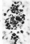
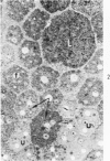
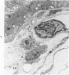
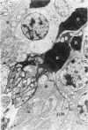
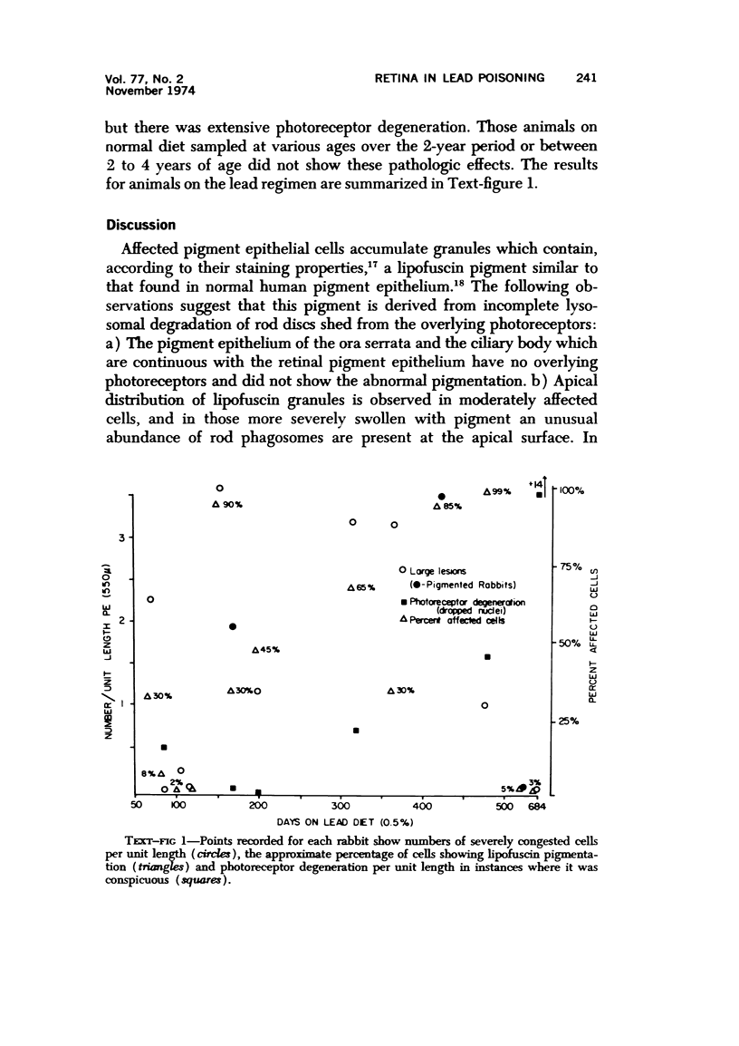
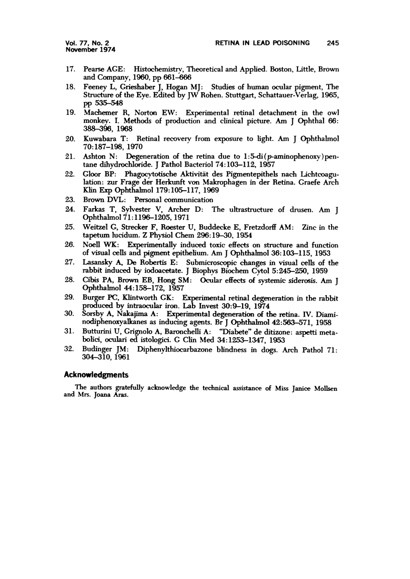
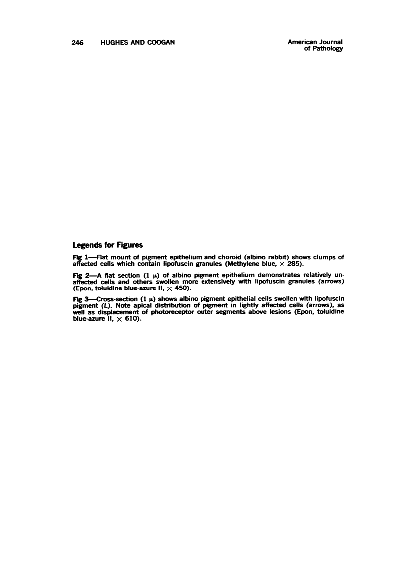
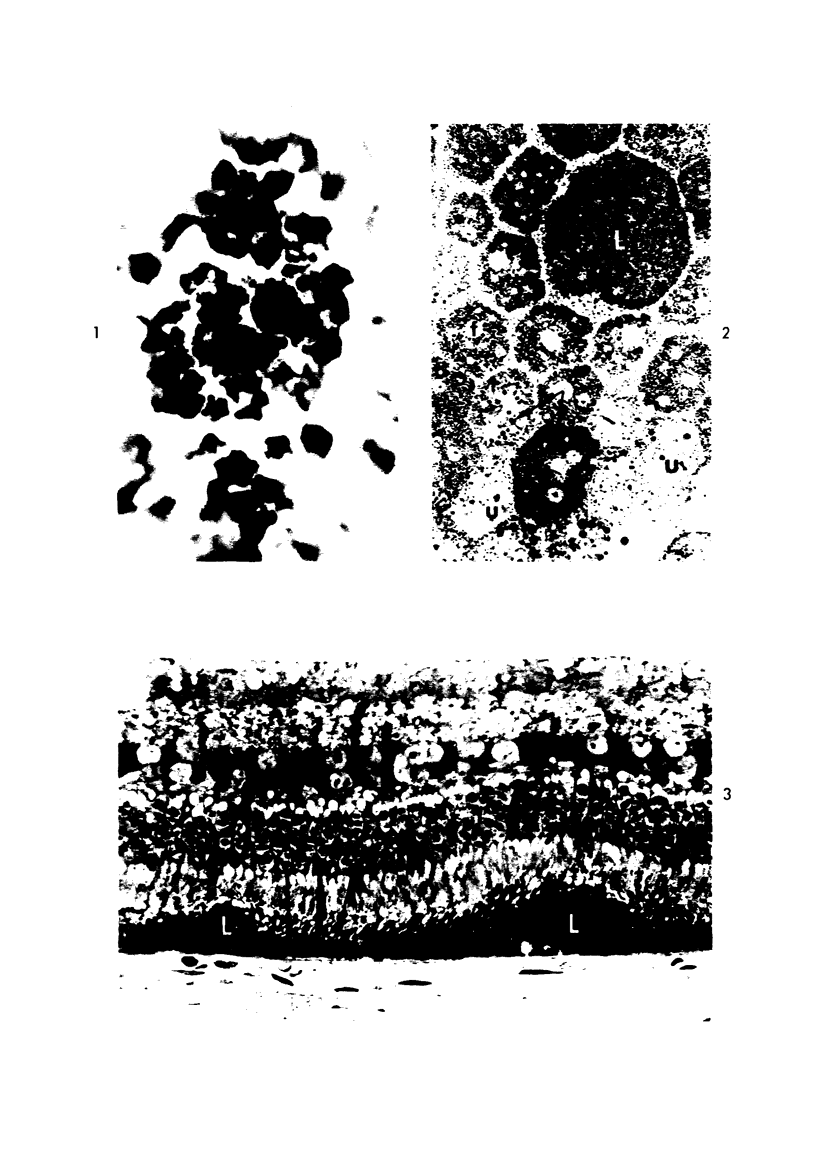
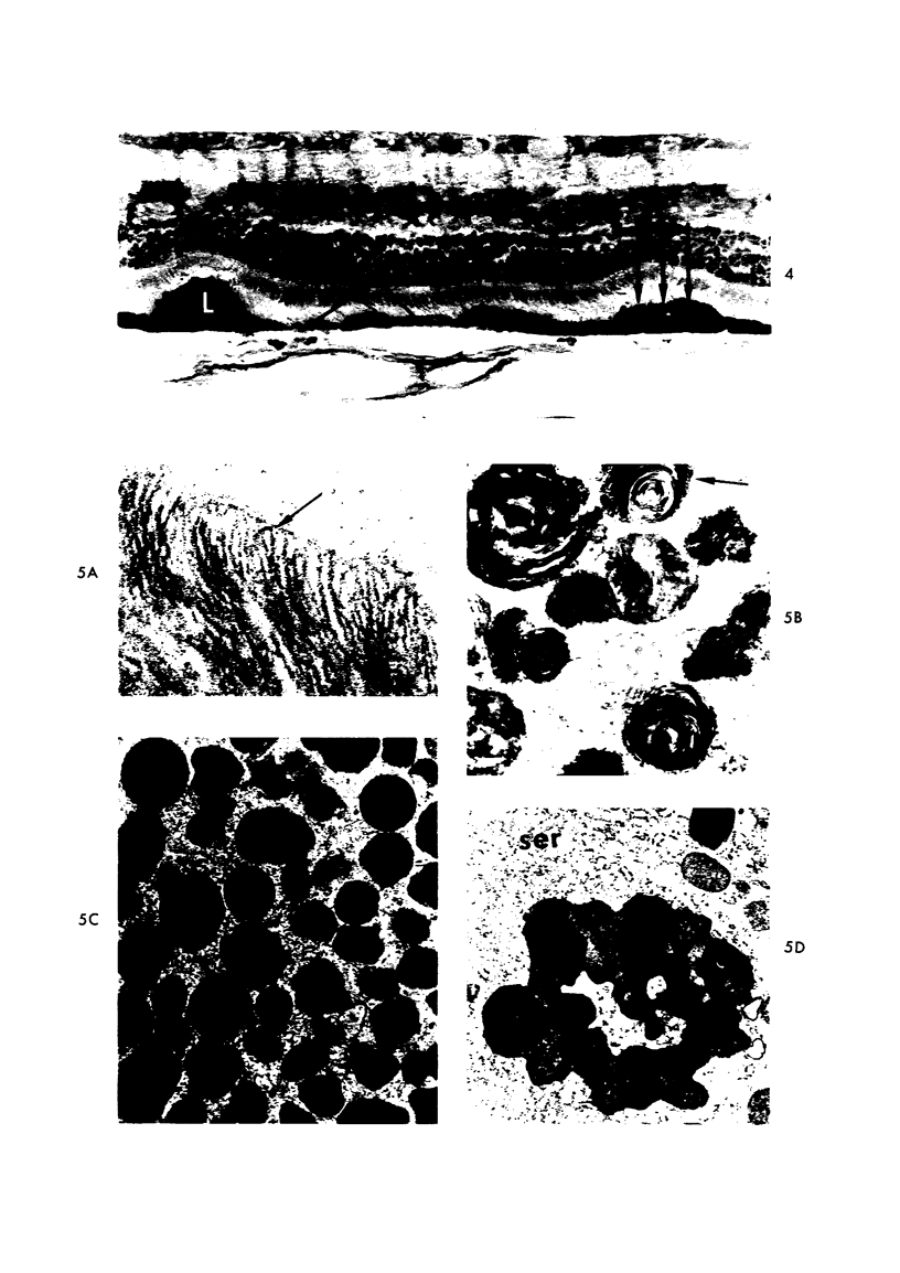
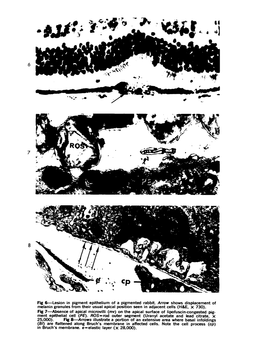
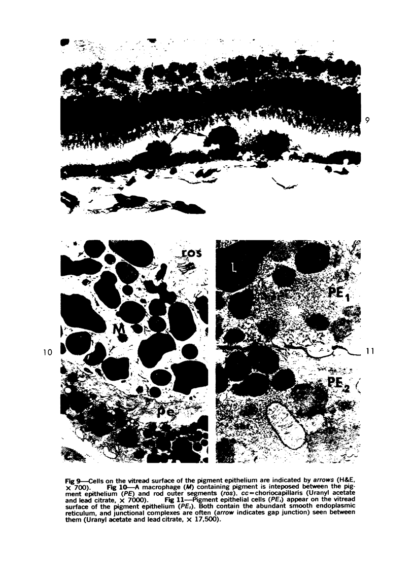
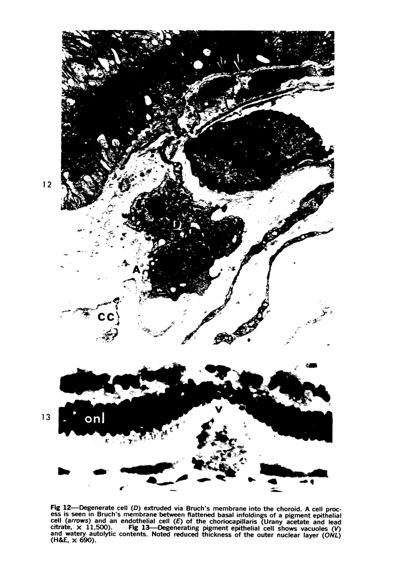
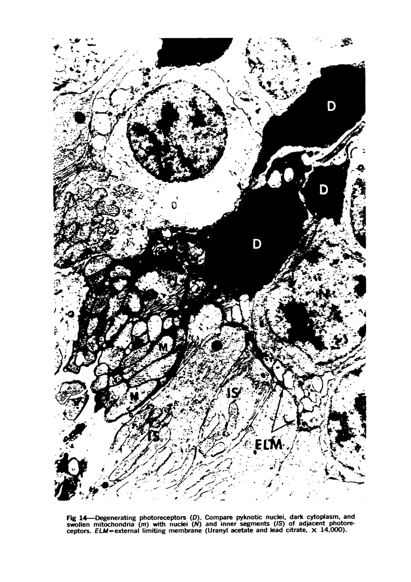
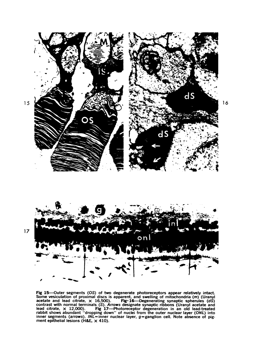
Multifocal lesions of the retinal pigment epithelium were observed in rabbits fed a diet contaning 0.5% lead subacetate for periods of up to 2 years. Groups of pigment epithelial cells became congested with a lipofuscin pigment which was apparently derived from phagosomes of rod outer segments. Lipofuscin granules displaced melanin granules from the apical surface of the retinal pigment epithelial cells and resulted in conspicuous brown pigmentation of these cells in albino animals. Migration of macrophages and pigment epithelial cells into the subretinal space was common in affected areas. This pathology was not observed in the pigment epithelium of the ora serrata, or ciliary body. At the latest time periods, the abnormal lipofuscin pigmentation subsided, and degeneration of photoreceptors occurred. The pathogenesis of the lesions is discussed.
Full text
PDF
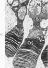
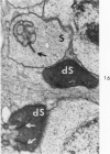
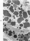
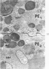
Images in this article
Selected References
These references are in PubMed. This may not be the complete list of references from this article.
- BUTTURINI U., GRIGNOLO A., BARONCHELLI A. Diabete da ditizone: aspetti metabolici, oculari ed istologici. G Clin Med. 1953 Nov;34(11):1253–1347. [PubMed] [Google Scholar]
- Bok D., Hall M. O. The role of the pigment epithelium in the etiology of inherited retinal dystrophy in the rat. J Cell Biol. 1971 Jun;49(3):664–682. doi: 10.1083/jcb.49.3.664. [DOI] [PMC free article] [PubMed] [Google Scholar]
- Burger P. C., Klintworth G. K. Experimental retinal degeneration in the rabbit produced by intraocular iron. Lab Invest. 1974 Jan;30(1):9–19. [PubMed] [Google Scholar]
- CIBIS P. A., BROWN E. B., HONG S. M. Ocular effects of systemic siderosis. Am J Ophthalmol. 1957 Oct;44(4 Pt 2):158–172. doi: 10.1016/0002-9394(57)90444-0. [DOI] [PubMed] [Google Scholar]
- Caley D. W., Johnson C., Liebelt R. A. The postnatal development of the retina in the normal and rodless CBA mouse: a light and electron microscopic study. Am J Anat. 1972 Feb;133(2):179–212. doi: 10.1002/aja.1001330205. [DOI] [PubMed] [Google Scholar]
- DOWLING J. E. Chemistry of visual adaptation in the rat. Nature. 1960 Oct 8;188:114–118. doi: 10.1038/188114a0. [DOI] [PubMed] [Google Scholar]
- DOWLING J. E. NUTRITIONAL AND INHERITED BLINDNESS IN THE RAT. Exp Eye Res. 1964 Dec;3:348–356. doi: 10.1016/s0014-4835(64)80042-7. [DOI] [PubMed] [Google Scholar]
- DOWLING J. E., SIDMAN R. L. Inherited retinal dystrophy in the rat. J Cell Biol. 1962 Jul;14:73–109. doi: 10.1083/jcb.14.1.73. [DOI] [PMC free article] [PubMed] [Google Scholar]
- Farkas T. G., Sylvester V., Archer D. The ultrastructure of drusen. Am J Ophthalmol. 1971 Jun;71(6):1196–1205. doi: 10.1016/0002-9394(71)90963-9. [DOI] [PubMed] [Google Scholar]
- Friedman E., Kuwabara T. The retinal pigment epithelium. IV. The damaging effects of radiant energy. Arch Ophthalmol. 1968 Aug;80(2):265–279. doi: 10.1001/archopht.1968.00980050267022. [DOI] [PubMed] [Google Scholar]
- Gloor B. P. Phagocytotische Aktivität des Pigmentepithels nach Lichtcoagulation. (Zur Frage der Herkunft von Makrophagen in der Retina) Albrecht Von Graefes Arch Klin Exp Ophthalmol. 1969;179(2):105–117. doi: 10.1007/BF00410377. [DOI] [PubMed] [Google Scholar]
- HASS G. M., BROWN D. V., EISENSTEIN R., HEMMENS A. RELATIONS BETWEEN LEAD POISONING IN RABBIT AND MAN. Am J Pathol. 1964 Nov;45:691–727. [PMC free article] [PubMed] [Google Scholar]
- Herron W. L., Jr, Riegel B. W. Production rate and removal of rod outer segment material in vitamin A deficiency. Invest Ophthalmol. 1974 Jan;13(1):46–53. [PubMed] [Google Scholar]
- Kuwabara T., Gorn R. A. Retinal damage by visible light. An electron microscopic study. Arch Ophthalmol. 1968 Jan;79(1):69–78. doi: 10.1001/archopht.1968.03850040071019. [DOI] [PubMed] [Google Scholar]
- Kuwabara T. Retinal recovery from exposure to light. Am J Ophthalmol. 1970 Aug;70(2):187–198. doi: 10.1016/0002-9394(70)90001-2. [DOI] [PubMed] [Google Scholar]
- LASANSKY A., DE ROBERTIS E. Submicroscopic changes in visual cells of the rabbit induced by iodoacetate. J Biophys Biochem Cytol. 1959 Mar 25;5(2):245–250. doi: 10.1083/jcb.5.2.245. [DOI] [PMC free article] [PubMed] [Google Scholar]
- Machemer R., Norton E. W. Experimental retinal detachment in the owl monkey. I. Methods of producation and clinical picture. Am J Ophthalmol. 1968 Sep;66(3):388–396. doi: 10.1016/0002-9394(68)91522-5. [DOI] [PubMed] [Google Scholar]
- NASSAR T. K., ISSIDORIDES M., SHANKLIN W. M. Concentric layers in the granules of human nervous lipofuscin demonstrated by silver impregnation. Stain Technol. 1960 Jan;35:15–18. doi: 10.3109/10520296009114709. [DOI] [PubMed] [Google Scholar]
- NOELL W. K. Experimentally induced toxic effects on structure and function of visual cells and pigment epithelium. Am J Ophthalmol. 1953 Jun;36(6 2):103–116. doi: 10.1016/0002-9394(53)90159-7. [DOI] [PubMed] [Google Scholar]
- Noell W. K., Walker V. S., Kang B. S., Berman S. Retinal damage by light in rats. Invest Ophthalmol. 1966 Oct;5(5):450–473. [PubMed] [Google Scholar]
- SORSBY A., NAKAJIMA A. Experimental degeneration of the retina. IV. Dia-minodiphenoxyalkanes as inducing agents. Br J Ophthalmol. 1958 Sep;42(9):563–571. doi: 10.1136/bjo.42.9.563. [DOI] [PMC free article] [PubMed] [Google Scholar]
- Spitznas M., Hogan M. J. Outer segments of photoreceptors and the retinal pigment epithelium. Interrelationship in the human eye. Arch Ophthalmol. 1970 Dec;84(6):810–819. doi: 10.1001/archopht.1970.00990040812022. [DOI] [PubMed] [Google Scholar]
- WEITZEL G., STRECKER F. J., ROESTER U., BUDDECKE E., FRETZDORFF A. M. Zink im Tapetum lucidum. Hoppe Seylers Z Physiol Chem. 1954;296(1-2):19–30. doi: 10.1515/bchm2.1954.296.1.19. [DOI] [PubMed] [Google Scholar]
- Young R. W., Bok D. Autoradiographic studies on the metabolism of the retinal pigment epithelium. Invest Ophthalmol. 1970 Jul;9(7):524–536. [PubMed] [Google Scholar]
- Young R. W., Bok D. Participation of the retinal pigment epithelium in the rod outer segment renewal process. J Cell Biol. 1969 Aug;42(2):392–403. doi: 10.1083/jcb.42.2.392. [DOI] [PMC free article] [PubMed] [Google Scholar]
- Young R. W. Shedding of discs from rod outer segments in the rhesus monkey. J Ultrastruct Res. 1971 Jan;34(1):190–203. doi: 10.1016/s0022-5320(71)90014-1. [DOI] [PubMed] [Google Scholar]


