Abstract
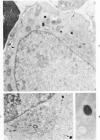
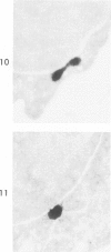
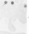
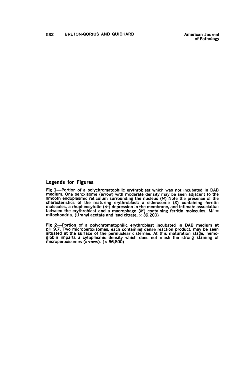
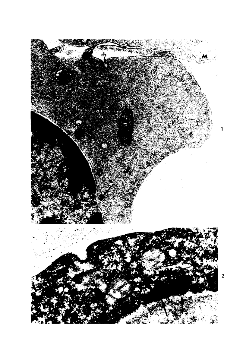
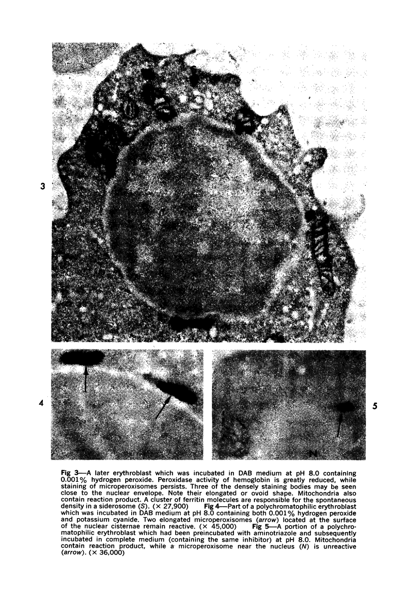
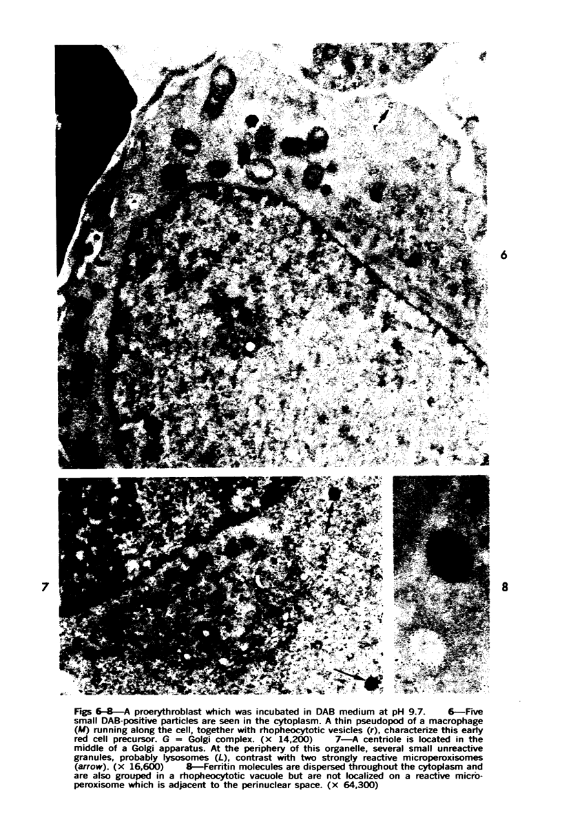
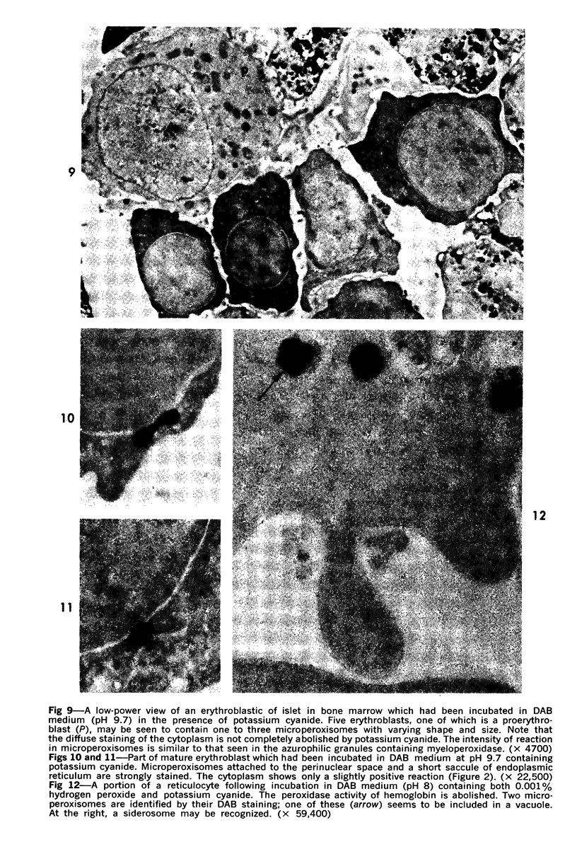
An alkaline diaminobenzidine (DAB) medium has been used to identify peroxidase activity in small granules (0.09 to 0.2 mu in diameter) present in all forms of maturing erythrocytic cells with the exception of erythrocytes. These granules, which were more frequent in proerythroblasts (from two to seven by thin section), were distinct from pleomorphic granules present in the close proximity to the Golgi apparatus. They were also distinct from ferritin molecules which were seen as aggregates in siderosomes of polychromatophilic erythroblasts. They often appeared in close association with the smooth membrane of the nuclear envelope. Optimal conditions for the visualization of these granules by incubation in alkaline DAB were obtained when the peroxidase activity of hemoglobin was reduced by addition of low concentrations of potassium cyanide. Lack of hydrogen peroxide in the incubation media completely inhibited the staining reaction of hemoglobin, while the positive reaction persisted in the granules. Aminotriazole in the incubation media prevented the staining of these organelles. These findings suggest that small granules seen in maturing erythroblasts contain catalase and that they correspond to microperoxisomes described in other tissues. The mechanism of their disappearance during reticulocyte maturation is unknown. The relationship between particulate catalase of erythroblasts and soluble erythrocytic catalase has not been elucidated.
Full text
PDF
Images in this article
Selected References
These references are in PubMed. This may not be the complete list of references from this article.
- Ackerman G. A. Ultrastructural localization of glycogen in erythrocytes and developing erythrocytic cells in normal human bone marrow. Z Zellforsch Mikrosk Anat. 1973 Jul 16;140(4):433–444. doi: 10.1007/BF00306670. [DOI] [PubMed] [Google Scholar]
- Aebi H. La catalase érythrocytaire. Expos Annu Biochim Med. 1969;29:139–166. [PubMed] [Google Scholar]
- Breton-Gorius J., Guichard J. Ultrastructural localization of peroxidase activity in human platelets and megakaryocytes. Am J Pathol. 1972 Feb;66(2):277–293. [PMC free article] [PubMed] [Google Scholar]
- Breton-Gorius J. Utilisation de la diaminobenzidine pour la mise en évidence, au microscope électronique, de l'hémoglobine intracellulaire. La réactivité des différents organelles des érythroblastes. Nouv Rev Fr Hematol. 1970 Mar-Apr;10(2):243–256. [PubMed] [Google Scholar]
- De Duve C., Baudhuin P. Peroxisomes (microbodies and related particles). Physiol Rev. 1966 Apr;46(2):323–357. doi: 10.1152/physrev.1966.46.2.323. [DOI] [PubMed] [Google Scholar]
- Dvorak A. M., Dvorak H. F., Karnovsky M. J. Cytochemical localization of peroxidase activity in the developing erythrocyte. Am J Pathol. 1972 May;67(2):303–326. [PMC free article] [PubMed] [Google Scholar]
- Fahimi H. D. Cytochemical localization of peroxidatic activity of catalase in rat hepatic microbodies (peroxisomes). J Cell Biol. 1969 Nov;43(2):275–288. doi: 10.1083/jcb.43.2.275. [DOI] [PMC free article] [PubMed] [Google Scholar]
- Graham R. C., Jr, Karnovsky M. J. The early stages of absorption of injected horseradish peroxidase in the proximal tubules of mouse kidney: ultrastructural cytochemistry by a new technique. J Histochem Cytochem. 1966 Apr;14(4):291–302. doi: 10.1177/14.4.291. [DOI] [PubMed] [Google Scholar]
- Hand A. R. Morphologic and cytochemical identification of peroxisomes in the rat parotid and other exocrine glands. J Histochem Cytochem. 1973 Feb;21(2):131–141. doi: 10.1177/21.2.131. [DOI] [PubMed] [Google Scholar]
- Herzog V., Fahimi H. D. The effect of glutaraldehyde on catalase. Biochemical and cytochemical studies with beef liver catalase and rat liver peroxisomes. J Cell Biol. 1974 Jan;60(1):303–311. doi: 10.1083/jcb.60.1.303. [DOI] [PMC free article] [PubMed] [Google Scholar]
- Hruban Z., Vigil E. L., Slesers A., Hopkins E. Microbodies: constituent organelles of animal cells. Lab Invest. 1972 Aug;27(2):184–191. [PubMed] [Google Scholar]
- Morikawa S., Harada T. Immunohistochemical localization of catalase in mammalian tissues. J Histochem Cytochem. 1969 Jan;17(1):30–35. doi: 10.1177/17.1.30. [DOI] [PubMed] [Google Scholar]
- Novikoff A. B., Goldfischer S. Visualization of peroxisomes (microbodies) and mitochondria with diaminobenzidine. J Histochem Cytochem. 1969 Oct;17(10):675–680. doi: 10.1177/17.10.675. [DOI] [PubMed] [Google Scholar]
- Novikoff A. B., Novikoff P. M., Davis C., Quintana N. Studies on microperoxisomes. II. A cytochemical method for light and electron microscopy. J Histochem Cytochem. 1972 Dec;20(12):1006–1023. doi: 10.1177/20.12.1006. [DOI] [PubMed] [Google Scholar]
- Novikoff A. B., Novikoff P. M., Davis C., Quintana N. Studies on microperoxisomes. V. Are microperoxisomes ubiquitous in mammalian cells? J Histochem Cytochem. 1973 Aug;21(8):737–755. doi: 10.1177/21.8.737. [DOI] [PubMed] [Google Scholar]
- Novikoff A. B., Novikoff P. M. Microperoxisomes. J Histochem Cytochem. 1973 Nov;21(11):963–966. doi: 10.1177/21.11.963. [DOI] [PubMed] [Google Scholar]
- Novikoff P. M., Novikoff A. B. Peroxisomes in absorptive cells of mammalian small intestine. J Cell Biol. 1972 May;53(2):532–560. doi: 10.1083/jcb.53.2.532. [DOI] [PMC free article] [PubMed] [Google Scholar]
- Roels F., Wisse E. Distinction cytochimique entre catalase et peroxydases. C R Acad Sci Hebd Seances Acad Sci D. 1973 Jan 15;276(3):391–393. [PubMed] [Google Scholar]
- Shio H., Farquhar M. G., de Duve C. Lysosomes of the arterial wall. IV. Cytochemical localization of acid phosphatase and catalase in smooth muscle cells and foam cells from rabbit atheromatous aorta. Am J Pathol. 1974 Jul;76(1):1–16. [PMC free article] [PubMed] [Google Scholar]


