Full text
PDF

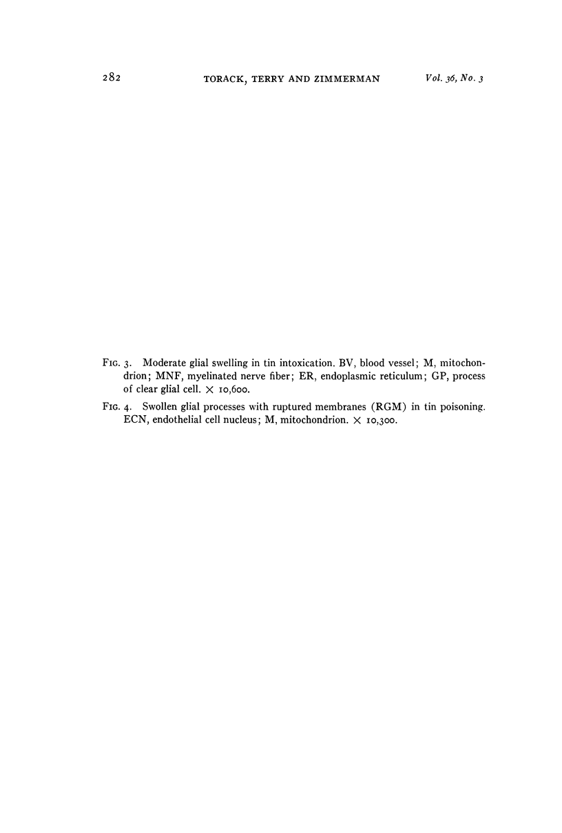
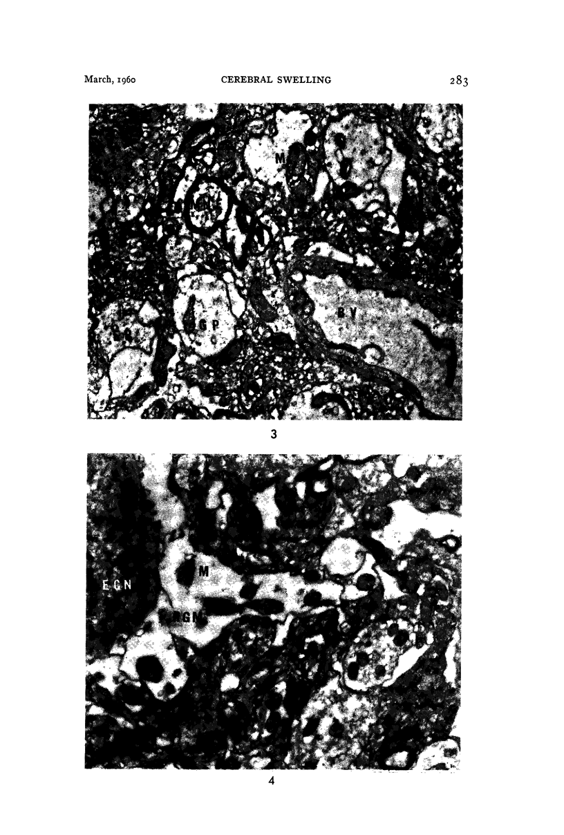
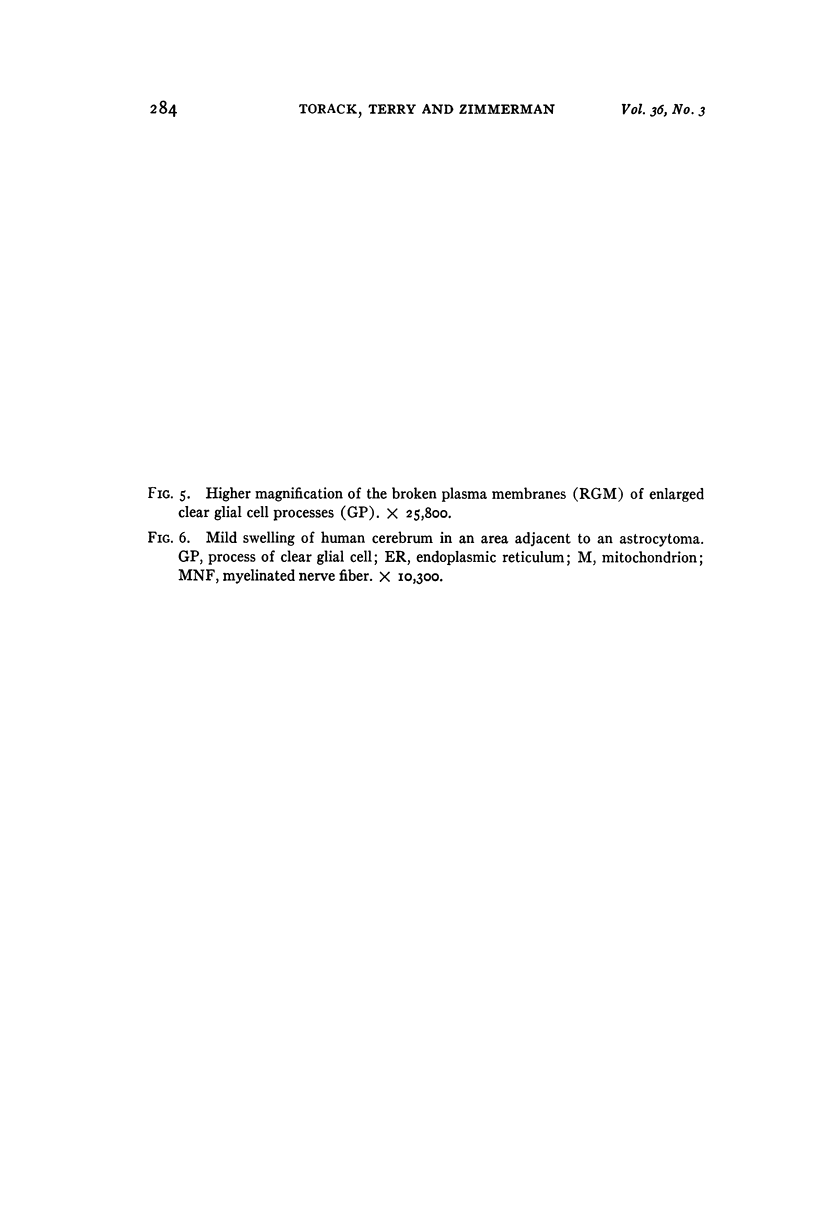
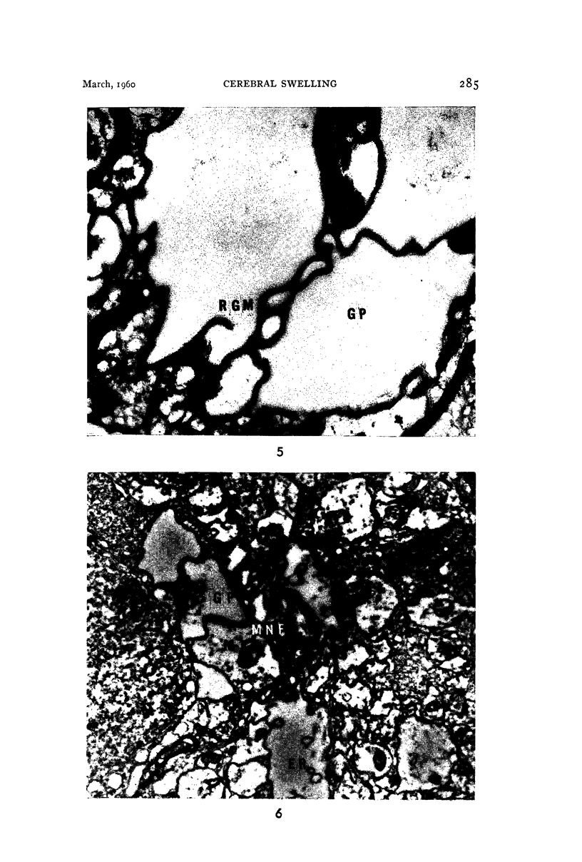
Images in this article
Selected References
These references are in PubMed. This may not be the complete list of references from this article.
- FARQUHAR M. G., HARTMANN J. F. Neuroglial structure and relationships as revealed by electron microscopy. J Neuropathol Exp Neurol. 1957 Jan;16(1):18–39. doi: 10.1097/00005072-195701000-00003. [DOI] [PubMed] [Google Scholar]
- GERSCHENFELD H. M., WALD F., ZADUNAISKY J. A., DE ROBERTIS E. D. Function of astroglia in the water-ion metabolism of the central nervous system: an electron microscope study. Neurology. 1959 Jun;9(6):412–425. doi: 10.1212/wnl.9.6.412. [DOI] [PubMed] [Google Scholar]
- PALADE G. E. A study of fixation for electron microscopy. J Exp Med. 1952 Mar;95(3):285–298. doi: 10.1084/jem.95.3.285. [DOI] [PMC free article] [PubMed] [Google Scholar]
- SCHULTZ R. L., MAYNARD E. A., PEASE D. C. Electron microscopy of neurons and neuroglia of cerebral cortex and corpus callosum. Am J Anat. 1957 May;100(3):369–407. doi: 10.1002/aja.1001000305. [DOI] [PubMed] [Google Scholar]


