Full text
PDF
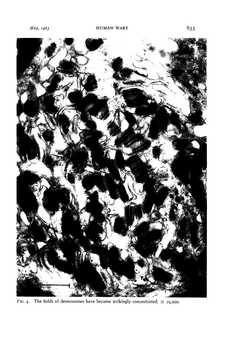
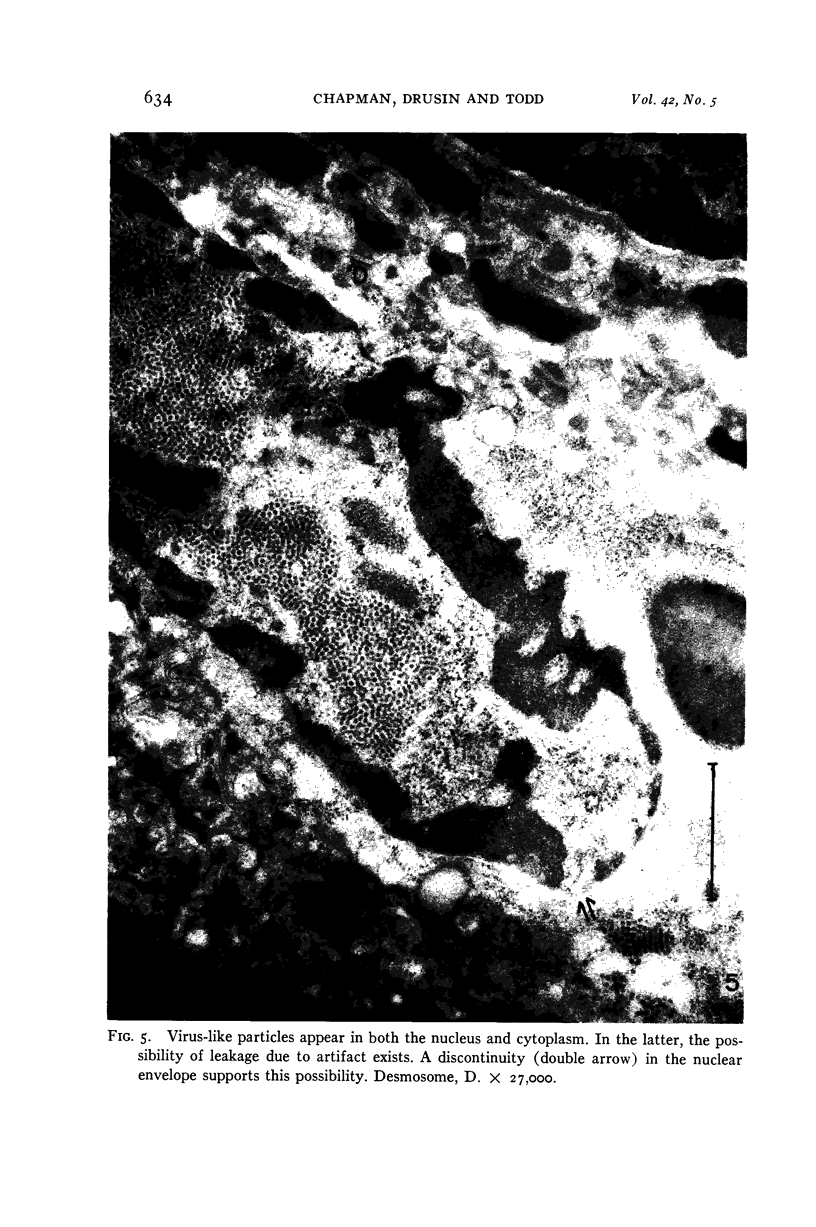
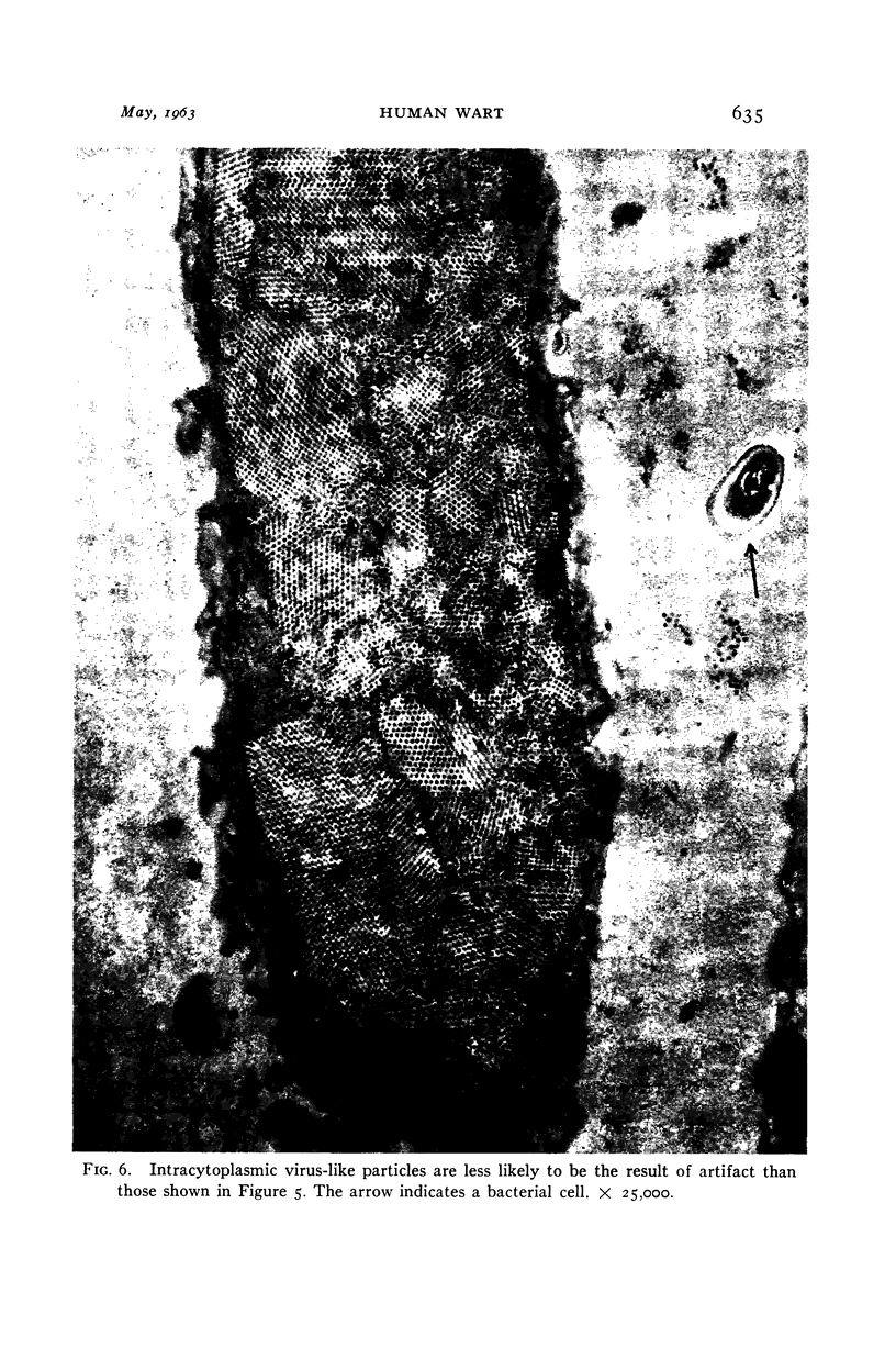
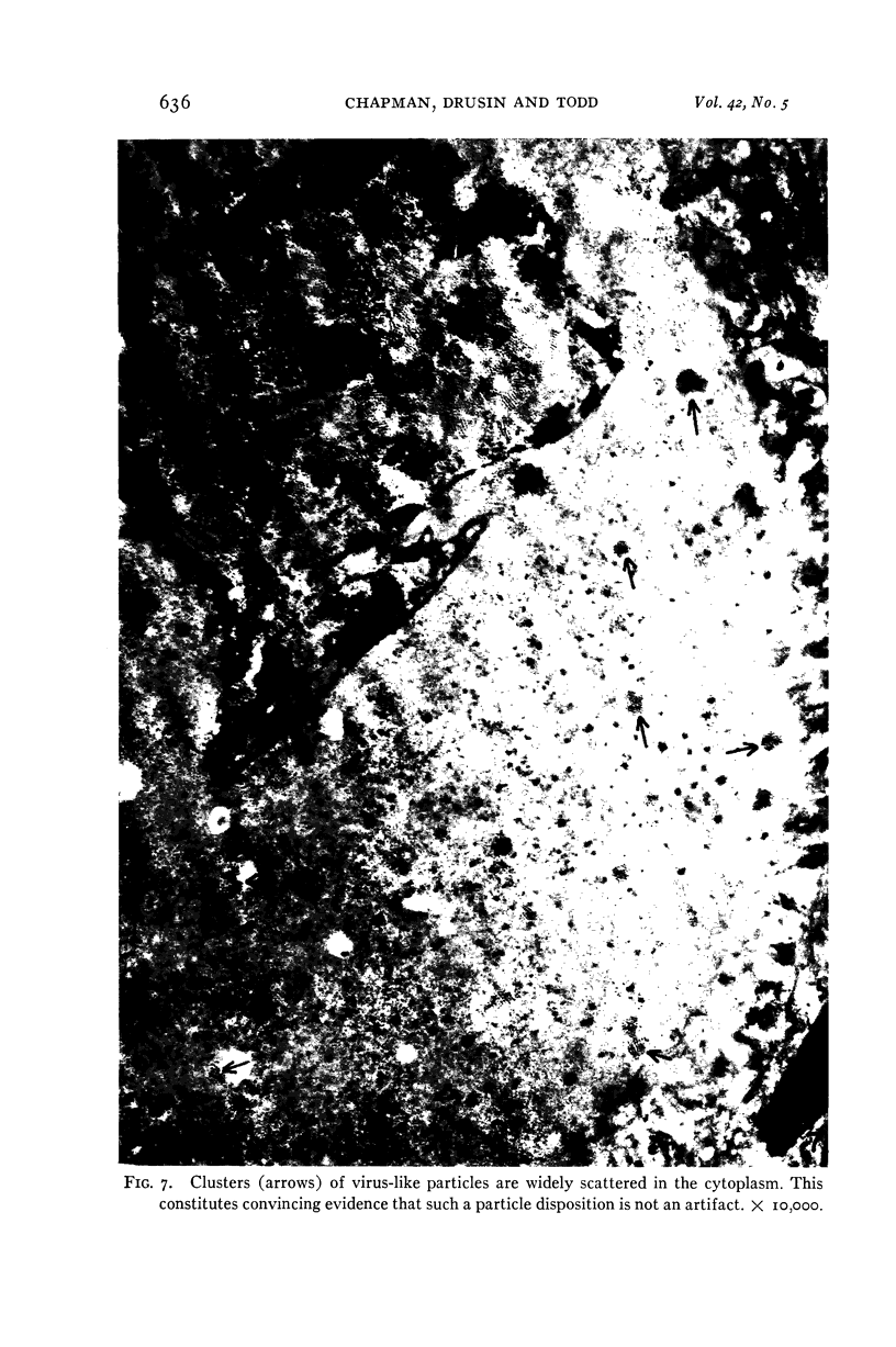
Images in this article
Selected References
These references are in PubMed. This may not be the complete list of references from this article.
- BLOCH D. P., GODMAN G. C. A cytological and cytochemical investigation of the development of the viral papilloma of human skin. J Exp Med. 1957 Feb 1;105(2):161–176. doi: 10.1084/jem.105.2.161. [DOI] [PMC free article] [PubMed] [Google Scholar]
- BUNTING H. Close-packed array of virus-like particles within cells of a human skin papilloma. Proc Soc Exp Biol Med. 1953 Nov;84(2):327–332. doi: 10.3181/00379727-84-20636. [DOI] [PubMed] [Google Scholar]
- BUSH G. L., CHAPMAN G. B. Electron microscopy of symbiotic bacteria in developing oocytes of the American cockroach, Periplaneta americana. J Bacteriol. 1961 Feb;81:267–276. doi: 10.1128/jb.81.2.267-276.1961. [DOI] [PMC free article] [PubMed] [Google Scholar]
- CHAPMAN G. B. Electron microscope observations on the behavior of the bacterial cytoplasmic membrane during cellular division. J Biophys Biochem Cytol. 1959 Oct;6:221–224. doi: 10.1083/jcb.6.2.221. [DOI] [PMC free article] [PubMed] [Google Scholar]
- CHAPMAN G. B., HANKS J. H., WALLACE J. H. An electron microscope study of the disposition and fine structure of Mycobacterium lepraemurium in mouse spleen. J Bacteriol. 1959 Feb;77(2):205–211. doi: 10.1128/jb.77.2.205-211.1959. [DOI] [PMC free article] [PubMed] [Google Scholar]
- CHAPMAN G. B., HILLIER J. Electron microscopy of ultra-thin sections of bacteria I. Cellular division in Bacillus cereus. J Bacteriol. 1953 Sep;66(3):362–373. doi: 10.1128/jb.66.3.362-373.1953. [DOI] [PMC free article] [PubMed] [Google Scholar]
- CHARLES A. Electron microscope observations on the human wart. Dermatologica. 1960 Oct;121:193–203. doi: 10.1159/000255270. [DOI] [PubMed] [Google Scholar]
- FRAJOLA W. J., GREIDER M. H., KLEINFELD R. G. Electron microscopy of intranuclear inclusions found in human and rat liver parenchymal cells. J Biophys Biochem Cytol. 1956 Jul 25;2(4 Suppl):435–438. doi: 10.1083/jcb.2.4.435. [DOI] [PMC free article] [PubMed] [Google Scholar]
- LUFT J. H. Improvements in epoxy resin embedding methods. J Biophys Biochem Cytol. 1961 Feb;9:409–414. doi: 10.1083/jcb.9.2.409. [DOI] [PMC free article] [PubMed] [Google Scholar]
- ORNSTEIN L. Mitochondrial and nuclear interaction. J Biophys Biochem Cytol. 1956 Jul 25;2(4 Suppl):351–352. doi: 10.1083/jcb.2.4.351. [DOI] [PMC free article] [PubMed] [Google Scholar]
- RICHARDSON K. C., JARETT L., FINKE E. H. Embedding in epoxy resins for ultrathin sectioning in electron microscopy. Stain Technol. 1960 Nov;35:313–323. doi: 10.3109/10520296009114754. [DOI] [PubMed] [Google Scholar]
- SIEGEL B. V. Electron microscopic studies of the virus of human warts. J Invest Dermatol. 1960 Aug;35:91–93. [PubMed] [Google Scholar]
- STONE R. S., SHOPE R. E., MOORE D. H. Electron microscope study of the development of the papilloma virus in the skin of the rabbit. J Exp Med. 1959 Oct 1;110:543–546. doi: 10.1084/jem.110.4.543. [DOI] [PMC free article] [PubMed] [Google Scholar]
- WATSON M. L. Staining of tissue sections for electron microscopy with heavy metals. J Biophys Biochem Cytol. 1958 Jul 25;4(4):475–478. doi: 10.1083/jcb.4.4.475. [DOI] [PMC free article] [PubMed] [Google Scholar]
- WILLIAMS M. G., HOWATSON A. F., ALMEIDA J. D. Morphological characterization of the virus of the human common wart (verruca vulgaris). Nature. 1961 Mar 18;189:895–897. doi: 10.1038/189895a0. [DOI] [PubMed] [Google Scholar]
- ZELICKSON A. S. Normal human keratinization processes as demonstrated by electron microscopy. J Invest Dermatol. 1961 Nov;37:369–379. [PubMed] [Google Scholar]


