

Abstract
We used the killing of Galleria mellonella (Lepidoptera: Pyralidae; the greater wax moth) caterpillar by the live vaccine strain (LVS) of Francisella tularensis to develop an invertebrate host system that can be used to study F. tularensis infection and the in vivo effects of antibacterial compounds on F. tularensis LVS. After injection into the insect hemocoel, F. tularensis LVS, killed caterpillars despite the association of LVS with hemocytes. The rate of killing depended on the number of bacteria injected. Antibiotic therapy with ciprofloxacin, levofloxacin or streptomycin administered before or after inoculation prolonged survival and decreased the tissue burden of F. tularensis in the hemocoel. Delayed drug treatment reduced the efficacy of antibacterials and especially streptomycin. The G. mellonella – F. tularensis LVS system may facilitate the in vivo study of F. tularensis, efficacy with antibacterial agents.
Keywords: Francisella tularensis, Galleria mellonella, tularemia, phagocytosis
1. Introduction
Francisella tularensis, the cause of tularemia, is one of the most infectious bacterial pathogens known. Two subspecies of major clinical importance have been identified, type A (subspecies tularensis) and type B (subspecies holarctica); type A is more virulent in humans [1]. Infection with F. tularensis subspecies tularensis (Type A) if not treated is associated with a high mortality rate (80% mortality for the pneumonic form and 10% for the ulceroglandular form) [2]. Due to the various routes of transmission and dissemination and the high virulence, F. tularensis is considered a category A bioterrorism agent by the Center for Disease Control and Prevention [3, 4].
The availability of genome sequence data for F. tularensis, new genetic tools and animal models, as well as studies in cell lines have aided in the study of this organism [5]. Mice have been used as a model system to study F. tularensis, while studies have shown that this pathogenic bacteria is also able to survive within amoebae [6] and is able to survive and grow within macrophages [7, 8]. An attenuated live vaccine strain (LVS) has been developed from F. tularensis subspecies holarctica [3]. The LVS confers some protection against tularemia infection in humans and in a number of animal models [9]; however, the basis of attenuation of LVS strain is not known. The vaccine is no longer available in the United States and its future is undetermined [3].
We propose to use the greater wax mother Galleria mellonella as a model host for F. tularensis LVS. A number of human pathogens have been studied in the G. mellonella pathosystem [10-16]. For example, the gram-negative bacterium Pseudomonas aeruginosa has been extensively studied in this system. Strain PA14, a human clinical isolate of P. aeruginosa, is pathogenic in mice and G. mellonella. Analysis of 32 different PA14 mutants in mice and G. mellonella showed a positive correlation in virulence patterns [11]. Pathogen defense is facilitated by the hemolymph system. G. mellonella is an attractive alternative host that offers the benefit of being able to maintained at a temperature range of 15 °C to 37 °C. It can be inoculated with a pathogen by means of injection [10] or by topical application [17].
We used the host-pathogen interaction between the G. mellonella larvae and the LVS of F. tularensis to develop an invertebrate host model to further study LVS. We are particularly interested in developing this model to study antibacterial activity to LVS as a model system for further exploration of antibacterial agents to F. tularensis virulent strains. We found that F. tularensis LVS killed G. mellonella larvae and killing of caterpillars depended on the number of bacteria in the inoculum and the incubation temperature following inoculation. The G. mellonella host response to F. tularensis involved melanization and association of bacteria with hemocytes. Antimicrobial therapy with streptomycin, levofloxacin or ciprofloxacin, but not ampicillin, administered before or after inoculation was effective in prolonging survival of caterpillars. However, in the case of streptomycin treatment, a delay in therapy by 24 hours neutralized the efficacy of this agent in G. mellonella survival.
2. Materials and Methods
2.1. Preparation of inoculum
The LVS of F. tularensis was used in all experiments. Bacterial cultures of LVS were maintained on Cysteine Heart Agar with 2% hemoglobin (CHA-H) and as frozen stocks. Frozen stocks of LVS were maintained on Brain Heart Infusion with 10% glycerol (BHI-G) at −80 °C. The concentration of LVS in the frozen stocks was 3×10⁸ Colony Forming Units (cfu)/ml, established by reading the Optical Density (OD) on a spectrophotometer and confirmed by plating serial dilutions on CHA-H plates and counting the cfu 72-96 hours later. For the heat-killing assay, LVS were exposed to 65 °C for 90 minutes. Heat killed LVS cells were washed in Phosphate Buffered Saline (PBS), resuspended and diluted to the appropriate concentration. For experiments utilizing heat-killed bacteria, the same concentration of the heat killed F. tularensis LVS was plated on CHA-H plates, to confirm the absence of colonies.
2.2. G. mellonella injection
G. mellonella caterpillars in the final larval stage (Vanderhorst Wholesale, St. Marys, Ohio) were stored in the dark. Caterpillars 0.30-0.35 g in weight were employed in all assays. A 10-μl Hamilton syringe was used to inject 10-μl aliquots of the inoculum into the hemocoel of each caterpillar via the last left proleg. Before injection, the area was cleaned using an alcohol swab. After injection, caterpillars were incubated in plastic containers. Sixteen randomly chosen caterpillars of the required weight were used per group and the number of dead caterpillars was scored daily. Caterpillars were considered dead when they displayed no movement in response to touch. For all the killing assays, we plotted killing curves and analyzed differences in survival with the Kaplan-Meier method using STATA 6 statistical software (Stata). A P value of less than 0.05 was considered significant.
The following control groups were used in all experiments. The first group included caterpillars that received doses of PBS. This group was used to assess the mortality related to trauma. A second group of larvae received no injection. In the studies evaluating antimicrobial efficacy, a third control group received the bacterial inoculum, but instead of antibacterial agents, received injection(s) with PBS. This control group was used as a control in the evaluation of the efficacy of the antimicrobial agents.
2.3. Administration of antimicrobials
A 10-μl Hamilton syringe was used to inject aliquots of ciprofloxacin 20 mg/kg, levofloxacin 15 mg/kg, streptomycin 15 mg/kg, or ampicillin 150 mg/kg. For experiments that required multiple injections, a different proleg was used for each injection, starting from the left last proleg and rotating left to right and moving proximally (i.e., injecting through the left last proleg, right last proleg, etc., as needed).
2.4. Tissue-burden culture studies
For the evaluation of the tissue burden of F. tularensis in caterpillars over time, the hemolymph from five caterpillars per group was collected into 1.5 ml Eppendorf tubes and serial dilutions were plated on CHA-H agar plates with 7.5 mg of colistin, 2.5 mg of amphotericin, 0.5 mg of lincomycin, 4 mg of trimethoprim, and 10 mg of ampicillin per liter [18]. Four groups (with 5 larvae each) were used for each condition. Plates were incubated at 37 °C for at least 72 hours before colonies were counted. Plotting was performed using Microsoft Excel, while the software program STATA 6 (Stata) was used for the statistical analysis of the cfu of G. mellonella in the hemocoel (Mann-Whitney and Kruskal-Wallis tests). A P value of less than 0.05 was considered significant.
2.5. Microscopy
Caterpillars after injection were maintained at 37 °C and 48 hours after inoculation, we collected their hemolymph into 1.5 ml Eppendorf tubes and fixed using 4% paraformaldehyde (Electron Microscopy Sciences, Fort Washington, Pennsylvania). Following fixation, the hemolymph was centrifuged and the supernatant was discarded. The cells were diluted in Grace's insect medium (Invitrogen, Carlsbad, California) and stained with mouse monoclonal antibody to F. tularensis lipopolysaccharide (LPS) as primary antibody (Abcam, Cambridge, Massachusetts), followed by staining with secondary monoclonal IgG2a Alexa Fluor 488 goat anti-mouse (Molecular Probes, Eugene, Oregon). Light and fluorescence microscopy were performed using an Olympus BX51 microscope and Vectashield mounting medium (Vector Laboratories, Youngstown, Ohio).
3. Results
3.1. Killing of G. mellonella by F. tularensis LVS
As shown in Fig. 1, inoculation of G. mellonella caterpillars with F. tularensis LVS at 37°C resulted in killing of the caterpillars, and killing depended on the number of bacteria injected. For example, after injection of 3×10⁵ cfu/larva, the median time to death was 3 days, while after injection of 3×10³ cfu/larva, the median time to death was >10 days. Injection of 3×10⁵ resulted in significantly faster killing compared to the control group of caterpillars that received sham injections with PBS (P<0.0001) and to the groups that received 3×10³ and 3×10⁴ cfus/larva (P<0.0001 for both). Moreover, injection of 3×10⁴ resulted in significantly faster killing compared to the group that received 3×10³ cfu/larva (P=0.0017). Of note, non-pathogenic microbial species do not kill G. mellonella. For example, killing of caterpillars by inocula of Escherichia coli (strain E. coli OP50), Saccharomyces cerevisiae or non-pathogenic strains of A. fumigatus as high as 1×10⁷ cfu/larva was not different previously than a control group that received PBS [10].
Fig. 1. Killing of G. mellonella caterpillars by F. tularensis LVS depends on the number of bacteria inoculated.

Kaplan-Meier plots of G. mellonella survival after injection of different inocula of F. tularensis LVS. Injection with 3×10⁵ cfu/larva resulted in significantly higher death rate, compared to injection with 3×10⁴ cfu/larva (P<0.0001), or 3×10³ cfu/larva (P<0.0001). Injection with 3×10⁴ cfu/larva resulted in significantly higher death rate compared to injection with 3×10³ cfu/larva (P=0.0017). There was no killing of caterpillars that received heat killed bacterial cells of the same strain (3×10⁵ cells/larva). The caterpillars were maintained at 37°C.
We also evaluated killing of G. mellonella at 30 °C. Similar to our findings at 37 °C, inoculation of G. mellonella with F. tularensis LVS resulted in killing of the caterpillars and killing depended on the number of bacterial cells injected. Again, in both cases, killing was significantly faster compared to a control group of caterpillars that received placebo injections with PBS (P<0.0001; Fig. 2). For example, injection of 3×10⁵ cfu/larva did not result in killing of all larvae at 30 °C, while it resulted in 100% mortality at 37 °C. Killing appeared to be more effective at 37 °C, a more effective growth temperature for F. tularensis.
Fig. 2. Killing of G. mellonella caterpillars by F. tularensis LVS depends on the temperature after inoculation.

Kaplan-Meier plots of G. mellonella survival after injection of different inocula of F. tularensis LVS. The caterpillars were maintained at 30°C. Injection with 3×10⁶ cfu/larva resulted in significantly higher death rate compared to controls and to injection with 3×10⁵ cfu/larvae. However, the killing rate at 30°C was slower than at 37°C (Figure 1), and at 30°C injection with 3×10⁵ cfu/larva was associated with significant survival (50% on Day 15).
3.2. Hemocytes are part of the G. mellonella response to F. tularensis
We found that the hemocytes are part of the G. mellonella response to F. tularensis and, by day 2 after inoculation, more than 90% of hemocytes are associated with F. tularensis bacteria (Fig. 3). Of note is that “nodulation” (encapsulation of large invading pathogens by layers of hemocytes) is part of the G. mellonella response to infection by large pathogens, such as fungi or parasites [10]. We did not find nodulation to be part of the insect response to F. tularensis and even when hemocytes adhered to each other, bacteria were bound to or phagocytosed by individual hemocytes and not placed between hemocytes. Melanization is also part of the insect reaction to infection [19] and over the progress of the F. tularensis infection, caterpillars demonstrated obvious signs of progressive melanization (Fig. 4).
Fig. 3. Association of F. tularensis LVS cells with G. mellonella hemocytes.

Hemocytes from caterpillars infected with F. tularensis LVS. After injection, caterpillars were maintained at 37°C and 48 hours after inoculation we collected their hemolymph. Following fixation, cells were diluted in Grace's insect medium and stained with mouse monoclonal antibody to F. tularensis lipopolysaccharide (LPS), as described under Materials and Methods. Left panel: Arrows point to FITC-labeled F. tularensis bacteria associated with a G. mellonella hemocyte. Right panel: Group of G. mellonella hemocytes with FITC-labeled associated F. tularensis bacteria.
Fig. 4. Melanization is a part of the insect response to infection by F. tularensis LVS.
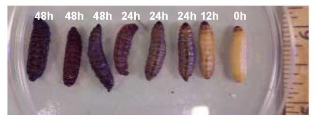
Over the progress of the F. tularensis infection, caterpillars demonstrated obvious signs of progressive melanization. From right to left: G. mellonella at different time points after challenge with an inoculum of the LVS of F. tularensis 2 × 10⁶ cfu/larva. Infected larvae show progressively increasing melanization. From right to left larvae are at 0, 12, 24 (three larvae) and 48 hours (the last three larvae) after inoculation. Petri dish diameter is 8.5 cm.
3.3. Study of antimicrobial agents in the G. mellonella - F. tularensis system
In order to evaluate if the G. mellonella - F. tularensis system could be used to study the effect of antibacterial agents, we investigated the role of antibacterials by administering single doses of streptomycin (15 mg/kg), ciprofloxacin (20 mg/kg), levofloxacin (15 mg/kg), or ampicillin (150 mg/kg), two hours after the inoculation of caterpillars with 3×10⁶ cfu/larva of F. tularensis LVS. Of note is that streptomycin is the most widely used treatment for management of severe human tularemia, while ciprofloxacin is an alternative treatment that is used in treatment or post-exposure prophylaxis [20].
Interestingly, streptomycin, ciprofloxacin and levofloxacin prolonged the survival of G. mellonella caterpillars at 37 °C compared to an infected control group that received ampicillin, an antibacterial that has no efficacy against LVS (Fig. 5; P values <0.0001 for all groups compared to controls and to the group received ampicillin). The doses of antibiotics used in these experiments were similar to the widely accepted doses of the same antibiotics in humans. The difference between ciprofloxacin, levofloxacin and streptomycin was not statistically significant.
Fig. 5. Antibacterial agents prolong the survival of G. mellonella caterpillars after challenge with F. tularensis LVS.

We examined the role of the most commonly used agents for F. tularensis infection by administering single doses of ciprofloxacin (CIP) 20 mg/kg, levofloxacin (LEV) 15 mg/kg, streptomycin (STR) 15 mg/kg or ampicillin (AMP) 150 mg/kg after the inoculation of caterpillars with 3×10⁶ cfu of F. tularensis LVS per larva. A control group received the F. tularensis inoculum and PBS instead of antibacterials. Treatment with ciprofloxacin, levofloxacin or streptomycin prolonged the survival of G. mellonella caterpillars (P< 0.0001 compared to control). However, ampicillin was not effective against F. tularensis LVS. The caterpillars were maintained at 37°C.
We also evaluated the efficacy of ciprofloxacin (20 mg/kg), levofloxacin (15mg/kg) or streptomycin (15 mg/kg) given one hour before inoculation with F. tularensis (3×10⁶ cfu/larva) (“prophylaxis” group). Again ciprofloxacin, levofloxacin and streptomycin were effective in prolonging the life span of caterpillars compared to a control group that received ampicillin 150 mg/kg (P<0.0001; Fig. 6A). In order to rule out any killing of larvae by ciprofloxacin, levofloxacin or streptomycin, we administered the same doses of these antibiotics alone to control groups of caterpillars. There were no deaths in these groups (data not shown).
Figure 6. Antibacterial agents before challenge with F. tularensis LVS prolong the survival of G. mellonella caterpillars.

A. We examined the role of the most commonly used agents for F. tularensis infection by administering a single dose of ciprofloxacin (CIP) 20 mg/kg, levofloxacin (LEV) 15 mg/kg, or streptomycin (STR) 15 mg/kg, one hour before infection with F. tularensis LVS 3×10⁶ cfu/larva. A control group received F. tularensis LVS and PBS instead of these drugs. Treatment with CIP, LEV or STR prolonged the survival of G. mellonella caterpillars (P<0.001 compared to control). Ampicillin (AMP) 150mg/kg given 1 hour before the infection with F. tularensis LVS 3×10⁶ cfu/larva did not protect G. mellonella caterpillars. B. We also examined the bacterial burden in the hemolymph after 48 hours using a single dose of ampicillin, ciprofloxacin, levofloxacin or streptomycin. A control group received treatment with PBS. CFU were counted per five larvae. The caterpillars were maintained at 37°C.
For the evaluation of the effectiveness of ciprofloxacin, levofloxacin and streptomycin in the reduction of the bacterial burden in the hemolymph, we administered the above noted doses of antibiotics after infection with 3×10⁴ cfu/larva; another group received PBS after infection. Ciprofloxacin, levofloxacin and streptomycin resulted in statistically significant reductions in bacterial counts compared to ampicillin and controls (P=0.02). There were no statistically significant differences between the groups that received ciprofloxacin, levofloxacin or streptomycin (Fig. 6B).
We further evaluated the efficacy of streptomycin (15mg/kg), ciprofloxacin (20mg/kg) and levofloxacin (15mg/kg) when administered for two doses at a delayed time point from the time of inoculation. G. mellonella infected with LVS were treated at 6 hours post infection followed by an additional drug dose at 24 hours. Treatment with streptomycin, ciprofloxacin and levofloxacin all significantly increased survival when drugs were administered 6 hours and 24 hours after infection with LVS compared to control groups (P=0.0075 for streptomycin and P<0.0001 for ciprofloxacin and levofloxacin respectively).
In another delayed drug dose assay, we delivered a dose of the antibacterial agent at 24 hours post infection, followed by an additional dose at 48 hours post infection. Treatment with ciprofloxacin and levofloxacin remained significant compared to treatment with PBS at 24 and 48 hours with P<0.0001 and P=0.0001 respectively. However, streptomycin was not significantly better compared to LVS infected G. mellonella that received placebo treatment (P=0.1071).
4. Discussion
In this report, we describe the use of the G. mellonella larvae and the LVS of F. tularensis to develop an invertebrate host system for the study of tularemia and the efficacy of antibacterial agents. We found that the LVS of F. tularensis killed G. mellonella larvae. Killing of larvae depended on the number of bacteria in the inoculum and the incubation temperature following inoculation. The G. mellonella host response to F. tularensis infection involved melanization and phagocytosis. Antimicrobial therapy with ciprofloxacin, levofloxacin or streptomycin administered before or after inoculation prolonged survival of larvae.
G. mellonella (the greater wax moth) is a unique invertebrate host model because it can survive at mammalian temperatures and can be used for the study of microbial pathogenesis, including phagocytosis. The host response of G. mellonella to infection consists of structural and passive barriers, as well as cellular and humoral responses that are mediated by hemocytes within the hemolymph. Different types of hemocytes have been identified in G. mellonella and the insect response includes phagocytosis, “nodulation” (encapsulation of large invading pathogens by layers of hemocytes), and melanization [21].
In addition to in vivo defense responses, the use of drug treatment for LVS can be evaluated in the G. mellonella model system. Drug treatment with ciprofloxacin or levofloxacin significantly prolongs survival of LVS infected larvae under each condition. Streptomycin significantly prolonged survival of G. mellonella larvae when administered at 2 or 6 hours after infection. However, when streptomycin treatment was delayed to 24 hours post infection, there was no significant difference compared to G. mellonella infected with LVS that received PBS treatment. Delayed drug treatment has also been shown to decrease survival in a mouse model system infected with F. tularensis [22].
The aminoglycosides streptomycin and gentamicin are bactericidal against F. tularensis and are currently the drugs of choice for the treatment of tularemia infection in humans [20]. Fluoroquinolones have good bactericidal activity against F. tularensis in vitro [23]. In contrast, beta-lactams, macrolides, lincosamides, and co-trimoxazole are not reliable for treatment of tularemia. Our results with antibacterials demonstrated efficacy in the G. mellonella system that was similar to results from clinical trials [3, 23].
Important limitations of the G. mellonella model should be considered. The genome of this organism has not been sequenced; this means that G. mellonella has limited genetic tractability compared to other invertebrate models such as Caenorhabditis elegans and Drosophila melanogaster [10]. Also, administration of inocula by injection makes high throughput screening difficult. Finally, the G. mellonella-F. tularensis LVS model requires the inoculation of larvae with a significant number of bacteria. This suggests that the hemocytes are able to clear a smaller number of F. tularensis LVS bacteria. It is anticipated that virulent bacterial strains such as F. tularensis Schu 4 will be able to kill G. mellonella larvae at significantly lower inocula than the LVS strain.
In conclusion, G. mellonella is a facile model that complements more cumbersome and expensive mammalian models and may minimize mammalian suffering for the study of F. tularensis pathogenesis. This host model provides a novel approach to the study of F. tularensis host-pathogen interactions at both mammalian and lower temperatures, study of the interaction of F. tularensis with insects, and evaluation of drug sensitivity profiles in vivo. Future studies are needed to investigate the use strains such as F. tularensis Schu 4, in order to establish the usefulness of this model in the study of virulent strains.
Acknowledgments
We appreciate the support of the New England Regional Center of Excellence for Biodefense/Emerging Infectious Diseases Research; grant number AI057159. We thank Drs. Andrew B. Onderdonk and Gerald A. Beltz for providing the F. tularensis LVS, advice and critical review of the manuscript. We also acknowledge support provided by a K08 award AI63084 from NIH and a New Scholar Award in Global Infectious Diseases of the Ellison Medical Foundation to E.M.
Abbreviations
- BHI-G
Brain Heart Infusion with 10% Glycerol
- CFU
Colony Forming Units
- CHA-H
Cysteine Heart Agar with 2% Hemoglobin
- LVS
Live Vaccine Strain
- PBS
phosphate-buffered saline
Footnotes
Publisher's Disclaimer: This is a PDF file of an unedited manuscript that has been accepted for publication. As a service to our customers we are providing this early version of the manuscript. The manuscript will undergo copyediting, typesetting, and review of the resulting proof before it is published in its final citable form. Please note that during the production process errors may be discovered which could affect the content, and all legal disclaimers that apply to the journal pertain.
References
- 1.Tarnvik A. Nature of protective immunity to Francisella tularensis. Rev. Infect. Dis. 1989;11:440–451. [PubMed] [Google Scholar]
- 2.Prior RG, Klasson L, Larsson P, Williams K, Lindler L, Sjostedt A, Svensson T, Tamas I, Wren BW, Oyston PC, Andersson SG, Titball RW. Preliminary analysis and annotation of the partial genome sequence of Francisella tularensis strain Schu 4. J. Appl. Microbiol. 2001;91:614–620. doi: 10.1046/j.1365-2672.2001.01499.x. [DOI] [PubMed] [Google Scholar]
- 3.Dennis DT, Inglesby TV, Henderson DA, Bartlett JG, Ascher MS, Eitzen E, Fine AD, Friedlander AM, Hauer J, Layton M, Lillibridge SR, McDade JE, Osterholm MT, O'Toole T, Parker G, Perl TM, Russell PK, Tonat K. Tularemia as a biological weapon: medical and public health management. Jama. 2001;285:2763–2773. doi: 10.1001/jama.285.21.2763. [DOI] [PubMed] [Google Scholar]
- 4.Ellis J, Oyston PC, Green M, Titball RW. Tularemia. Clin. Microbiol. Rev. 2002;15:631–646. doi: 10.1128/CMR.15.4.631-646.2002. [DOI] [PMC free article] [PubMed] [Google Scholar]
- 5.Hernychova L, Stulik J, Halada P, Macela A, Kroca M, Johansson T, Malina M. Construction of a Francisella tularensis two-dimensional electrophoresis protein database. Proteomics. 2001;1:508–515. doi: 10.1002/1615-9861(200104)1:4<508::AID-PROT508>3.0.CO;2-K. [DOI] [PubMed] [Google Scholar]
- 6.Lauriano CM, Barker JR, Yoon SS, Nano FE, Arulanandam BP, Hassett DJ, Klose KE. MglA regulates transcription of virulence factors necessary for Francisella tularensis intraamoebae and intramacrophage survival. Proc. Natl. Acad. Sci. U. S. A. 2004;101:4246–4249. doi: 10.1073/pnas.0307690101. [DOI] [PMC free article] [PubMed] [Google Scholar]
- 7.Clemens DL, Lee BY, Horwitz MA. Francisella tularensis enters macrophages via a novel process involving pseudopod loops. Infect. Immun. 2005;73:5892–5902. doi: 10.1128/IAI.73.9.5892-5902.2005. [DOI] [PMC free article] [PubMed] [Google Scholar]
- 8.Golovliov I, Baranov V, Krocova Z, Kovarova H, Sjostedt A. An attenuated strain of the facultative intracellular bacterium Francisella tularensis can escape the phagosome of monocytic cells. Infect. Immun. 2003;71:5940–5950. doi: 10.1128/IAI.71.10.5940-5950.2003. [DOI] [PMC free article] [PubMed] [Google Scholar]
- 9.Burke DS. Immunization against tularemia: analysis of the effectiveness of live Francisella tularensis vaccine in prevention of laboratory-acquired tularemia. J. Infect. Dis. 1977;135:55–60. doi: 10.1093/infdis/135.1.55. [DOI] [PubMed] [Google Scholar]
- 10.Mylonakis E, Moreno R, El Khoury JB, Idnurm A, Heitman J, Calderwood SB, Ausubel FM. A.Diener Galleria mellonella as a model system to study Cryptococcus neoformans pathogenesis. Infect. Immun. 2005;73:3842–50. doi: 10.1128/IAI.73.7.3842-3850.2005. [DOI] [PMC free article] [PubMed] [Google Scholar]
- 11.Jander G, Rahme LG, Ausubel FM. Positive correlation between virulence of Pseudomonas aeruginosa mutants in mice and insects. J. Bacteriol. 2000;182:3843–3845. doi: 10.1128/jb.182.13.3843-3845.2000. [DOI] [PMC free article] [PubMed] [Google Scholar]
- 12.Miyata S, Casey M, Frank DW, Ausubel FM, Drenkard E. Use of the Galleria mellonella caterpillar as a model host to study the role of the type III secretion system in Pseudomonas aeruginosa pathogenesis. Infect. Immun. 2003;71:2404–2413. doi: 10.1128/IAI.71.5.2404-2413.2003. [DOI] [PMC free article] [PubMed] [Google Scholar]
- 13.Morton DB, Barnett RI, Chadwick JS. Structural alterations to Proteus mirabilis as a result of exposure to haemolymph from the larvae of Galleria mellonella. Microbios. 1984;39:177–185. [PubMed] [Google Scholar]
- 14.Morton DB, Dunphy GB, Chadwick JS. Reactions of hemocytes of immune and non-immune Galleria mellonella larvae to Proteus mirabilis. Dev. Comp Immunol. 1987;11:47–55. doi: 10.1016/0145-305x(87)90007-3. [DOI] [PubMed] [Google Scholar]
- 15.Wang C, Typas MA, Butt TM. Detection and characterisation of pr1 virulent gene deficiencies in the insect pathogenic fungus Metarhizium anisopliae. FEMS Microbiol. Lett. 2002;213:251–255. doi: 10.1111/j.1574-6968.2002.tb11314.x. [DOI] [PubMed] [Google Scholar]
- 16.Leger RJ, Screen SE, Shams-Pirzadeh B. Lack of host specialization in Aspergillus flavus. Appl. Environ. Microbiol. 2000;66:320–324. doi: 10.1128/aem.66.1.320-324.2000. [DOI] [PMC free article] [PubMed] [Google Scholar]
- 17.Scully LR, Bidochka MJ. Serial passage of the opportunistic pathogen Aspergillus flavus through an insect host yields decreased saprobic capacity. Can. J. Microbiol. 2005;51:185–189. doi: 10.1139/w04-124. [DOI] [PubMed] [Google Scholar]
- 18.Petersen JM, Schriefer ME, Gage KL, Montenieri JA, Carter LG, Stanley M, Chu MC. Methods for enhanced culture recovery of Francisella tularensis. Appl. Environ. Microbiol. 2004;70:3733–3735. doi: 10.1128/AEM.70.6.3733-3735.2004. [DOI] [PMC free article] [PubMed] [Google Scholar]
- 19.Slepneva IA, Komarov DA, Glupov VV, Serebrov VV, Khramtsov VV. Influence of fungal infection on the DOPA-semiquinone and DOPA-quinone production in haemolymph of Galleria mellonella larvae. Biochem. Biophys. Res. Commun. 2003;300:188–191. doi: 10.1016/s0006-291x(02)02766-3. [DOI] [PubMed] [Google Scholar]
- 20.Anonymous Tularemia: Current, comprehensive information on pathogenesis, microbiology, epidemiology, diagnosis, treatment, and prophylaxis. http://www.cidrap.umn.edu/idsa/bt/tularemia/biofacts/tularemiafactsheet.html. access confirmed on August 18, 2005
- 21.Kavanagh K, Reeves EP. Exploiting the potential of insects for in vivo pathogenicity testing of microbial pathogens. FEMS Microbiol. Rev. 2004;28:101–12. doi: 10.1016/j.femsre.2003.09.002. [DOI] [PubMed] [Google Scholar]
- 22.Piercy T, Steward J, Lever MS, Brooks TJ. In vivo efficacy of fluoroquinolones against systemic tularaemia infection in mice. J. Antimicrob. Chemother. 2005;56:1069–1073. doi: 10.1093/jac/dki359. [DOI] [PubMed] [Google Scholar]
- 23.Perez-Castrillon JL, Bachiller-Luque P, Martin-Luquero M, Mena-Martin FJ, Herreros V. Tularemia epidemic in northwestern Spain: clinical description and therapeutic response. Clin. Infect. Dis. 2001;33:573–576. doi: 10.1086/322601. [DOI] [PubMed] [Google Scholar]