Abstract
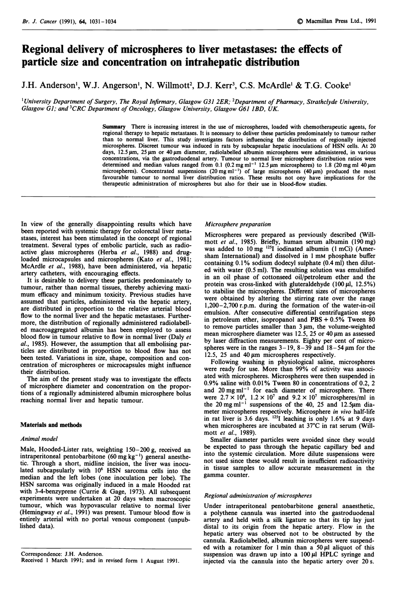
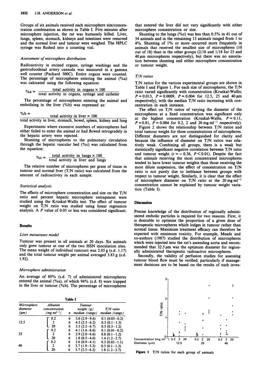
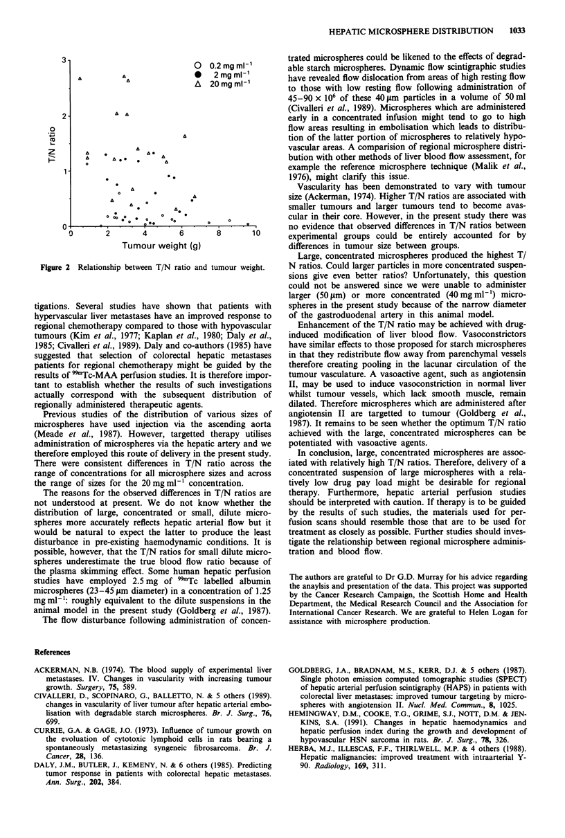
There is increasing interest in the use of microspheres, loaded with chemotherapeutic agents, for regional therapy to hepatic metastases. It is necessary to deliver these particles predominately to tumour rather than to normal liver. This study investigates factors influencing the distribution of regionally injected microspheres. Discreet tumour was induced in rats by subcapsular hepatic inoculations of HSN cells. At 20 days, 12.5 microns, 25 microns or 40 microns diameter, radiolabelled albumin microspheres were administered, in various concentrations, via the gastroduodenal artery. Tumour to normal liver microsphere distribution ratios were determined and median values ranged from 0.1 (0.2 mg ml-1 12.5 microns microspheres) to 1.8 (20 mg ml 40 microns microspheres). Concentrated suspensions (20 mg ml-1) of large microspheres (40 microns) produced the most favourable tumour to normal liver distribution ratios. These results not only have implications for the therapeutic administration of microspheres but also for their use in blood-flow studies.
Full text
PDF


Selected References
These references are in PubMed. This may not be the complete list of references from this article.
- Ackerman N. B. The blood supply of experimental liver metastases. IV. Changes in vascularity with increasing tumor growth. Surgery. 1974 Apr;75(4):589–596. [PubMed] [Google Scholar]
- Civalleri D., Scopinaro G., Balletto N., Claudiani F., De Cian F., DeCian F., Camerini G., DePaoli M., Bonalumi U. Changes in vascularity of liver tumours after hepatic arterial embolization with degradable starch microspheres. Br J Surg. 1989 Jul;76(7):699–703. doi: 10.1002/bjs.1800760716. [DOI] [PubMed] [Google Scholar]
- Currie G. A., Gage J. O. Influence of tumour growth on the evolution of cytotoxic lymphoid cells in rats bearing a spontaneously metastasizing syngeneic fibrosarcoma. Br J Cancer. 1973 Aug;28(2):136–146. doi: 10.1038/bjc.1973.131. [DOI] [PMC free article] [PubMed] [Google Scholar]
- Daly J. M., Butler J., Kemeny N., Yeh S. D., Ridge J. A., Botet J., Bading J. R., DeCosse J. J., Benua R. S. Predicting tumor response in patients with colorectal hepatic metastases. Ann Surg. 1985 Sep;202(3):384–393. doi: 10.1097/00000658-198509000-00017. [DOI] [PMC free article] [PubMed] [Google Scholar]
- Goldberg J. A., Bradnam M. S., Kerr D. J., McKillop J. H., Bessent R. G., McArdle C. S., Willmott N., George W. D. Single photon emission computed tomographic studies (SPECT) of hepatic arterial perfusion scintigraphy (HAPS) in patients with colorectal liver metastases: improved tumour targetting by microspheres with angiotensin II. Nucl Med Commun. 1987 Dec;8(12):1025–1032. doi: 10.1097/00006231-198712000-00012. [DOI] [PubMed] [Google Scholar]
- Hemingway D. M., Cooke T. G., Grime S. J., Nott D. M., Jenkins S. A. Changes in hepatic haemodynamics and hepatic perfusion index during the growth and development of hypovascular HSN sarcoma in rats. Br J Surg. 1991 Mar;78(3):326–330. doi: 10.1002/bjs.1800780319. [DOI] [PubMed] [Google Scholar]
- Herba M. J., Illescas F. F., Thirlwell M. P., Boos G. J., Rosenthall L., Atri M., Bret P. M. Hepatic malignancies: improved treatment with intraarterial Y-90. Radiology. 1988 Nov;169(2):311–314. doi: 10.1148/radiology.169.2.3174978. [DOI] [PubMed] [Google Scholar]
- Kaplan W. D., Ensminger W. D., Come S. E., Smith E. H., D'Orsi C. J., Levin D. C., Takvorian R. W., Steele G. D., Jr Radionuclide angiography to predict patient response to hepatic artery chemotherapy. Cancer Treat Rep. 1980;64(12):1217–1222. [PubMed] [Google Scholar]
- Kato T., Nemoto R., Mori H., Takahashi M., Harada M. Arterial chemoembolization with mitomycin C microcapsules in the treatment of primary or secondary carcinoma of the kidney, liver, bone and intrapelvic organs. Cancer. 1981 Aug 1;48(3):674–680. doi: 10.1002/1097-0142(19810801)48:3<674::aid-cncr2820480303>3.0.co;2-e. [DOI] [PubMed] [Google Scholar]
- Kim D. K., Watson R. C., Pahnke L. D., Fortner J. G. Tumor vascularity as a prognostic factor for hepatic tumors. Ann Surg. 1977 Jan;185(1):31–34. doi: 10.1097/00000658-197701000-00005. [DOI] [PMC free article] [PubMed] [Google Scholar]
- Malik A. B., Kaplan J. E., Saba T. M. Reference sample method for cardiac output and regional blood flow determinations in the rat. J Appl Physiol. 1976 Mar;40(3):472–475. doi: 10.1152/jappl.1976.40.3.472. [DOI] [PubMed] [Google Scholar]
- McArdle C. S., Lewi H., Hansell D., Kerr D. J., McKillop J., Willmott N. Cytotoxic-loaded albumin microspheres: a novel approach to regional chemotherapy. Br J Surg. 1988 Feb;75(2):132–134. doi: 10.1002/bjs.1800750214. [DOI] [PubMed] [Google Scholar]
- Meade V. M., Burton M. A., Gray B. N., Self G. W. Distribution of different sized microspheres in experimental hepatic tumours. Eur J Cancer Clin Oncol. 1987 Jan;23(1):37–41. doi: 10.1016/0277-5379(87)90416-0. [DOI] [PubMed] [Google Scholar]
- Willmott N., Chen Y., Goldberg J., Mcardle C., Florence A. T. Biodegradation rate of embolized protein microspheres in lung, liver and kidney of rats. J Pharm Pharmacol. 1989 Jul;41(7):433–438. doi: 10.1111/j.2042-7158.1989.tb06496.x. [DOI] [PubMed] [Google Scholar]
- Willmott N., Cummings J., Stuart J. F., Florence A. T. Adriamycin-loaded albumin microspheres: preparation, in vivo distribution and release in the rat. Biopharm Drug Dispos. 1985 Jan-Mar;6(1):91–104. doi: 10.1002/bdd.2510060111. [DOI] [PubMed] [Google Scholar]


