Abstract
Living organisms are continually under attack from a vast array of DNA-damaging agents that imperils their genomic integrity. As a consequence, cells posses an army of enzymes to repair their damaged chromosomes. However, DNA lesions often persist and pose a considerable threat to survival, because they can block the cell's replicase and its ability to complete genome duplication. It has been clear for many years that cells must possess a mechanism whereby the DNA lesion could be tolerated and physically bypassed. Yet it was only within the past decade that specialized DNA polymerases for “translesion DNA synthesis” or “TLS” were identified and characterized. Many of the TLS enzymes belong to the recently described “Y-family” of DNA polymerases. By possessing a spacious preformed active site, these enzymes can physically accommodate a variety of DNA lesions and facilitate their bypass. Flexible DNA-binding domains and a variable binding pocket for the replicating base pair further allow these TLS polymerases to select specific lesions to bypass and favor distinct non-Watson–Crick base pairs. Consequently, TLS polymerases tend to exhibit much lower fidelity than the cell's replicase when copying normal DNA, which results in a dramatic increase in mutagenesis. Occasionally this can be beneficial, but it often speeds the onset of cancer in humans. Cells use both transcriptional and posttranslational regulation to keep these low-fidelity polymerases under strict control and limit their access to a replication fork. Our perspective focuses on the mechanistic insights into TLS by the Y-family polymerases, how they are regulated, and their effects on genomic (in)stability that have been described in the past decade.
Keywords: catalysis, regulation, ubiquitylation, Y-family polymerases
Discovery of the Y-Family of DNA Polymerases
It was clear from genetic studies carried out in the mid-1970s that damage-induced mutagenesis was not a passive process but that it required the active participation of several key proteins. Of particular relevance to our current story was the observation that mutations in the Escherichia coli umu (UV-induced mutability) locus (1, 2) and Saccharomyces cerevisiae Rev1 (UV reversion) locus (3, 4) greatly reduced the level of cellular mutagenesis observed after the respective organism was exposed to a variety of DNA-damaging agents. At that time, insights into the mutagenic process were gained largely through genetic experiments, and, for many years, it was hypothesized that the Umu and Rev1 proteins were simply accessory factors to other polymerases. In the case of the Umu proteins, translesion DNA synthesis (TLS) was thought to occur in a two-step process, in which E. coli DNA polymerase III first inserted a base opposite the lesion, and, in the second step, the (mis)inserted base was extended with the help of the Umu proteins (5). The idea that such a mutagenic process was conserved throughout evolution was strengthened with the discovery that the N-terminal portion of S. cerevisiae Rev1 shared limited sequence homology with the E. coli UmuC protein (6). Indeed, as the genomes of more organisms were deciphered, additional orthologs were identified, including the E. coli DinB protein (7), the archael Dbh protein (8), and the S. cerevisiae Rad30 protein (9), leading to the conclusion that a “superfamily” of so-called “mutagenesis” proteins existed in many organisms. Their mutagenic mechanism remained unknown however, because the sequence motifs were unique and not homologous to any protein of known function.
The first biochemical clue as to how these proteins might actually facilitate TLS came with the observation that the highly purified S. cerevisiae Rev1 protein exhibited a dCMP transferase activity (10), which correlated well with the mutagenic specificity observed during the bypass of abasic sites in vivo (11). The proverbial “floodgates” opened early in 1999 with the discovery that the S. cerevisiae Rad30 protein could actually use all four dNTPs for synthesis and was able to bypass a thymine–thymine cyclobutane dimer with the same efficiency and accuracy as undamaged thymines. As the seventh eukaryotic DNA polymerase described in the literature at the time, the enzyme was called polη (12). Using a completely independent approach, Masutani et al. developed an elegant in vitro lesion-bypass assay that allowed them to identify and purify human polη (13). Perhaps more importantly, defects in the gene encoding human polη were shown to cause the sunlight-sensitive and cancer-prone Xeroderma pigmentosum variant (XP-V) syndrome (14, 15).
1999 turned out to be a decisive turning point in our understanding of TLS, with reports showing that the related E. coli DinB (16) and UmuD'2C proteins (17, 18) were also bona fide DNA polymerases called E. coli polIV and E. coli polV, respectively. By the end of the year, two more orthologs had been identified in humans. One shared similarity to S. cerevisiae Rad30 and was initially called RAD30B (19). The other shared greatest similarity to E. coli dinB and was called DINB1 (20, 21). Both genes were subsequently shown to encode DNA polymerases. As the ninth eukaryotic polymerase described in the literature, the Rad30B protein was accordingly named polι (22–24). Unfortunately, the rapid pace at which eukaryotic polymerases were discovered in 1999 and 2000 resulted in some confusion in the literature with regard to the name of the polymerase encoded by the human DINB1 gene, with it being referred to as both polθ (25) and polκ (26, 27). The nomenclature issue was subsequently resolved upon the acceptance of a proposal for a revised procedure of naming of eukaryotic polymerases (28) and resulted in the DINB1 protein being formally identified as polκ.
Thus, within the span of just a few months, the earlier genetic model proposing an accessory role for the mutagenesis proteins fell by the wayside, as it became obvious that the novel polymerases posses intrinsic lesion-bypassing capabilities. The fact that the lesion-bypass enzymes had been identified in bacteria and both lower and higher eukaryotes suggested that the mechanism of TLS was likely to be conserved throughout evolution (29). The TLS polymerases were initially referred to as belonging to the UmuC/DinB/Rev1/Rad30 superfamily of proteins (19, 20), but this terminology was cumbersome, and it was generally agreed by scientists studying the enzymes at that time that these phylogenetically related proteins should be collectively known as the Y-family of DNA polymerases (30). Recent database searches suggest that well over 300 members of this ever growing family have now been identified in bacteria, archaea, and eukaryotes. Each organism often encodes more than one Y-family polymerase. For example, E. coli has two (polIV and polV), and S. cerevisiae has two (polη and Rev1), whereas humans have a complement of four (polη, polι, polκ, and Rev1) (30, 31). Multiple Y-family members in an organism often have different substrate preference and TLS efficiency.
The Primary Structure of Y-Family Polymerases
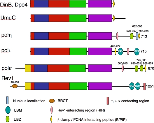
All Y-family members share a conserved N-terminal polymerase domain of 350–450 residues containing the catalytic active site, and a C-terminal appendage of varying size that appears crucial for regulatory protein–protein interactions (31) (Fig. 1). Mouse and human Rev1 are unique in that they have an additional N-terminal BRCT domain preceding the catalytic domain of the enzyme (32) (Fig. 1). The C-terminal appendage in archaeal and certain bacterial Y-family polymerases is often no more than a dozen residues in length, but it can be >70 residues in certain bacterial enzymes, such as E. coli UmuC, and is up to 350 residues long in most eukaryotic Y-family polymerases (Fig. 1).
Fig. 1.
Structural domains of the Y-family polymerases. The polymerase domain is labeled in red (palm), blue (finger), green (thumb), purple (LF), and yellow (N-terminal addition in polκ and Rev1). The regulatory units are color- and shape-coded as indicated at the bottom of the figure. UBM stands for ubiquitin-binding motif, UBZ for ubiquitin-binding zinc finger, and BRCT for Brca1 C-terminal domain.
General Features of the Polymerase Domain
Structural Comparison with Replicative DNA Polymerases.
The Y-family polymerases are best characterized by translesion synthesis and low fidelity when copying normal DNA. Their error rate during normal DNA synthesis is 10−2 to 10−4, which is ≈1–2 orders of magnitude higher than those of replicases in the A- and B-family even when the intrinsic proofreading function (the 3′–5′ exonuclease activity) is removed (33). Crystal structures of the polymerase domain of two archaeal (Dbh and Dpo4) (34–36) and four eukaryotic Y-family members (REV1, polη, ι, and κ) (37–40) have been determined with or without bound substrates. These structures collectively provide a molecular basis for understanding their unusual biochemical properties.
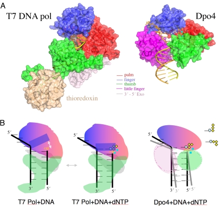
Four structural subdomains are found in each polymerase domain (Fig. 2A). The first 250–350 residues including the five signature motifs (8) constitute the catalytic core of the polymerases and form the thumb, palm, and finger subdomains as found in all known DNA and RNA polymerases. Despite a lack of apparent sequence homology, the palm domain of the A-, B-, and Y-family polymerases, as well as reverse transcriptases, are highly conserved, and three carboxylates essential for the catalysis are located on identical structural elements (41). Although the secondary structures vary broadly in the thumb and finger domains across the different polymerase families, their location in the tertiary structure and roles in interaction with DNA and nucleotide substrate are conserved among all polymerases. Yet, the thumb and finger are distinctively smaller in Y-family polymeraes (Fig. 2A).
Fig. 2.
Structural comparison of T7 DNA polymerase (A-family) (PDB ID code 1T7P) and Dpo4 (Y-family) (PDB ID code 2AGQ). (A) The polymerase domain is shown in the same colors as in Fig. 1. Thioredoxin (wheat) enhances the processivity of T7 DNA pol. DNA is shown in yellow (primer) and brown (template) tubes, the two metal ions as cyan spheres, and the incoming nucleotide (only visible in Dpo4) as silver and multicolored sticks. (B) Diagrams of the conformational change of a helix (solid blue rectangle) in the finger domain of T7 DNA pol upon binding of a correct incoming nucleotide (dNTP). Movement of the helix is indicated by a gray arrow. The reactants and catalysts are snug in the closed active site. (C) Illustration of the flexible LF and thumb domains of Y-family polymerases, which facilitate the movement of the template–primer duplex. The spacious and open active site also allows multiple conformations of the dNTP (as diagramed in the upper right corner) and makes it difficult to align the 3′-OH of the primer strand, dNTP, metal ions, and catalytic carboxylates.
At the C terminus of the catalytic core, ≈100 residues form a structurally conserved domain unique to the Y-family polymerases. It has been called either the little finger (LF) domain, based on the analogy to a right hand (in addition to palm, thumb, and finger) and its role in DNA binding (36) (Fig. 2A), or the polymerase-associate domain (PAD) (37). In contrast to replicases, where the finger domain that interacts with the replicating base pair undergoes the largest conformational changes upon substrate binding, it is the LF and thumb domains that sandwich the upstream DNA that are the most mobile in Y-family polymerases (Fig. 2B). In the absence of DNA substrate, the LF can be closely associated with the catalytic core through a tether as observed in Dbh (35), or wildly flexible as in polκ (42). The mobility of the LF and thumb can alter the positions of DNA substrate relative to the catalytic core and, consequently, the activity of the polymerase (43, 44). Indeed, switching the LF domains between archaeal Dbh and Dpo4 reverses the catalytic efficiency of the two homologous enzymes (45).
Kinetic Properties and Substrate Specificity.
The active site of Y-family polymerases is preformed before substrate binding, in contrast to that of the A-, B-, and RT-family polymerases (34, 46). Because of the small finger and thumb, the active site is also remarkably solvent-exposed and not as geometrically constrained to reject non-Watson–Crick base pairs (Fig. 2B). The “spaciousness” of the active site gives grounds for erroneous base pairing and the ability to accommodate bulky DNA lesions (36).
For the polymerization reaction, the 3′-OH of a primer strand and the α-phosphate of a dNTP have to be placed adjacent to each other and oriented for the phosphoryl transfer reaction. With high-fidelity polymerases, in the presence of a correct dNTP for the template–primer duplex, the finger domain undergoes a large conformational change (Fig. 2B), and the active site becomes “closed.” In this closed structure, the 3′-OH and α-phosphate of dNTP are then aligned with the catalytically essential metal ions and carboxylates for the chemical bond formation. The fidelity is thus achieved mainly in two steps: a large conformational change of the finger domain and alignment of reactants and catalysts in the active site. A wrong incoming dNTP and, hence, mismatched replicating base pair inhibits both steps (47–49). Despite a preformed and spacious active site, Y-family polymerases are selective in nucleotide incorporation and exhibit fidelity exceeding what is warranted by Watson–Crick base pairing alone (50, 51). The fidelity of the Y-family polymerase is likely achieved in the substrate alignment step. The flexible LF domain and spacious active site, which readily accepts damaged or mispaired DNA, actually make the alignment of a dNTP, DNA and two metal ions for catalysis difficult and even more difficult in the presence of mismatched and damaged substrates (52) (Fig. 2C). Consequently, the Y-family polymerases depend more on hydrogen bonds between a replicating base pair than its size or shape (50, 53) and are catalytically less efficient than the A- and B-family replicases (54).
A series of structural studies of Dpo4 complexed with abnormal DNA templates, including oxidative damage (44, 55–57), UV cross-linking (43), benzo[a]pyrene diol epoxide (BPDE) adduct (58), and abasic lesions (59) (Fig. 3A) have been reported. Dpo4 can accommodate almost every lesion in its active site by a multitude of contortions in DNA template, primer, or incoming dNTP, but it efficiently catalyzes only the bypass of abasic lesions (59, 60). The 3′-OH of the primer strand, the α-phosphate of the incoming dNTP and the catalytically essential metal ions are often found to deviate from the ideals for catalysis when an unfavorable lesion is present (Fig. 2C) (52, 61). Not surprisingly, the Y-family polymerases differ in the active site geometry and flexibility of the LF, which gives rise to their differences in spectrum of mutations and TLS efficiency.
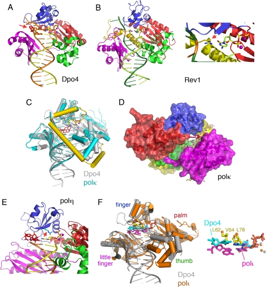
Fig. 3.
Structural and biochemical features of individual Y-family polymerases. (A) Dpo4 bypassing an abasic lesion (PDB ID code 1S0N). The polymerase domain is colored as in Fig. 1, and the red arrow points at the looped-out abasic site analog. The nucleotide 5′ to the abasic site serves as the template base to direct nucleotide incorporation. The dNTP is shown in sticks, and the two metal ions are shown as purple spheres. (B) Rev1 complexed with DNA (PDB ID code 2AQ4). The overall structures of Rev1 and Dpo4 are superimposable, including the incoming dNTP and metal ions. But the N-terminal region (shown in yellow) of Rev1 displaces the template base (highlighted in orange), and an Arg side chain is inserted in its place. A close-up view of the active site is shown on the right. The red arrow points at the Arg that forms two hydrogen bonds with dCTP. (C) Superposition of the structures of Dpo4 complexed with BPDE-dA-adducted DNA (PDB ID code 1S0M) (shown in silver with the BPDE-dA highlighted in red) and polκ complexed with a normal DNA (PDB ID code 2OH2) (shown in cyan with the N-terminal three-helix insertion highlighted in yellow). The two proteins (in which the α-helices are represented by cylinders), DNAs, and dNTPs in particular are superimposable. The N-terminal addition of polκ (yellow) can partially shield the BPDE adduct in the major groove, which otherwise is exposed to solvent as in the complex with Dpo4. (D) The backside of the polκ–DNA ternary complex structure. The polymerase domain is shown in a molecular surface representation and colored as in Fig. 1. The crevice separates the LF (purple) and finger domains (blue) and also extends to the palm domain. The normal template base is shown as red sticks. If it were a BPDE-dG adduct, the BPDE moiety in the minor groove could be accommodated in the large crevice during the nucleotide insertion step as well as the subsequent primer extension. (E) A close-up view of the polη active site (PDB ID code 1JIH). A CPD-containing DNA, dATP paired with the 3′ T of the CPD (shown as red sticks) and two metal ion (purple spheres) are borrowed from the Dpo4–CPD complex structure (PDB ID code 1RYR) after superimposing the palm and finger domains of the two proteins. The R73 in yeast polη (R61 in human polη), which is proposed to stabilize the incoming dATP, is shown as cyan and blue sticks (with the red arrow pointing at it). (F) Comparison of polι (PDB ID code 2FLL, colored in orange) and Dpo4 (PDB ID code 2AGQ, colored in silver). The overall structures of the two polymerase–substrate ternary complexes are quite similar. The replicating base pair in Dpo4 (shown in cyan) is superimposable with those in polκ and Rev1 (Fig. 4 B and C), but it differs from that in polι (magenta) because of potential clashes with the large aliphatic side chains (L62, V64, and L78, shown in yellow) present in the finger domain of polι. Interestingly, the triphosphate moieties of dNTP (orange and red) are more or less superimposable between Dpo4 and polι. A close-up view of the superimposed active sites is shown on the right.
Unique Features and TLS Specificity of Individual Y-Family Polymerases
Rev1.
The first identified Y-family member, REV1, possesses the unique ability to incorporate only dCMP. The crystal structure of the polymerase domain of S. cerevisiae REV1 complexed with a primer–template and dCTP reveals the molecular mechanism (40). In comparison with Dpo4 (Fig. 3A), REV1 has an N-terminal extension, which forms a long helix that fills the gap between the finger and LF (Fig. 3B). This N-terminal helix approaches the template–primer duplex from the minor groove side and forces the template base to flip out of the double helix, and, in its place, it supplies an arginine side chain to hydrogen bond with the incoming dCTP, thus making REV1 a dCMP polymerase. The use of protein residues to direct nucleotide incorporation is reminiscent of the CCA-adding enzyme in tRNA synthesis (62).
E. coli DNA Polymerase IV and Eukaryotic Polκ.
PolIV (DinB) and its eukaryotic ortholog, polκ, are reported to be particularly adept at bypassing deoxyguanosines with adducts at the N2 position, such as furfuryl and benzo[a]pyrene diol epoxide (BPDE), efficiently and accurately (63–68). Mouse embryo fibroblasts (Mefs) lacking polκ are correspondingly sensitive to the killing effects of benzo[a]pyrene (69). Polκ, which is highly expressed in cells enriched in steroid hormones, may have evolved to specifically bypass endogenous polyhydrocarbon lesions, such as those derived from estrogen (70). The role of polκ in maintaining genomic stability of germ-line cells is highlighted by the observation that male mice lacking polκ exhibit an elevated spontaneous mutator phenotype (71). PolIV and polκ are also similar in their preponderance to make template misalignments resulting in −1 frameshifts and missense mutations when replicating undamaged DNA (72, 73).
Among the crystal structures of polymerase–DNA–dNTP ternary complexes, Dpo4 (an ortholog of polIV) and polκ are strikingly similar (Fig. 3C). All four protein domains, the template–primer duplex, and incoming dNTP can be superimposed. The mechanism of looping-out an abasic lesion (59) (Fig. 3A) or skipping a template base (36) as observed for Dpo4, may be used by polIV and polκ to make −1 frameshift. Although Dpo4 and polκ are closely related (20, 30), polκ is far more efficient than Dpo4 in bypassing bulky aromatic adducts like BPDE (58, 74–77). In the structure of Dpo4 complexed with a BPDE-dA adduct, the bulky hydrophobic adduct was placed in the solvent-exposed major groove to allow the chemical bond formation (58). But exposing the hydrophobic benzo[a]pyrene moiety is energetically unfavorable. Superposition of the ternary complexes reveals two features in polκ, which may account for its unique TLS activity (Fig. 3 C and D). First, polκ has a large N-terminal extension, as does Rev1, but in polκ it forms a lid that partially covers the otherwise exposed major groove of the replicating base pair. This N-terminal lid in polκ, which is absent in Dpo4, Rev1, polη, and polι, may alleviate the unfavorable exposure of the bulky adduct for efficient polymerization (Fig. 3C). Second, because of the truncation of a connecting loop, the finger domain of polκ no longer interacts with the LF domain even in the substrate ternary complex, and the gap between LF and the catalytic core is enlarged and extended because of amino acid alterations (Fig. 3D). The equivalent cleft in Dpo4 and polι is much smaller and is nonexistent in REV1 (Fig. 3B). In contrast, the crevice in polκ appears to be large enough to accommodate a bulk adduct in the minor groove, e.g., BPDE-dG, in successive steps of nucleotide incorporation and primer extension opposite the lesion.
E. coli DNA Polymerase V and Eukaryotic Polη.
E. coli polV and eukaryotic polη are similar in being able to bypass a broad spectrum of DNA lesions and are hypothesized to be close structural relatives (78). Polη is especially efficient at bypassing a thymine–thymine cyclobutane pyrimidine dimer (CPD) (79–81). Although the in vitro misincorporation frequency opposite the CPD is in the range of 10−2 to 10−3, which is normally considered error-prone, the in vivo bypass of CPDs by polη must be more efficient and accurate than by other DNA polymerases, because defects in polη lead to a dramatic increase in mutagenesis and carcinogenesis in mammals (14, 15, 82, 83). As indicated by the XPV syndrome, a pivotal cellular role for polη is to protect mammals from the deleterious consequences of prolonged exposure to UV light. Human polη also appears to bypass intrastrand cisplatin de oxy guano sine adducts rather efficiently (84–86). Although the ability to bypass a CPD clearly protects us from UV-induced cancers, the concomitant ability of polη to bypass cisplatin adducts may actually reduce the efficacy of certain chemotherapeutic agents like cisplatin and gemcitabine, a combination of which is commonly used to treat a wide spectrum of cancers, thereby facilitating tumor progression, rather than suppressing it (87, 88).
A crystal structure of either polV or polη complexed with a lesion-containing DNA substrate is currently unavailable. Homologous modeling and unpublished results from T. Carell's laboratory (personal communication), however, suggest that an arginine in the finger subdomain uniquely conserved among polη homologs (R73 in S. cerevisiae and R61 in human) likely stabilizes dNTP and two metal ions in the active conformation before binding of template–primer (Fig. 3E). The “fixed” dNTP, metal ions, and catalytic residues may enable these polymerases to capture a broad spectrum of lesions transiently and at the same time promoting catalysis.
Polι.
Polι is related to polη in sequence, but exhibits very different TLS properties in vitro. Whereas polη bypasses a T–T CPD efficiently and accurately, polι does so inefficiently and inaccurately (89, 90). Clues to polι's role in the TLS of UV induced lesions in vivo are beginning to emerge from studies with mice carrying a naturally occurring nonsense mutation near the 5′ end of the Poli gene (91). Mice lacking polι develop mesenchymal cancers when exposed to UV light (82), and Mefs lacking polι also exhibit an altered spectrum of UV-induced mutations (83), suggesting that polι may, under certain circumstances, facilitate TLS of UV photoproducts. Indeed, it seems likely that polι is the enzyme that substitutes for polη in X. pigmentosum variant patients and is responsible for the high frequency and altered spectrum of mutations characteristic of the XPV phenotype (92).
The murine Poli gene is located on chromosome 18q22 (19) and lies within the boundaries of the previously described Pulmonary adenoma resistance 2 (Par2) locus, and it has been suggested that defects and/or alterations in polι may be responsible for the susceptibility of mice to urethane induced pulmonary adenomas (93, 94). Consistent with this hypothesis, mice carrying the Poli nonsense mutation have a high susceptibility to urethane-induced lung tumors (95).
Polι's fidelity when replicating undamaged DNA is most unique, because both the human and murine enzymes posses the remarkable ability to misincorporate G opposite T, 3- to 10-fold more frequently than the correct base A. In contrast, when replicating template A, the enzyme is reasonably accurate, with a misincorporation frequency of ≈10−4 (22–24). Thus, the fidelity of the enzyme can vary by 105-fold, depending on the template base being replicated.
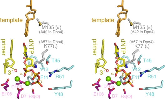
Multiple crystal structure of human polι complexed with normal DNA substrate and dNTP have been reported (38, 96, 97). One striking feature is that its active site is most different from Dpo4, REV1, polη, and polκ by the presence of several large aliphatic residues, which forbid the template–primer and dNTP to bind in the normal positions (Fig. 3F). In the polι complexes, the replicating base pair is shifted by several Ångstroms away from the finger domain and often assumes Hoogsteen conformation. Despite the distortions, the dNTP and two metal ions are in the catalytically active position and are likely stabilized by the Lys residue (K77 in human polι) (Fig. 4), which is equivalent to R73 of S. cerevisaea polη. The distortion of base pairing and dNTP stabilization may lead to the skewed preference of incorporating dGMP opposite template dT by polι.
Fig. 4.
A composite active site of the Y-family polymerases in stereoview. The 3′-end nucleotide of the primer strand (pale yellow), the template nucleotide (orange), the incoming dNTP [yellow (C)/blue (N)/red (O)], the three catalytic carboxylates [magenta (C)/red (O)], the nearby carbonyl group [F8(O)] that coordinates one metal ion, and the conserved residues interacting with dNTP [light blue (C)/blue (N)/red (O)] are shown as sticks, and the two metal ions are shown as green spheres. These conserved residues are labeled according to Dpo4 for convenience. K77 of polι that stabilizes the incoming dNTP and M135 of polκ that stabilizes the template base are shown in gray/blue (N)/brown (S) sticks. These residues are replaced by Ala's in Dpo4. The 3′-OH group (indicated as a red “o”) is usually absent in the crystal structures for the purpose of capturing enzyme–substrate complexes.
DNA Repair, Mutagenesis, and Other Activities of Y-Family Polymerases
In addition to TLS, it is becoming increasingly clear that, under certain circumstances, the Y-family polymerases also have access to undamaged DNA. First, although the molecular mechanisms underlying the somatic hypermutation of variable Ig genes are still being unraveled, it is clear that defects in human and murine polη lead to a dramatic and specific decrease in somatic mutations at A/T base pairs (98–100), whereas defects in murine Rev1 causes a reduction in G/C somatic mutations (101, 102). In contrast, polι and polκ appear to play no role in somatic hypermutation (91, 103–105).
Very recently, it has been suggested that polη may participate in recombinational repair, because chicken cells lacking polη cannot undergo recombination-induced gene conversion (106), and human polη appears particularly efficient at extending D-loop recombination intermediates in vitro (107).
Ogi and Lehmann (108) have recently reported that polκ deficient Mefs are also UV-sensitive. Polκ is unable to insert a base opposite CPDs in vitro (25–27) but can extend mispairs across from the lesion if it is inserted by another polymerase (109). However, the UV-sensitivity of the polκ Mefs is unlikely to be attributed to an inability to facilitate TLS but is more likely to result from an unexpected role for polκ in nucleotide excision repair (108).
Regulation of Y-Family Polymerases by Transcription and Protein Degradation
Given that the Y-family polymerases are intrinsically error-prone, it makes teleological sense that they are strictly regulated to minimize any spurious mutagenesis and ensure that they are only used at specific times and/or locations. It is now evident that each organism uses a myriad of mechanisms to keep Y-family polymerases under control. Perhaps the best studied is E. coli polV, whose subunits (UmuD′ and UmuC) are temporally regulated via DNA damage-induced transcription (110) and targeted proteolysis (111–113) to keep the intracellular levels of the proteins to a minimum. In addition to low cellular levels, polV activity is regulated by means of a number of protein–protein interactions, most notably with RecA, because certain missense mutations in recA also render E. coli nonmutable (114). Recent data suggest that RecA protein stimulates the catalytic activity of polV in vitro by forming a nucleoprotein filament on DNA in trans to the lesion-containing DNA substrate bypassed by polV (115).
Regulation of the eukaryotic Y-family polymerases is equally complex. Although the S. cerevisiae Rad30 transcript is induced ≈3-fold in response to UV-damage (9), the activity/cellular level of most eukaryotic Y-family polymerases appear to be primarily regulated via posttranslational pathways either involving direct modification of the polymerase, or by means of key protein–protein interactions. For example, S. cerevisiae REV1 protein has recently been shown to become phosphorylated in a Mec1 and cell cycle-dependent manner (116) and, somewhat unexpectedly, exhibits highest expression in G2/M rather than S-phase (116, 117). Like E. coli polV, the basal levels of S. cerevisiae polη appear to be kept to a minimum by targeted proteolysis. In the case of polη, the protein is ubiquitinated and subsequently degraded by the cell's proteasome (118, 119). This clearly helps reduce the level of spontaneous mutagenesis in S. cerevisiae, because mutations in the proteasome lead to a 3- to 5-fold increase in polη-dependent mutagenesis (118). Interestingly, upon UV-irradiation, when one could easily imagine that polη's TLS activities are most desirable, the enzyme is transiently stabilized with the estimated half-life of the protein increasing from 20 to 120 min. As a consequence, the intracellular concentration of polη increases, with maximal levels observed 90 min after UV treatment (118).
Access to Replication Fork by Interactions with PCNA/β Clamp, Ubiquitin, and Interactions Among the Y-Family Polymerases
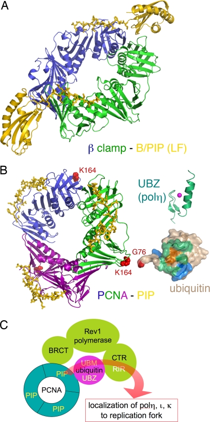
Y-family polymerases interact with the cell's replication processivity factor, the β-sliding clamp in E. coli (17, 120, 121), and PCNA in archaea and eukaryotes (122, 123). Structural studies of E. coli proteins reveal that these interactions are mediated by the β-clamp/PCNA-interaction peptide (B/PIP) occurring after the LF domain in the polymerase and the canonical B/PIP-binding surface on the β-clamp/PCNA (124) (Fig. 5A). Such interactions are required for the biological functions of Y-family polymerase in vivo (123, 125). Given that the β-clamp is a homodimer with one potential polymerase-binding site in each protomer, it was hypothesized that one clamp might be able to physically accommodate two different polymerases (126). Indeed, support for such a hypothesis comes from the fact that both polIII and polIV appear able to interact simultaneously with the β-clamp (121, 127).
Fig. 5.
Access of Y-family polymerases to a replication fork is regulated by posttranslational modification and protein–protein interactions. (A) Ribbon diagram of E. coli polIV C-terminal region (including the LF domain and B/PIP) complexed with the β-clamp (PDB ID code 1UNN). The two subunits of β clamp are shown in green and blue, and polIV is shown in yellow. The B/PIP of polIV is represented by a stick model. (B) Interactions among PCNA–PIP (represented by p21, PDB ID code 1AXC), ubiquitin, UBZ, and UBM. The trimeric PCNA is shown in blue, green, and purple ribbon diagram. The PIP peptide from p21 is shown as yellow sticks. When ubiquitinated, PCNA is covalently linked through its K164 (represented by red spheres) with G76 (highlighted in red) of ubiquitin (PDB ID code 2G45). Ubiquitin is shown in molecular surface representation, the conserved I44 is highlighted in orange, and the surrounding areas that have been mapped to interact with UBZ and UBM are highlighted in green and blue, respectively. The NMR structure of the polη UBZ (PDB ID code 2I5O) is shown in green ribbon diagrams, and a magenta sphere represents the zinc ion. (C) A cartoon summarizing the protein–protein interactions of eukaryotic Y-family polymerases. Rev1 (pea green), polη, polι, and polκ each interact with PCNA (cyan) and ubiquitin (magenta), and the C-terminal region of Rev1 interacts with polη, polι, and polκ (collectively represented by the curvy red arrow). The multilayered interactions occur in response to DNA damage and may allow polη, polι, and polκ to be recruited to replication forks.
Human polη and polκ contain amino acid residues with a good match to the PIP consensus motif near their C termini (123, 128) (Fig. 1). The PIP box in human polι is noncanonical but, nevertheless, is hypothesized to adopt the same structure as classical PCNA-binding proteins, such as the p21 protein (129) (Fig. 5B). Interestingly, and in contrast to the other eukaryotic Y-family polymerases, polι's PIP box is located immediately downstream of the LF domain of the polymerase (129, 130) (Fig. 1), and the enzyme's ability to interact with PCNA is essential for it to accumulate into damage-induced replication foci (129, 131).
Eukaryotic PCNA is a homotrimer, so, in theory, up to three different polymerases could bind to the clamp at any given time. However, some 50 or 60 cellular proteins are known to physically interact with PCNA, and, clearly, not all can interact with the clamp at one time (132, 133). Eukaryotic cells appear to rely heavily on the posttranslational modification of PCNA by ubiquitin and/or SUMO proteins as a means to discriminate between the various TLS polymerases and other repair proteins (134). In the case of S. cerevisiae, monoubiquitination of PCNA at K164 by Rad6/Rad18 helps promote both polη- and polζ-dependent TLS of damaged DNA (135), possibly by stimulating the catalytic activity of polη and Rev1 (136). Further extension of the ubiquitin moieties through the actions of Mms2-Ubc13 and Rad5 results in the damaged DNA being funneled into an error-free damage-avoidance pathway (134). In contrast, SUMOylation of PCNA at K164 or K127 appears to be required for polζ-dependent spontaneous mutagenesis (135).
Although SUMOylation of mammalian PCNA has yet to be observed, both mono- and polyubiquitination of human PCNA has been reported (137–141). All four human Y-family polymerases can bind unmodified and monoubiquitinated PCNA, but the affinity with which they do so varies. In the case of polη, the interaction between monoubiquitinated PCNA is much stronger than with unmodified PCNA. Thus, monoubiquitination of PCNA at the site of damaged DNA may physically target the polymerase to lesions in DNA and help facilitate a switch between the cells replicase and polη (137, 138, 142–144).
The ability of the Y-family polymerases to bind ubiquitinated PCNA can be attributed to the fact that they posses a ubiquitin-binding motif (UBM) or ubiquitin-binding zinc-finger motif (UBZ) in their respective C termini (145–147). The interactions of UBM and UBZ with free ubiquitin and monoubiquitylated PCNA have been demonstrated by NMR (145, 148) (Fig. 5B). The biological importance of ubiquitin binding is highlighted by the fact that, unlike wild-type polη, a polη UBZ mutant fails to restore UV-resistance to normally UV-sensitive XPV cells (145). Similarly, the ability of polι to accumulate in UV-induced replication foci is greatly reduced in a polι UBM mutant compared with the wild-type protein (146).
As noted above, Rev1 possesses the unique ability to use dCMP. However, such an activity may not be its “raison d'être”, because the S. cerevisiae Rev1–1 mutant is defective for damage-induced mutagenesis despite the retention of considerable dCMP transferase activity (149). The critical role of Rev1 in TLS therefore appears to be structural rather than catalytic. Indeed, both the human and murine Rev1 protein have been shown to interact with polymerases η, ι, and κ (150–152). Moreover, the Rev1 BRCT domain also interacts with PCNA independent of PIP (153) and the Rev1 UBMs with ubiquitin (147). Thus, Rev1, PCNA and ubiquitin can interact with one another, and, meanwhile, all three can interact with polymerases η, ι, or κ. Last but not least, a physical interaction between polη and polι has been reported to guide polι to replication foci (154). The multilayered interactions among the various TLS polymerases and with ubiquitinated PCNA may therefore provide a structural platform for polymerase switching during TLS (144) (Fig. 5C).
Outlook of Future Research
It is truly remarkable how our understanding of translesion synthesis has been transformed over the past 10 years. Although our knowledge has expanded exponentially during this time period, there is still much to be learned. For example, what are the primary cellular roles of polι, polκ, and Rev1, and are there any human diseases associated with defects, or up-regulation of these polymerases? Perhaps the biggest challenge will be our ability to decipher the molecular mechanisms that regulate controlled access of the Y-family polymerases to a replication fork, gap, or D-loop, where they participate in TLS, nucleotide excision repair, or recombination repair, respectively. Recent studies indicate that eukaryotic cells largely achieve this goal through a multitude of posttranslational modifications that alter the relative binding affinity of the polymerases to their protein partners. It is hoped that continued studies in this area will revolutionize our understanding of TLS and its effects on genomic stability, just as the initial discovery and characterization of the Y-family polymerases has during the past decade.
Acknowledgments
We thank Dr. T. Carell for sharing research results with us before publication. W.Y. and R.W. are supported by the Intramural Research Program of the National Institutes of Health.
Footnotes
The authors declare no conflict of interest.
References
- 1.Kato T, Shinoura Y. Mol Gen Genet. 1977;156:121–131. doi: 10.1007/BF00283484. [DOI] [PubMed] [Google Scholar]
- 2.Steinborn G. Mol Gen Genet. 1978;165:87–93. doi: 10.1007/BF00270380. [DOI] [PubMed] [Google Scholar]
- 3.Lemontt JF. Genetics. 1971;68:21–33. doi: 10.1093/genetics/68.1.21. [DOI] [PMC free article] [PubMed] [Google Scholar]
- 4.Lawrence CW, Christensen R. Genetics. 1976;82:207–232. doi: 10.1093/genetics/82.2.207. [DOI] [PMC free article] [PubMed] [Google Scholar]
- 5.Bridges BA, Woodgate R. Proc Natl Acad Sci USA. 1985;82:4193–4197. doi: 10.1073/pnas.82.12.4193. [DOI] [PMC free article] [PubMed] [Google Scholar]
- 6.Larimer FW, Perry JR, Hardigree AA. J Bacteriol. 1989;171:230–237. doi: 10.1128/jb.171.1.230-237.1989. [DOI] [PMC free article] [PubMed] [Google Scholar]
- 7.Ohmori H, Hatada E, Qiao Y, Tsuji M, Fukuda R. Mutat Res. 1995;347:1–7. doi: 10.1016/0165-7992(95)90024-1. [DOI] [PubMed] [Google Scholar]
- 8.Kulaeva OI, Koonin EV, McDonald JP, Randall SK, Rabinovich N, Connaughton JF, Levine AS, Woodgate R. Mutat Res. 1996;357:245–253. doi: 10.1016/0027-5107(96)00164-9. [DOI] [PubMed] [Google Scholar]
- 9.McDonald JP, Levine AS, Woodgate R. Genetics. 1997;147:1557–1568. doi: 10.1093/genetics/147.4.1557. [DOI] [PMC free article] [PubMed] [Google Scholar]
- 10.Nelson JR, Lawrence CW, Hinkle DC. Nature. 1996;382:729–731. doi: 10.1038/382729a0. [DOI] [PubMed] [Google Scholar]
- 11.Gibbs PE, Lawrence CW. J Mol Biol. 1995;251:229–236. doi: 10.1006/jmbi.1995.0430. [DOI] [PubMed] [Google Scholar]
- 12.Johnson RE, Prakash S, Prakash L. Science. 1999;283:1001–1004. doi: 10.1126/science.283.5404.1001. [DOI] [PubMed] [Google Scholar]
- 13.Masutani C, Araki M, Yamada A, Kusumoto R, Nogimori T, Maekawa T, Iwai S, Hanaoka F. EMBO J. 1999;18:3491–3501. doi: 10.1093/emboj/18.12.3491. [DOI] [PMC free article] [PubMed] [Google Scholar]
- 14.Masutani C, Kusumoto R, Yamada A, Dohmae N, Yokoi M, Yuasa M, Araki M, Iwai S, Takio K, Hanaoka F. Nature. 1999;399:700–704. doi: 10.1038/21447. [DOI] [PubMed] [Google Scholar]
- 15.Johnson RE, Kondratick CM, Prakash S, Prakash L. Science. 1999;285:263–265. doi: 10.1126/science.285.5425.263. [DOI] [PubMed] [Google Scholar]
- 16.Wagner J, Gruz P, Kim SR, Yamada M, Matsui K, Fuchs RP, Nohmi T. Mol Cell. 1999;4:281–286. doi: 10.1016/s1097-2765(00)80376-7. [DOI] [PubMed] [Google Scholar]
- 17.Tang M, Shen X, Frank EG, O'Donnell M, Woodgate R, Goodman MF. Proc Natl Acad Sci USA. 1999;96:8919–8924. doi: 10.1073/pnas.96.16.8919. [DOI] [PMC free article] [PubMed] [Google Scholar]
- 18.Reuven NB, Arad G, Maor-Shoshani A, Livneh Z. J Biol Chem. 1999;274:31763–31766. doi: 10.1074/jbc.274.45.31763. [DOI] [PubMed] [Google Scholar]
- 19.McDonald JP, Rapic-Otrin V, Epstein JA, Broughton BC, Wang X, Lehmann AR, Wolgemuth DJ, Woodgate R. Genomics. 1999;60:20–30. doi: 10.1006/geno.1999.5906. [DOI] [PubMed] [Google Scholar]
- 20.Gerlach VL, Aravind L, Gotway G, Schultz RA, Koonin EV, Friedberg EC. Proc Natl Acad Sci USA. 1999;96:11922–11927. doi: 10.1073/pnas.96.21.11922. [DOI] [PMC free article] [PubMed] [Google Scholar]
- 21.Ogi T, Kato T, Jr, Kato T, Ohmori H. Genes Cells. 1999;4:607–618. doi: 10.1046/j.1365-2443.1999.00289.x. [DOI] [PubMed] [Google Scholar]
- 22.Tissier A, McDonald JP, Frank EG, Woodgate R. Genes Dev. 2000;14:1642–1650. [PMC free article] [PubMed] [Google Scholar]
- 23.Johnson RE, Washington MT, Haracska L, Prakash S, Prakash L. Nature. 2000;406:1015–1019. doi: 10.1038/35023030. [DOI] [PubMed] [Google Scholar]
- 24.Zhang Y, Yuan F, Wu X, Wang Z. Mol Cell Biol. 2000;20:7099–7108. doi: 10.1128/mcb.20.19.7099-7108.2000. [DOI] [PMC free article] [PubMed] [Google Scholar]
- 25.Johnson RE, Prakash S, Prakash L. Proc Natl Acad Sci USA. 2000;97:3838–3843. doi: 10.1073/pnas.97.8.3838. [DOI] [PMC free article] [PubMed] [Google Scholar]
- 26.Ohashi E, Ogi T, Kusumoto R, Iwai S, Masutani C, Hanaoka F, Ohmori H. Genes Dev. 2000;14:1589–1594. [PMC free article] [PubMed] [Google Scholar]
- 27.Zhang Y, Yuan F, Wu X, Wang M, Rechkoblit O, Taylor JS, Geacintov NE, Wang Z. Nucleic Acids Res. 2000;28:4138–4146. doi: 10.1093/nar/28.21.4138. [DOI] [PMC free article] [PubMed] [Google Scholar]
- 28.Burgers PM, Koonin EV, Bruford E, Blanco L, Burtis KC, Christman MF, Copeland WC, Friedberg EC, Hanaoka F, Hinkle DC, et al. J Biol Chem. 2001;276:43487–43490. doi: 10.1074/jbc.R100056200. [DOI] [PubMed] [Google Scholar]
- 29.Woodgate R. Genes Dev. 1999;13:2191–2195. doi: 10.1101/gad.13.17.2191. [DOI] [PubMed] [Google Scholar]
- 30.Ohmori H, Friedberg EC, Fuchs RPP, Goodman MF, Hanaoka F, Hinkle D, Kunkel TA, Lawrence CW, Livneh Z, Nohmi T, et al. Mol Cell. 2001;8:7–8. doi: 10.1016/s1097-2765(01)00278-7. [DOI] [PubMed] [Google Scholar]
- 31.Goodman MF. Annu Rev Biochem. 2002;71:17–50. doi: 10.1146/annurev.biochem.71.083101.124707. [DOI] [PubMed] [Google Scholar]
- 32.Lawrence CW. Adv Protein Chem. 2004;69:167–203. doi: 10.1016/S0065-3233(04)69006-1. [DOI] [PubMed] [Google Scholar]
- 33.Bebenek K, Kunkel TA. Adv Protein Chem. 2004;69:137–165. doi: 10.1016/S0065-3233(04)69005-X. [DOI] [PubMed] [Google Scholar]
- 34.Zhou BL, Pata JD, Steitz TA. Mol Cell. 2001;8:427–437. doi: 10.1016/s1097-2765(01)00310-0. [DOI] [PubMed] [Google Scholar]
- 35.Silvian LF, Toth EA, Pham P, Goodman MF, Ellenberger T. Nat Struct Biol. 2001;8:984–989. doi: 10.1038/nsb1101-984. [DOI] [PubMed] [Google Scholar]
- 36.Ling H, Boudsocq F, Woodgate R, Yang W. Cell. 2001;107:91–102. doi: 10.1016/s0092-8674(01)00515-3. [DOI] [PubMed] [Google Scholar]
- 37.Trincao J, Johnson RE, Escalante CR, Prakash S, Prakash L, Aggarwal AK. Mol Cell. 2001;8:417–426. doi: 10.1016/s1097-2765(01)00306-9. [DOI] [PubMed] [Google Scholar]
- 38.Nair DT, Johnson RE, Prakash S, Prakash L, Aggarwal AK. Nature. 2004;430:377–380. doi: 10.1038/nature02692. [DOI] [PubMed] [Google Scholar]
- 39.Lone S, Townson SA, Uljon SN, Johnson RE, Brahma A, Nair DT, Prakash S, Prakash L, Aggarwal AK. Mol Cell. 2007;25:601–614. doi: 10.1016/j.molcel.2007.01.018. [DOI] [PubMed] [Google Scholar]
- 40.Nair DT, Johnson RE, Prakash L, Prakash S, Aggarwal AK. Science. 2005;309:2219–2222. doi: 10.1126/science.1116336. [DOI] [PubMed] [Google Scholar]
- 41.Yang W. FEBS Lett. 2005;579:868–872. doi: 10.1016/j.febslet.2004.11.047. [DOI] [PubMed] [Google Scholar]
- 42.Uljon SN, Johnson RE, Edwards TA, Prakash S, Prakash L, Aggarwal AK. Structure (London) 2004;12:1395–1404. doi: 10.1016/j.str.2004.05.011. [DOI] [PubMed] [Google Scholar]
- 43.Ling H, Boudsocq F, Plosky BS, Woodgate R, Yang W. Nature. 2003;424:1083–1087. doi: 10.1038/nature01919. [DOI] [PubMed] [Google Scholar]
- 44.Rechkoblit O, Malinina L, Cheng Y, Kuryavyi V, Broyde S, Geacintov NE, Patel DJ. PLoS Biol. 2006;4:e11. doi: 10.1371/journal.pbio.0040011. [DOI] [PMC free article] [PubMed] [Google Scholar]
- 45.Boudsocq F, Kokoska RJ, Plosky BS, Vaisman A, Ling H, Kunkel TA, Yang W, Woodgate R. J Biol Chem. 2004;279:32932–32940. doi: 10.1074/jbc.M405249200. [DOI] [PubMed] [Google Scholar]
- 46.Yang W. Curr Opin Struct Biol. 2003;13:23–30. doi: 10.1016/s0959-440x(02)00003-9. [DOI] [PubMed] [Google Scholar]
- 47.Rothwell PJ, Mitaksov V, Waksman G. Mol Cell. 2005;19:345–355. doi: 10.1016/j.molcel.2005.06.032. [DOI] [PubMed] [Google Scholar]
- 48.Showalter AK, Tsai MD. Biochemistry. 2002;41:10571–10576. doi: 10.1021/bi026021i. [DOI] [PubMed] [Google Scholar]
- 49.Tsai YC, Johnson KA. Biochemistry. 2006;45:9675–9687. doi: 10.1021/bi060993z. [DOI] [PMC free article] [PubMed] [Google Scholar]
- 50.Mizukami S, Kim TW, Helquist SA, Kool ET. Biochemistry. 2006;45:2772–2778. doi: 10.1021/bi051961z. [DOI] [PubMed] [Google Scholar]
- 51.Fiala KA, Suo Z. Biochemistry. 2004;43:2106–2115. doi: 10.1021/bi0357457. [DOI] [PubMed] [Google Scholar]
- 52.Vaisman A, Ling H, Woodgate R, Yang W. EMBO J. 2005;24:2957–2967. doi: 10.1038/sj.emboj.7600786. [DOI] [PMC free article] [PubMed] [Google Scholar]
- 53.Potapova O, Chan C, DeLucia AM, Helquist SA, Kool ET, Grindley ND, Joyce CM. Biochemistry. 2006;45:890–898. doi: 10.1021/bi051792i. [DOI] [PMC free article] [PubMed] [Google Scholar]
- 54.Beard WA, Shock DD, Vande Berg BJ, Wilson SH. J Biol Chem. 2002;277:47393–47398. doi: 10.1074/jbc.M210036200. [DOI] [PubMed] [Google Scholar]
- 55.Zang H, Irimia A, Choi JY, Angel KC, Loukachevitch LV, Egli M, Guengerich FP. J Biol Chem. 2006;281:2358–2372. doi: 10.1074/jbc.M510889200. [DOI] [PubMed] [Google Scholar]
- 56.Eoff RL, Irimia A, Egli M, Guengerich FP. J Biol Chem. 2007;282:1456–1467. doi: 10.1074/jbc.M609661200. [DOI] [PubMed] [Google Scholar]
- 57.Eoff RL, Angel KC, Egli M, Guengerich FP. J Biol Chem. 2007;282:13573–13584. doi: 10.1074/jbc.M700656200. [DOI] [PubMed] [Google Scholar]
- 58.Ling H, Sayer JM, Plosky BS, Yagi H, Boudsocq F, Woodgate R, Jerina DM, Yang W. Proc Natl Acad Sci USA. 2004;101:2265–2269. doi: 10.1073/pnas.0308332100. [DOI] [PMC free article] [PubMed] [Google Scholar]
- 59.Ling H, Boudsocq F, Woodgate R, Yang W. Mol Cell. 2004;13:751–762. doi: 10.1016/s1097-2765(04)00101-7. [DOI] [PubMed] [Google Scholar]
- 60.Fiala KA, Hypes CD, Suo Z. J Biol Chem. 2007;282:8188–8198. doi: 10.1074/jbc.M610718200. [DOI] [PubMed] [Google Scholar]
- 61.Yang W, Lee JY, Nowotny M. Mol Cell. 2006;22:5–13. doi: 10.1016/j.molcel.2006.03.013. [DOI] [PubMed] [Google Scholar]
- 62.Xiong Y, Steitz TA. Nature. 2004;430:640–645. doi: 10.1038/nature02711. [DOI] [PubMed] [Google Scholar]
- 63.Jarosz DF, Godoy VG, Delaney JC, Essigmann JM, Walker GC. Nature. 2006;439:225–228. doi: 10.1038/nature04318. [DOI] [PubMed] [Google Scholar]
- 64.Neeley WL, Delaney S, Alekseyev YO, Jarosz DF, Delaney JC, Walker GC, Essigmann JM. J Biol Chem. 2007;282:12741–12748. doi: 10.1074/jbc.M700575200. [DOI] [PubMed] [Google Scholar]
- 65.Shen X, Sayer JM, Kroth H, Ponten I, O'Donnell M, Woodgate R, Jerina DM, Goodman MF. J Biol Chem. 2002;277:5265–5274. doi: 10.1074/jbc.M109575200. [DOI] [PubMed] [Google Scholar]
- 66.Suzuki N, Ohashi E, Kolbanovskiy A, Geacintov NE, Grollman AP, Ohmori H, Shibutani S. Biochemistry. 2002;41:6100–6106. doi: 10.1021/bi020049c. [DOI] [PubMed] [Google Scholar]
- 67.Rechkoblit O, Zhang Y, Guo D, Wang Z, Amin S, Krzeminsky J, Louneva N, Geacintov NE. J Biol Chem. 2002;277:30488–30494. doi: 10.1074/jbc.M201167200. [DOI] [PubMed] [Google Scholar]
- 68.Zhang Y, Wu X, Guo D, Rechkoblit O, Wang Z. DNA Repair (Amsterdam) 2002;1:559–569. doi: 10.1016/s1568-7864(02)00055-1. [DOI] [PubMed] [Google Scholar]
- 69.Ogi T, Shinkai Y, Tanaka K, Ohmori H. Proc Natl Acad Sci USA. 2002;99:15548–15553. doi: 10.1073/pnas.222377899. [DOI] [PMC free article] [PubMed] [Google Scholar]
- 70.Suzuki N, Itoh S, Poon K, Masutani C, Hanaoka F, Ohmori H, Yoshizawa I, Shibutani S. Biochemistry. 2004;43:6304–6311. doi: 10.1021/bi0360298. [DOI] [PubMed] [Google Scholar]
- 71.Burr KL, Velasco-Miguel S, Duvvuri VS, McDaniel LD, Friedberg EC, Dubrova YE. DNA Repair (Amsterdam) 2006;5:860–862. doi: 10.1016/j.dnarep.2006.04.003. [DOI] [PubMed] [Google Scholar]
- 72.Kim SR, Maenhaut-Michel G, Yamada M, Yamamoto Y, Matsui K, Sofuni T, Nohmi T, Ohmori H. Proc Natl Acad Sci USA. 1997;94:13792–13797. doi: 10.1073/pnas.94.25.13792. [DOI] [PMC free article] [PubMed] [Google Scholar]
- 73.Wagner J, Nohmi T. J Bacteriol. 2000;182:4587–4595. doi: 10.1128/jb.182.16.4587-4595.2000. [DOI] [PMC free article] [PubMed] [Google Scholar]
- 74.Perlow-Poehnelt RA, Likhterov I, Scicchitano DA, Geacintov NE, Broyde S. J Biol Chem. 2004;279:36951–36961. doi: 10.1074/jbc.M404332200. [DOI] [PubMed] [Google Scholar]
- 75.Huang X, Kolbanovskiy A, Wu X, Zhang Y, Wang Z, Zhuang P, Amin S, Geacintov NE. Biochemistry. 2003;42:2456–2466. doi: 10.1021/bi026912q. [DOI] [PubMed] [Google Scholar]
- 76.Avkin S, Goldsmith M, Velasco-Miguel S, Geacintov N, Friedberg EC, Livneh Z. J Biol Chem. 2004;279:53298–53305. doi: 10.1074/jbc.M409155200. [DOI] [PubMed] [Google Scholar]
- 77.Choi JY, Angel KC, Guengerich FP. J Biol Chem. 2006;281:21062–21072. doi: 10.1074/jbc.M602246200. [DOI] [PubMed] [Google Scholar]
- 78.Lee CH, Chandani S, Loechler EL. J Mol Graphics Model. 2006;25:87–102. doi: 10.1016/j.jmgm.2005.10.009. [DOI] [PubMed] [Google Scholar]
- 79.Johnson RE, Washington MT, Prakash S, Prakash L. J Biol Chem. 2000;275:7447–7450. doi: 10.1074/jbc.275.11.7447. [DOI] [PubMed] [Google Scholar]
- 80.Masutani C, Kusumoto R, Iwai S, Hanaoka F. EMBO J. 2000;19:3100–3109. doi: 10.1093/emboj/19.12.3100. [DOI] [PMC free article] [PubMed] [Google Scholar]
- 81.McCulloch SD, Kokoska RJ, Masutani C, Iwai S, Hanaoka F, Kunkel TA. Nature. 2004;428:97–100. doi: 10.1038/nature02352. [DOI] [PubMed] [Google Scholar]
- 82.Ohkumo T, Kondo Y, Yokoi M, Tsukamoto T, Yamada A, Sugimoto T, Kanao R, Higashi Y, Kondoh H, Tatematsu M, et al. Mol Cell Biol. 2006;26:7696–7706. doi: 10.1128/MCB.01076-06. [DOI] [PMC free article] [PubMed] [Google Scholar]
- 83.Dumstorf CA, Clark AB, Lin Q, Kissling GE, Yuan T, Kucherlapati R, McGregor WG, Kunkel TA. Proc Natl Acad Sci USA. 2006;103:18083–18088. doi: 10.1073/pnas.0605247103. [DOI] [PMC free article] [PubMed] [Google Scholar]
- 84.Chaney SG, Campbell SL, Bassett E, Wu Y. Crit Rev Oncol Hematol. 2005;53:3–11. doi: 10.1016/j.critrevonc.2004.08.008. [DOI] [PubMed] [Google Scholar]
- 85.Vaisman A, Masutani C, Hanaoka F, Chaney SG. Biochemistry. 2000;39:4575–4580. doi: 10.1021/bi000130k. [DOI] [PubMed] [Google Scholar]
- 86.Bassett E, King NM, Bryant MF, Hector S, Pendyala L, Chaney SG, Cordeiro-Stone M. Cancer Res. 2004;64:6469–6475. doi: 10.1158/0008-5472.CAN-04-1328. [DOI] [PubMed] [Google Scholar]
- 87.Albertella MR, Green CM, Lehmann AR, O'Connor MJ. Cancer Res. 2005;65:9799–9806. doi: 10.1158/0008-5472.CAN-05-1095. [DOI] [PubMed] [Google Scholar]
- 88.Chen YW, Cleaver JE, Hanaoka F, Chang CF, Chou KM. Mol Cancer Res. 2006;4:257–265. doi: 10.1158/1541-7786.MCR-05-0118. [DOI] [PubMed] [Google Scholar]
- 89.Tissier A, Frank EG, McDonald JP, Iwai S, Hanaoka F, Woodgate R. EMBO J. 2000;19:5259–5266. doi: 10.1093/emboj/19.19.5259. [DOI] [PMC free article] [PubMed] [Google Scholar]
- 90.Vaisman A, Frank EG, Iwai S, Ohashi E, Ohmori H, Hanaoka F, Woodgate R. DNA Repair (Amsterdam) 2003;2:991–1006. doi: 10.1016/s1568-7864(03)00094-6. [DOI] [PubMed] [Google Scholar]
- 91.McDonald JP, Frank EG, Plosky BS, Rogozin IB, Masutani C, Hanaoka F, Woodgate R, Gearhart PJ. J Exp Med. 2003;198:635–643. doi: 10.1084/jem.20030767. [DOI] [PMC free article] [PubMed] [Google Scholar]
- 92.Wang Y, Woodgate R, McManus TP, Mead S, McCormick JJ, Maher VM. Cancer Res. 2007;67:3018–3026. doi: 10.1158/0008-5472.CAN-06-3073. [DOI] [PubMed] [Google Scholar]
- 93.Lee GH, Nishimori H, Sasaki Y, Matsushita H, Kitagawa T, Tokino T. Oncogene. 2003;22:2374–2382. doi: 10.1038/sj.onc.1206387. [DOI] [PubMed] [Google Scholar]
- 94.Wang M, Devereux TR, Vikis HG, McCulloch SD, Holliday W, Anna C, Wang Y, Bebenek K, Kunkel TA, Guan K, You M. Cancer Res. 2004;64:1924–1931. doi: 10.1158/0008-5472.can-03-3080. [DOI] [PubMed] [Google Scholar]
- 95.Lee GH, Matsushita H. Cancer Sci. 2005;96:256–259. doi: 10.1111/j.1349-7006.2005.00042.x. [DOI] [PMC free article] [PubMed] [Google Scholar]
- 96.Wang J. Nature. 2005;437:E6–E7. doi: 10.1038/nature04199. discussion, E7. [DOI] [PubMed] [Google Scholar]
- 97.Nair DT, Johnson RE, Prakash L, Prakash S, Aggarwal AK. Structure (London) 2005;13:1569–1577. doi: 10.1016/j.str.2005.08.010. [DOI] [PubMed] [Google Scholar]
- 98.Zeng X, Winter DB, Kasmer C, Kraemer KH, Lehmann AR, Gearhart PJ. Nat Immunol. 2001;2:537–541. doi: 10.1038/88740. [DOI] [PubMed] [Google Scholar]
- 99.Delbos F, De Smet A, Faili A, Aoufouchi S, Weill JC, Reynaud CA. J Exp Med. 2005;201:1191–1196. doi: 10.1084/jem.20050292. [DOI] [PMC free article] [PubMed] [Google Scholar]
- 100.Mayorov VI, Rogozin IB, Adkison LR, Gearhart PJ. J Immunol. 2005;174:7781–7786. doi: 10.4049/jimmunol.174.12.7781. [DOI] [PubMed] [Google Scholar]
- 101.Jansen JG, Langerak P, Tsaalbi-Shtylik A, van den Berk P, Jacobs H, de Wind N. J Exp Med. 2006;203:319–323. doi: 10.1084/jem.20052227. [DOI] [PMC free article] [PubMed] [Google Scholar]
- 102.Ross AL, Sale JE. Mol Immunol. 2006;43:1587–1594. doi: 10.1016/j.molimm.2005.09.017. [DOI] [PubMed] [Google Scholar]
- 103.Martomo SA, Yang WW, Vaisman A, Maas A, Yokoi M, Hoeijmakers JH, Hanaoka F, Woodgate R, Gearhart PJ. DNA Repair (Amsterdam) 2006;5:392–298. doi: 10.1016/j.dnarep.2005.12.006. [DOI] [PubMed] [Google Scholar]
- 104.Shimizu T, Azuma T, Ishiguro M, Kanjo N, Yamada S, Ohmori H. Immunol Lett. 2005;98:259–264. doi: 10.1016/j.imlet.2004.11.022. [DOI] [PubMed] [Google Scholar]
- 105.Schenten D, Gerlach VL, Guo C, Velasco-Miguel S, Hladik CL, White CL, Friedberg EC, Rajewsky K, Esposito G. Eur J Immunol. 2002;32:3152–3160. doi: 10.1002/1521-4141(200211)32:11<3152::AID-IMMU3152>3.0.CO;2-2. [DOI] [PubMed] [Google Scholar]
- 106.Kawamoto T, Araki K, Sonoda E, Yamashita YM, Harada K, Kikuchi K, Masutani C, Hanaoka F, Nozaki K, Hashimoto N, Takeda S. Mol Cell. 2005;20:793–799. doi: 10.1016/j.molcel.2005.10.016. [DOI] [PubMed] [Google Scholar]
- 107.McIlwraith MJ, Vaisman A, Liu Y, Fanning E, Woodgate R, West SC. Mol Cell. 2005;20:783–792. doi: 10.1016/j.molcel.2005.10.001. [DOI] [PubMed] [Google Scholar]
- 108.Ogi T, Lehmann AR. Nat Cell Biol. 2006;8:640–642. doi: 10.1038/ncb1417. [DOI] [PubMed] [Google Scholar]
- 109.Washington MT, Johnson RE, Prakash L, Prakash S. Proc Natl Acad Sci USA. 2002;99:1910–1914. doi: 10.1073/pnas.032594399. [DOI] [PMC free article] [PubMed] [Google Scholar]
- 110.Bagg A, Kenyon CJ, Walker GC. Proc Natl Acad Sci USA. 1981;78:5749–5753. doi: 10.1073/pnas.78.9.5749. [DOI] [PMC free article] [PubMed] [Google Scholar]
- 111.Frank EG, Ennis DG, Gonzalez M, Levine AS, Woodgate R. Proc Natl Acad Sci USA. 1996;93:10291–10296. doi: 10.1073/pnas.93.19.10291. [DOI] [PMC free article] [PubMed] [Google Scholar]
- 112.Gonzalez M, Frank EG, Levine AS, Woodgate R. Genes Dev. 1998;12:3889–3899. doi: 10.1101/gad.12.24.3889. [DOI] [PMC free article] [PubMed] [Google Scholar]
- 113.Gonzalez M, Rasulova F, Maurizi MR, Woodgate R. EMBO J. 2000;19:5251–5258. doi: 10.1093/emboj/19.19.5251. [DOI] [PMC free article] [PubMed] [Google Scholar]
- 114.Dutreix M, Moreau PL, Bailone A, Galibert F, Battista JR, Walker GC, Devoret R. J Bacteriol. 1989;171:2415–2423. doi: 10.1128/jb.171.5.2415-2423.1989. [DOI] [PMC free article] [PubMed] [Google Scholar]
- 115.Schlacher K, Cox MM, Woodgate R, Goodman MF. Nature. 2006;442:883–887. doi: 10.1038/nature05042. [DOI] [PubMed] [Google Scholar]
- 116.Sabbioneda S, Bortolomai I, Giannattasio M, Plevani P, Muzi-Falconi M. DNA Repair (Amsterdam) 2007;6:121–127. doi: 10.1016/j.dnarep.2006.09.002. [DOI] [PubMed] [Google Scholar]
- 117.Waters LS, Walker GC. Proc Natl Acad Sci USA. 2006;103:8971–8976. doi: 10.1073/pnas.0510167103. [DOI] [PMC free article] [PubMed] [Google Scholar]
- 118.Skoneczna A, McIntyre J, Skoneczny M, Policinska Z, Sledziewska-Gojska E. J Mol Biol. 2007;366:1074–1086. doi: 10.1016/j.jmb.2006.11.093. [DOI] [PubMed] [Google Scholar]
- 119.McIntyre J, Podlaska A, Skoneczna A, Halas A, Sledziewska-Gojska E. Mutat Res. 2006;593:153–163. doi: 10.1016/j.mrfmmm.2005.07.003. [DOI] [PubMed] [Google Scholar]
- 120.Dalrymple BP, Kongsuwan K, Wijffels G, Dixon NE, Jennings PA. Proc Natl Acad Sci USA. 2001;98:11627–11632. doi: 10.1073/pnas.191384398. [DOI] [PMC free article] [PubMed] [Google Scholar]
- 121.Indiani C, McInerney P, Georgescu R, Goodman MF, O'Donnell M. Mol Cell. 2005;19:805–815. doi: 10.1016/j.molcel.2005.08.011. [DOI] [PubMed] [Google Scholar]
- 122.Dionne I, Nookala RK, Jackson SP, Doherty AJ, Bell SD. Mol Cell. 2003;11:275–282. doi: 10.1016/s1097-2765(02)00824-9. [DOI] [PubMed] [Google Scholar]
- 123.Haracska L, Kondratick CM, Unk I, Prakash S, Prakash L. Mol Cell. 2001;8:407–415. doi: 10.1016/s1097-2765(01)00319-7. [DOI] [PubMed] [Google Scholar]
- 124.Bunting KA, Roe SM, Pearl LH. EMBO J. 2003;22:5883–5892. doi: 10.1093/emboj/cdg568. [DOI] [PMC free article] [PubMed] [Google Scholar]
- 125.Wagner J, Fujii S, Gruz P, Nohmi T, Fuchs RP. EMBO Rep. 2000;1:484–488. doi: 10.1093/embo-reports/kvd109. [DOI] [PMC free article] [PubMed] [Google Scholar]
- 126.Pages V, Fuchs RP. Oncogene. 2002;21:8957–8966. doi: 10.1038/sj.onc.1206006. [DOI] [PubMed] [Google Scholar]
- 127.Burnouf DY, Olieric V, Wagner J, Fujii S, Reinbolt J, Fuchs RP, Dumas P. J Mol Biol. 2004;335:1187–1197. doi: 10.1016/j.jmb.2003.11.049. [DOI] [PubMed] [Google Scholar]
- 128.Ogi T, Kannouche P, Lehmann AR. J Cell Sci. 2005;118:129–136. doi: 10.1242/jcs.01603. [DOI] [PubMed] [Google Scholar]
- 129.Vidal AE, Kannouche P, Podust VN, Yang W, Lehmann AR, Woodgate R. J Biol Chem. 2004;279:48360–48368. doi: 10.1074/jbc.M406511200. [DOI] [PubMed] [Google Scholar]
- 130.Prakash S, Johnson RE, Prakash L. Annu Rev Biochem. 2005;74:317–353. doi: 10.1146/annurev.biochem.74.082803.133250. [DOI] [PubMed] [Google Scholar]
- 131.Kannouche P, Lehmann A. Methods Enzymol. 2006;408:407–415. doi: 10.1016/S0076-6879(06)08025-6. [DOI] [PubMed] [Google Scholar]
- 132.Tsurimoto T. Front Biosci. 1999;4:D849–D858. doi: 10.2741/tsurimoto. [DOI] [PubMed] [Google Scholar]
- 133.Moldovan GL, Pfander B, Jentsch S. Cell. 2007;129:665–679. doi: 10.1016/j.cell.2007.05.003. [DOI] [PubMed] [Google Scholar]
- 134.Hoege C, Pfander B, Moldovan GL, Pyrowolakis G, Jentsch S. Nature. 2002;419:135–141. doi: 10.1038/nature00991. [DOI] [PubMed] [Google Scholar]
- 135.Stelter P, Ulrich HD. Nature. 2003;425:188–191. doi: 10.1038/nature01965. [DOI] [PubMed] [Google Scholar]
- 136.Garg P, Burgers PM. Proc Natl Acad Sci USA. 2005;102:18361–18366. doi: 10.1073/pnas.0505949102. [DOI] [PMC free article] [PubMed] [Google Scholar]
- 137.Kannouche PL, Wing J, Lehmann AR. Mol Cell. 2004;14:491–500. doi: 10.1016/s1097-2765(04)00259-x. [DOI] [PubMed] [Google Scholar]
- 138.Watanabe K, Tateishi S, Kawasuji M, Tsurimoto T, Inoue H, Yamaizumi M. EMBO J. 2004;23:3886–3896. doi: 10.1038/sj.emboj.7600383. [DOI] [PMC free article] [PubMed] [Google Scholar]
- 139.Motegi A, Sood R, Moinova H, Markowitz SD, Liu PP, Myung K. J Cell Biol. 2006;175:703–708. doi: 10.1083/jcb.200606145. [DOI] [PMC free article] [PubMed] [Google Scholar]
- 140.Unk I, Hajdu I, Fatyol K, Szakal B, Blastyak A, Bermudez V, Hurwitz J, Prakash L, Prakash S, Haracska L. Proc Natl Acad Sci USA. 2006;103:18107–18112. doi: 10.1073/pnas.0608595103. [DOI] [PMC free article] [PubMed] [Google Scholar]
- 141.Chiu RK, Brun J, Ramaekers C, Theys J, Weng L, Lambin P, Gray DA, Wouters BG. PLoS Genet. 2006;2:e116. doi: 10.1371/journal.pgen.0020116. [DOI] [PMC free article] [PubMed] [Google Scholar]
- 142.Plosky BS, Woodgate R. Curr Opin Genet Dev. 2004;14:113–119. doi: 10.1016/j.gde.2004.02.002. [DOI] [PubMed] [Google Scholar]
- 143.Friedberg EC, Lehmann AR, Fuchs RP. Mol Cell. 2005;18:499–505. doi: 10.1016/j.molcel.2005.03.032. [DOI] [PubMed] [Google Scholar]
- 144.Lehmann AR, Niimi A, Ogi T, Brown S, Sabbioneda S, Wing JF, Kannouche PL, Green CM. DNA Repair (Amsterdam) 2007;6:891–899. doi: 10.1016/j.dnarep.2007.02.003. [DOI] [PubMed] [Google Scholar]
- 145.Bienko M, Green CM, Crosetto N, Rudolf F, Zapart G, Coull B, Kannouche P, Wider G, Peter M, Lehmann AR, et al. Science. 2005;310:1821–1824. doi: 10.1126/science.1120615. [DOI] [PubMed] [Google Scholar]
- 146.Plosky BS, Vidal A, Fernandez de Henestrosa AR, McLenigan MP, McDonald JP, Mead S, Woodgate R. EMBO J. 2006;25:2847–2855. doi: 10.1038/sj.emboj.7601178. [DOI] [PMC free article] [PubMed] [Google Scholar]
- 147.Guo C, Tang TS, Bienko M, Parker JL, Bielen AB, Sonoda E, Takeda S, Ulrich HD, Dikic I, Friedberg EC. Mol Cell Biol. 2006;26:8892–8900. doi: 10.1128/MCB.01118-06. [DOI] [PMC free article] [PubMed] [Google Scholar]
- 148.Bomar MG, Pai MT, Tzeng SR, Li SS, Zhou P. EMBO Rep. 2007;8:247–251. doi: 10.1038/sj.embor.7400901. [DOI] [PMC free article] [PubMed] [Google Scholar]
- 149.Nelson JR, Gibbs PE, Nowicka AM, Hinkle DC, Lawrence CW. Mol Microbiol. 2000;37:549–554. doi: 10.1046/j.1365-2958.2000.01997.x. [DOI] [PubMed] [Google Scholar]
- 150.Guo C, Fischhaber PL, Luk-Paszyc MJ, Masuda Y, Zhou J, Kamiya K, Kisker C, Friedberg EC. EMBO J. 2003;22:6621–6630. doi: 10.1093/emboj/cdg626. [DOI] [PMC free article] [PubMed] [Google Scholar]
- 151.Ohashi E, Murakumo Y, Kanjo N, Akagi J, Masutani C, Hanaoka F, Ohmori H. Genes Cells. 2004;9:523–531. doi: 10.1111/j.1356-9597.2004.00747.x. [DOI] [PubMed] [Google Scholar]
- 152.Tissier A, Kannouche P, Reck MP, Lehmann AR, Fuchs RP, Cordonnier A. DNA Repair (Amsterdam) 2004;3:1503–1514. doi: 10.1016/j.dnarep.2004.06.015. [DOI] [PubMed] [Google Scholar]
- 153.Guo C, Sonoda E, Tang TS, Parker JL, Bielen AB, Takeda S, Ulrich HD, Friedberg EC. Mol Cell. 2006;23:265–271. doi: 10.1016/j.molcel.2006.05.038. [DOI] [PubMed] [Google Scholar]
- 154.Kannouche P, Fernandez de Henestrosa AR, Coull B, Vidal AE, Gray C, Zicha D, Woodgate R, Lehmann AR. EMBO J. 2002;21:6246–6256. doi: 10.1093/emboj/cdf618. [DOI] [PMC free article] [PubMed] [Google Scholar]


