

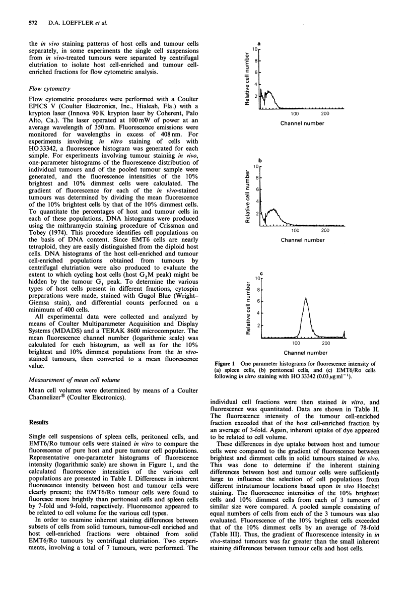
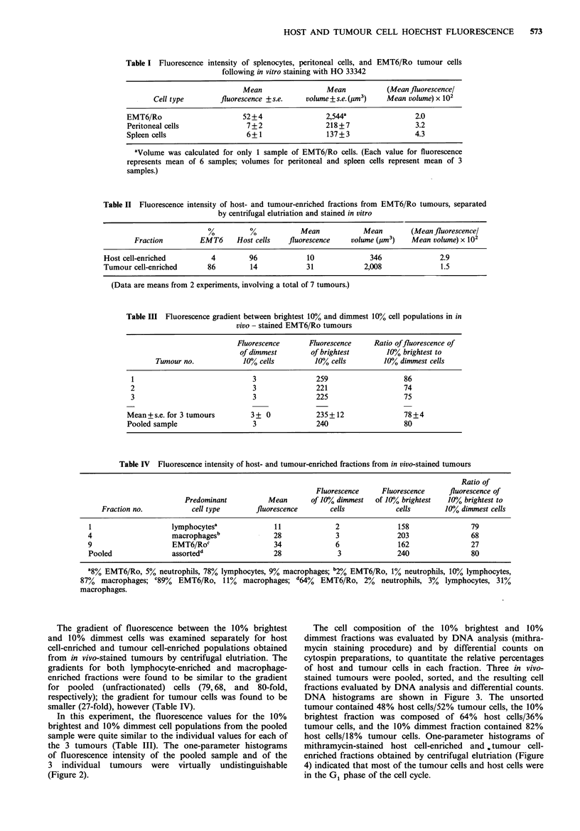
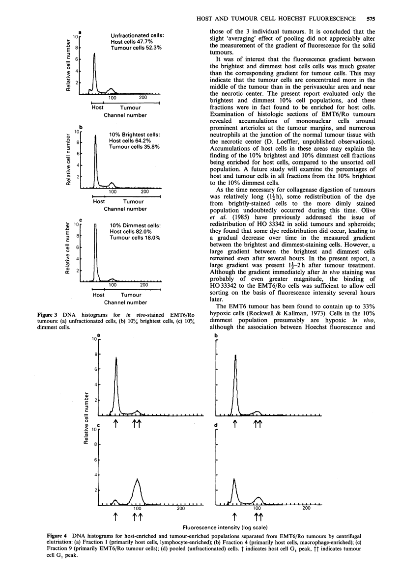
Abstract
Hoechst 33342 is a fluorescent dye used for cell selection from tumours based upon intratumour location. When the dye is administered i.v. to tumour-bearing animals, cellular fluorescence is directly related to the proximity of cells to blood vessels. The present study compared inherent Hoechst fluorescence between in vitro-stained EMT6/Ro (mouse mammary sarcoma) cells and host cells, to determine if these populations have different staining characteristics that may influence cell selection procedures. Tumour cell fluorescence exceeded host cell staining by 8-fold when pure cell populations (EMT6/Ro monolayer cells, mouse spleen and peritoneal cells) were compared, and 3-fold for tumour cell-enriched and host cell-enriched populations from solid tumours. Inherent uptake of HO 33342 appeared to be correlated with cell volume. These differences in inherent dye uptake between host and tumour cells were found to be minor in comparison to the fluorescence gradient between the 10% brightest and 10% dimmest (78-fold) cell populations from in vivo-stained tumours.
Full text
PDF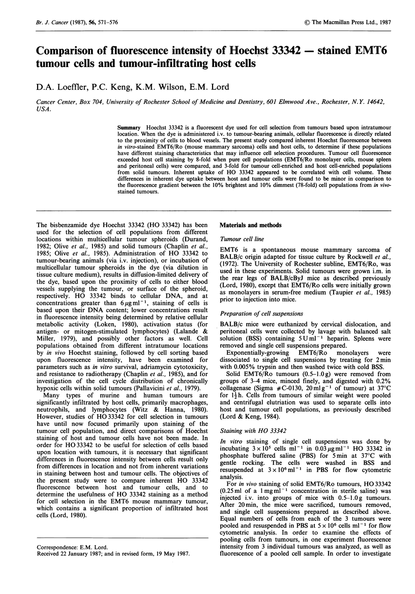
Selected References
These references are in PubMed. This may not be the complete list of references from this article.
- Chaplin D. J., Durand R. E., Olive P. L. Cell selection from a murine tumour using the fluorescent probe Hoechst 33342. Br J Cancer. 1985 Apr;51(4):569–572. doi: 10.1038/bjc.1985.79. [DOI] [PMC free article] [PubMed] [Google Scholar]
- Crissman H. A., Tobey R. A. Cell-cycle analysis in 20 minutes. Science. 1974 Jun 21;184(4143):1297–1298. doi: 10.1126/science.184.4143.1297. [DOI] [PubMed] [Google Scholar]
- Durand R. E. Use of Hoechst 33342 for cell selection from multicell systems. J Histochem Cytochem. 1982 Feb;30(2):117–122. doi: 10.1177/30.2.6174559. [DOI] [PubMed] [Google Scholar]
- Lalande M. E., Miller R. G. Fluorescence flow analysis of lymphocyte activation using Hoechst 33342 dye. J Histochem Cytochem. 1979 Jan;27(1):394–397. doi: 10.1177/27.1.86569. [DOI] [PubMed] [Google Scholar]
- Loken M. R. Separation of viable T and B lymphocytes using a cytochemical stain, Hoechst 33342. J Histochem Cytochem. 1980 Jan;28(1):36–39. doi: 10.1177/28.1.6153191. [DOI] [PubMed] [Google Scholar]
- Lord E. M. Comparison of in situ and peripheral host immunity to syngeneic tumours employing the multicellular spheroid model. Br J Cancer Suppl. 1980 Apr;4:123–127. [PMC free article] [PubMed] [Google Scholar]
- Lord E. M., Keng P. C. Methods for using centrifugal elutriation to separate malignant and lymphoid cell populations. J Immunol Methods. 1984 Mar 30;68(1-2):147–155. doi: 10.1016/0022-1759(84)90145-5. [DOI] [PubMed] [Google Scholar]
- Olive P. L., Chaplin D. J., Durand R. E. Pharmacokinetics, binding and distribution of Hoechst 33342 in spheroids and murine tumours. Br J Cancer. 1985 Nov;52(5):739–746. doi: 10.1038/bjc.1985.252. [DOI] [PMC free article] [PubMed] [Google Scholar]
- Pallavicini M. G., Lalande M. E., Miller R. G., Hill R. P. Cell cycle distribution of chronically hypoxic cells and determination of the clonogenic potential of cells accumulated in G2 + M phases after irradiation of a solid tumor in vivo. Cancer Res. 1979 Jun;39(6 Pt 1):1891–1897. [PubMed] [Google Scholar]
- Rockwell S. C., Kallman R. F., Fajardo L. F. Characteristics of a serially transplanted mouse mammary tumor and its tissue-culture-adapted derivative. J Natl Cancer Inst. 1972 Sep;49(3):735–749. [PubMed] [Google Scholar]
- Rockwell S., Kallman R. F. Cellular radiosensitivity and tumor radiation response in the EMT6 tumor cell system. Radiat Res. 1973 Feb;53(2):281–294. [PubMed] [Google Scholar]