Abstract
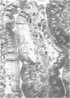
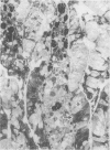
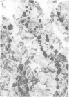
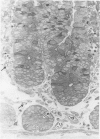
Previous studies have shown serious mucosal damage and destruction to be associated with intestinal ischaemia/reperfusion. As both destruction and concomitant regeneration can be observed together in this potentially lethal condition we have studied the development and sequence of events by evaluating morphological changes of the small intestine in an ischaemia/reperfusion model in anaesthetized rats. Forty-five minutes of ischaemia was followed by 4 hours of reperfusion. Tissue samples of the small intestine were examined by light microscopy in normal and semithin sections. Samples were collected at the end of ischaemia, at 10 min, and at 1, 2, 3 and 4 hours of reperfusion, respectively. Survival was assessed in a parallel group of anaesthetized rats. The morphological changes were described and they were analysed by a semi-quantitative method using five different markers of histological alteration. The mortality rate of a control survival group was 100%. Mucosal destruction at the end of ischaemia and during reperfusion was diffuse and steadily increased as a function of reperfusion time. At the same time the epithelium showed intensive regenerative growth which covered the denuded mucosal surface by the third hour of reperfusion. A secondary epithelial desquamation followed this process and was accompanied by heavy inflammatory cell infiltration. The infiltration may be the cause of the secondary epithelial injury.
Full text
PDF
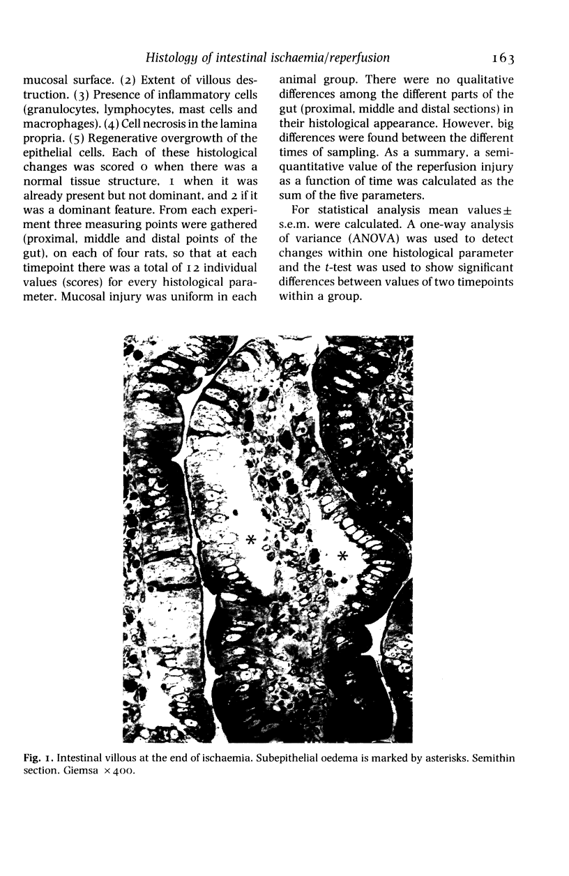
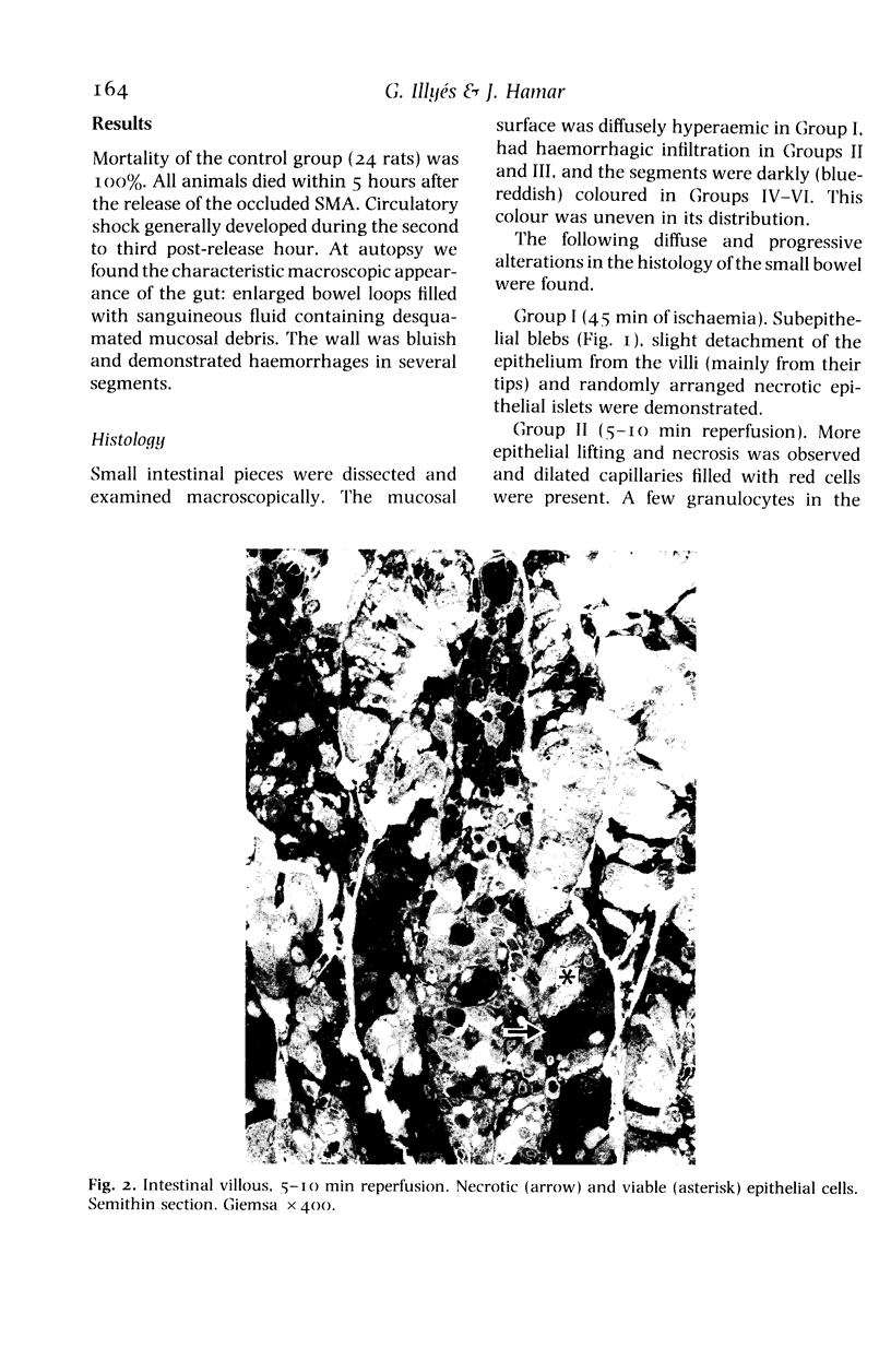
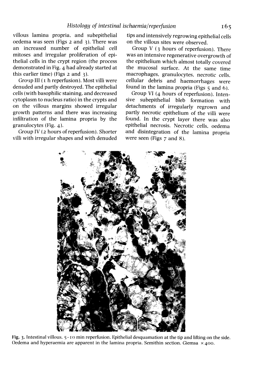
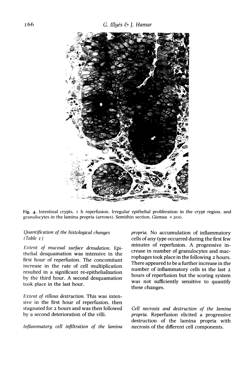
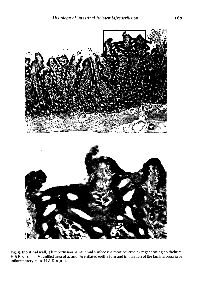
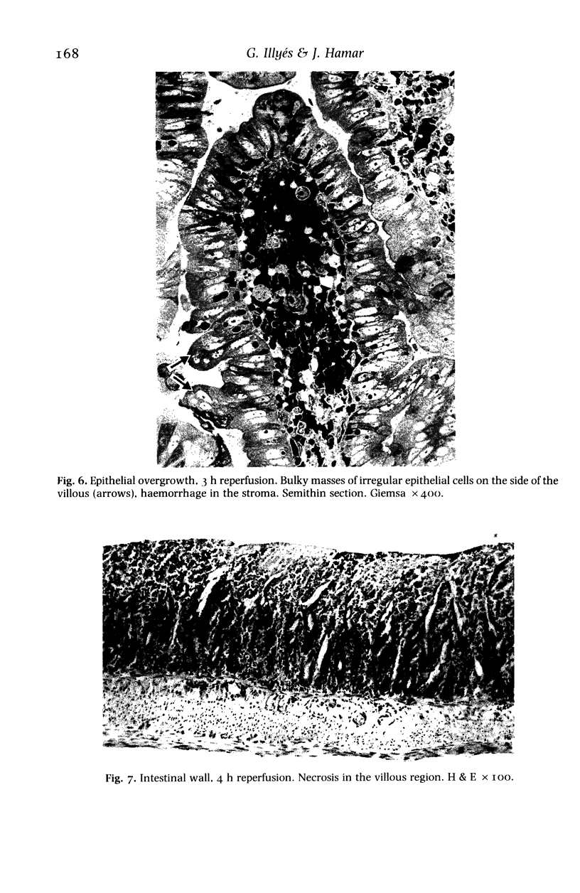
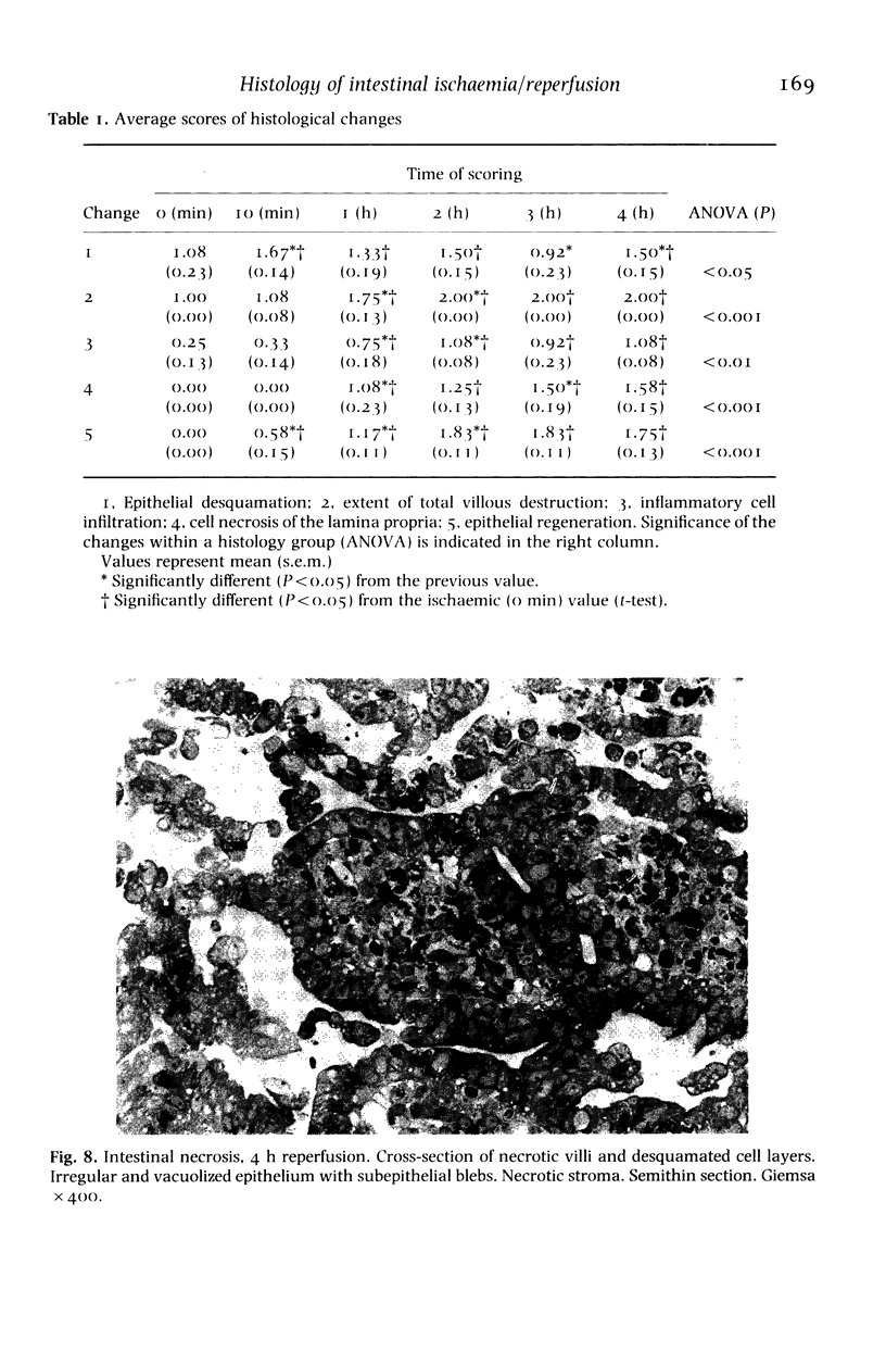
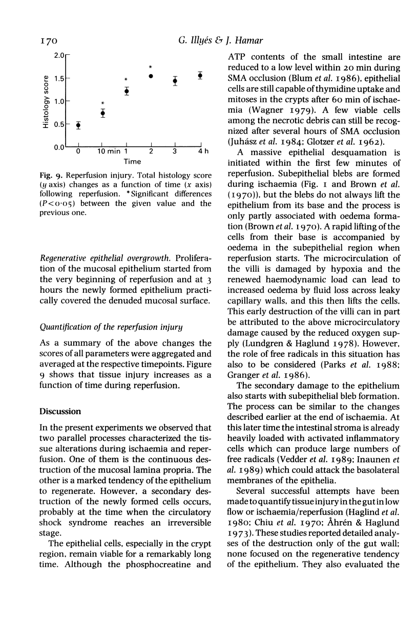
Images in this article
Selected References
These references are in PubMed. This may not be the complete list of references from this article.
- Andersson R., Pärsson H., Isaksson B., Norgren L. Acute intestinal ischemia. A 14-year retrospective investigation. Acta Chir Scand. 1984;150(3):217–221. [PubMed] [Google Scholar]
- Blum H., Summers J. J., Schnall M. D., Barlow C., Leigh J. S., Jr, Chance B., Buzby G. P. Acute intestinal ischemia studies by phosphorus nuclear magnetic resonance spectroscopy. Ann Surg. 1986 Jul;204(1):83–88. doi: 10.1097/00000658-198607000-00012. [DOI] [PMC free article] [PubMed] [Google Scholar]
- Brown R. A., Chiu C. J., Scott H. J., Gurd F. N. Ultrastructural changes in the canine ileal mucosal cell after mesenteric arterial occlusion. Arch Surg. 1970 Aug;101(2):290–297. doi: 10.1001/archsurg.1970.01340260194029. [DOI] [PubMed] [Google Scholar]
- Chiu C. J., McArdle A. H., Brown R., Scott H. J., Gurd F. N. Intestinal mucosal lesion in low-flow states. I. A morphological, hemodynamic, and metabolic reappraisal. Arch Surg. 1970 Oct;101(4):478–483. doi: 10.1001/archsurg.1970.01340280030009. [DOI] [PubMed] [Google Scholar]
- Granger D. N., Rutili G., McCord J. M. Superoxide radicals in feline intestinal ischemia. Gastroenterology. 1981 Jul;81(1):22–29. [PubMed] [Google Scholar]
- Inauen W., Suzuki M., Granger D. N. Mechanisms of cellular injury: potential sources of oxygen free radicals in ischemia/reperfusion. Microcirc Endothelium Lymphatics. 1989 Jun-Oct;5(3-5):143–155. [PubMed] [Google Scholar]
- McMINN R. M., MITCHELL J. E. The formation of villi following artificial lesions of the mucosa in the small intestine of the cat. J Anat. 1954 Jan;88(1):99–106. [PMC free article] [PubMed] [Google Scholar]


